 |
|
(Clicking
on the thumbnail images will launch a new window and a larger version
of the thumbnail.)
|
| Last updated: August 9, 2005 |
Hippotragus equinus
Order: Artiodactyla
Family: Bovidae
1) General Zoological Data
The genus Hippotragus contains three species: The extinct blue buck, the Sable antelope (H. niger) and the Roan antelope, Hippotragus equinus. Ill-defined subspecies of roan have been alluded to and their mitochondrial DNA was studied by Mathee & Robinson (1999). Information on the sable antelope is comprised in a separate chapter. The extinct "blue buck" ("Blaubock") found detailed description in the monograph by Erna Mohr (1967). These hippotragine species are distributed through Eastern Africa, but range to Ethiopia and South Africa. The roan antelope shares many features with the sable antelope but its horns are significantly shorter and its coat is somewhat lighter. Both sexes have horns and males are somewhat larger than females. Roan antelopes are found in more open environments than sable antelopes. Herds are dominated by a single male; their weight is between 150 and 300 kg (Nowak, 1999).
The population structure of roan and sable antelopes was studied with mitochondrial DNA control region analysis by Mathee & Robinson (1999). They concluded that the findings reflect the subspecific, locational origin of the roan populations, while in sable antelopes care should be taken during translocations of defined sable subpopulations. Wilson & Hirst (1977) presented a detailed monograph on ecology of South African Roan and Sable antelopes. Roan antelopes are not commonly seen in zoos according to Puschmann (1989).
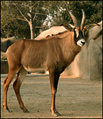 |
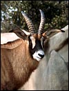
Roan antelope at the San Diego Zoo. |
Dittrich (1970) reported on the breeding of roan antelopes from the Hannover zoo. The length of gestation is from 268-280 days (Nowak, 1999). The single calf weighs 13-18 kg. The placenta weighed 1,900 g in the single specimen available.
3) Implantation
No details currently exist on early gestational stages in roan antelope placentation. Mossman (1987) described a mesometrial cord insertion of Hippotraginae and that both uterine horns are used for placentation. The uterus, however, is described a "duplex", with Y-shaped cervical canal.
4)
General Characterization of the Placenta
This is a polycotyledonary placenta with remarkable fusion of the large
number of large cotyledons. The cotyledons are much larger than those
of most other antelopes. The specimen available to me comes from a term
pregnancy with a live birth. It weighed 1,900 g and had 100 cotyledons,
although I admit, they were difficult to enumerate. The individual cotyledons
measured up to 10 cm in diameter but were flat (0.3 g) and were arranged
in four major rows. The umbilical cord was 25 cm long, had no twists and
inserted near the center. The placenta is similar in many ways to that
of the Sable antelope (Hippotragus niger) - click
here to see that chapter. A second specimen became available in August, 2005. It weighed 1,500 g, had four rows of 80 cotyledons, a number of them very small. The umbilical cord was 8 cm long.
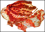 |
Gross appearance of the Roan antelope placenta with confluent cotyledons. |
The feto-maternal border is typically epithelio-chorial with very tall subchorionic trophoblast and cuboidal trophoblast covering the villi. There are numerous binucleate cells, presumably producing placental lactogen as in other ungulates. The villi are sparsely branched and have large fetal blood vessels. There is no pigment in the subchorionic trophoblast.
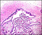 |
Chorionic membrane above, lined by cylindrical trophoblast below. |
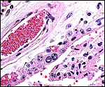 |
Surface of typical villus with cuboidal trophoblast and binucleate cells. The large vessel at left is a fetal villous vessel; below left the indentation of vessels into the trophoblast is evident. |
The umbilical cord contained 4 vessels and a large allantoic duct. It measured 25 cm in length, had no twists and was mostly the allantoic portion of the cord. The cord of the second specimen was only 8 cm long.
7) Uteroplacental circulation
I am not aware of any published reports.
8)
Extraplacental membranes
Numerous tiny, white amnionic foci of squamous metaplasia were present.
These are "pearls" of keratin within the amnion, just below
the flat epithelium of the amnion. One such pearl is shown below. The
allantoic epithelium was too poorly preserved for photography.
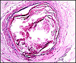 |
Amnion with squamous metaplasia, appearing grossly as a white "pearl". |
Lacking an implanted uterine placental specimen, nothing can be said about invasion of the uterus. It would be unusual, however, if in this ungulate endometrial infiltration occurs.
10)
Endometrium
I have not been able to observe this.
11)
Various features
None.
12)
Endocrinology
I am not aware of any publications that detail endocrine findings in roan
antelopes.
13)
Genetics
Roan antelopes have 60 acrocentric chromosomes that are essentially the
same as those of sable antelopes. Minor G-band and C-band differences
have been described to exist between these two species (Fordyce-Boyer
et al., 1995).
Hybrids have not been described. Mathee & Robinson (1999) examined
the mitochondrial DNA structure of different populations of roan and sable
antelopes (see above).
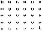 |
Karyotype of male Roan antelope. |
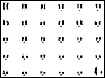 |
G-banded karyotype of male roan antelope. |
I am not aware of any studies.
15)
Pathological features
Malignant catarrhal fever has been reported to occur in a roan antelope
at Regent's Park by Gulland et al. (1989).Reid & Bridgen (1991) recovered
a herpes virus from cryopreserved roan antelope cells and established
its closer relationship to the alcelaphine herpesvirus than the ovine
virus (OHV-2). When injected into rabbits it produced malignant catarrh
and the authors concluded that the roan antelope needs to be considered
a potential carrier of this virus. Similarly, Flach et al. (2002) isolated
the virus from roan and other captive artiodactyl species. Kariuki et
al. (1989) identified Amblyomma and liver flukes at Ruma National
Park. The only pathological diagnosis made by Griner (1983) in his extensive
experience with zoo animal pathology is a case of aspiration pneumonia
whose ultimate cause could not be determined. Other data on blood parasites
may be found in Wilson & Hirst (1977).
16)
Physiologic data
Hematological studies to establish basic values were published by Pospisil
et al. (1984). Hattingh (1988) studied the biochemical responses to stress
in roan and impala. Citino et al. (2001) made extensive observations on
the effects of anesthetic agents in roan antelopes and prescribed a recommended
dose for immobilization. The gastric anatomy of eleven different Zambian
game animals, including the roan antelope, was described by Stafford & Stafford (1993).
17) Other resources
Fibroblast cell strains of several roan antelopes are available from CRES
at the San Diego Zoo and can be obtained by contacting Dr. Oliver Ryder
at oryder@ucsd.edu.
18)
Other remarks - What additional Information is needed?
No early embryological studies or early stages of implantation have been
described. Endocrine studies of gestations are needed.
Acknowledgement
The animal photograph in this chapter comes from the Zoological Society
of San Diego. I appreciate also very much the help of the pathologists
at the San Diego Zoo.
References
Citino, S.B., Bush, M., Grobler, D. and Lance, W.: Anaesthesia of roan
antelope (Hippotragus equinus) with a combination of A3080, medetomidine
and ketamine. J. S. Afr. Vet. Assoc. 72:29-32, 2001.
Dittrich, L.: Breeding the roan antelope Hippotragus equinus at Hannover zoo. Int. Zoo Yearb. 9:119, 1970.
Griner, L.A.: Pathology of Zoo Animals. Zoological Society of San Diego, San Diego, California, 1983.
Gulland, F.M., Reid, H.W., Buxton, D., Lewis, J.C., Kock, R.A. and Kirkwood, J.K.: Malignant catarrhal fever in a roan antelope (Hippotragus equinus) at Regent's Park. Vet. Rec. 124:42-43, 1989.
Flach, E.J., Reid, H., Pow, I. and Klemt, A.: Gamma herpesvirus carrier status of captive artiodactyls. Res. Vet. Sci. 73:93-99, 2002.
Fordyce-Boyer, R., Sanger, T., Loskutoff, N., Kumamoto, A.T., Johnston, L. and Armstrong, D.: Comparative cytogenetic study of the Roan and Sable antelope, Hippotragus equinus and Hippotragus niger. Applied Cytogenet. 21:189-191, 1995.
Hattingh, J.: Comparative quantitation of the physiological response to acute stress in impala and roan antelope. Comp. Biochem. Physiol. A 89:547-551, 1988.
Kariuki, D.P., Injairo, R., Boyce, W.L., Wellde, B.T. and Ngethe, S.: Parasite survey of eight wild animals in the Ruma National Park. Ann. Trop. Med. Parasitol. 83 Suppl.1:115-118, 1989.
Mathee, C.A. and Robinson, T.J.: Mitochondrial DNA population structure of roan and sable antelope: implications for the translocation and conservation of the species. Mol. Ecol. 8:227-238, 1999.
Mohr, E.: Der Blaubock. Hippotragus leucophaeus (Pallas, 1766). Eine Dokumentation Paul Parey, Hamburg, 1967.
Mossman, H.W.: Vertebrate Fetal Membranes. MacMillan, Houndmills, 1987.
Nowak, R.M.: Walker's Mammals of the World. 6th ed. The Johns Hopkins Press, Baltimore, 1999.
Pospisil, J., Kase, F., Vahala, J. and Mouchova, I.: Basic haematological values in antelopes - II. The Hippotraginae and the Tragelaphinae. Comp. Biochem. Physiol. A 78:799-807, 1984.
Puschmann, W.: Zootierhaltung. Vol. 2, Säugetiere. VEB Deutscher Landwirtschaftsverlag Berlin, 1989.
Reid, H.W. and Bridgen, A.: Recovery of a herpesvirus from a roan antelope (Hippotragus equinus) Vet. Microbiol. 28:269-278, 1991.
Stafford, K.J. and Stafford, Y.M.: The anatomy of the omasum of some Zambian game species. Anat. Histol. Embryol. 22:342-347, 1993.
Wilson, D.E. and Hirst, S.M.: Ecology and factors limiting Roan and Sable antelope populations in South Africa. Wildlife Monograph # 54 in, J. Wildl. Management 41:1-111, 1977.