The placenta is a neglected organ, considering how important its function is prior to birth. If this is true for human placentas, it is even more so true for animal placentas. They are not only difficult to obtain, but they are so varied in their appearance that many students find them difficult to examine. It is perhaps also for that reason that there is so little information on pathological features, of which there is a large number in human gestations. It stands to reason that similar abnormalities should occur in animal placentas. They might be more often identified, if we only understood the normality of these organs in the first place.
That is the reason for producing this "book". I have collected placentas from many different species, mostly at the San Diego Zoo, and I continue to be amazed at the variety of forms. To understand their evolutionary paths remains a major challenge for biologists also. Here I want to present the normal anatomy of animal placentas and add some features of gestation that appear relevant. Because there would be few buyers for such a book and because I am far from having completed it anyway, this will, therefore, be an ongoing production with new forms added continually. For that reason I have decided to write this for internet access. This is thus free to the interested student to inspect and copy. I would welcome it, however, if the material were being used for some publications, that credit be given to this "book". Furthermore, if there are readers who have placentas available and that are as yet unpublished, I would be interested to hear from them. Perhaps those placentas can be incorporated into this work. Also, if there are mistakes, additions or other criticisms, I would appreciate hearing from the reader. This is an easily corrected and updated publication. The reader should also appreciate that many placentas obtained in zoos are somewhat autolyzed; only the best ones were chosen for this summary. If better ones are available to anybody, please let me use them for updating.
I have prepared this writing originally in MS-Word. The text can easily be printed from the web. There is much literature attached but it is not a complete literature survey of the topic. This book will merely guide the interested reader to relevant sources.
I have subdivided the text into 18 headings and endeavor to make this as useful to the reader as possible. I thus welcome suggestions for improvement. If a reader submits a placenta, I would appreciate it then if the same format that is used here was followed.
There have been a few major texts on comparative placentation. I will list them here and make reference to them frequently throughout the text. These texts need to be consulted to get a complete overview of the placenta from different animal species. The difficulty for many pathologists with some of these texts, however, has been that they rely so heavily on drawings. Drawings are often more difficult to comprehend than histologic and macroscopic pictures. I endeavor here to provide both for an easier understanding of the various placentas. Because of the size of the individual image files, many are presented as "thumbnails"; clicking on them will open a new browser window with a larger version of the same image. (Note that this requires a browser--either Internet Explorer or Netscape--that is version 4 or newer.) If really better pictures were needed, I will be glad to supply them.
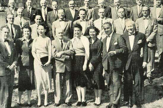
There
are a number of placental "luminaries" whom I have met in the
past at various meetings. Because they are so vital to this field, I have
placed two pictures of them next.

This
picture was taken at a regular "Gestation" Meeting in Princeton,
NJ, in 1958. There are some well-known placentologists or reproductive
physiologists in attendance. They are: Front row from the left:
Ed Dempsey, E. Ramsey, V. Wharton, Carl Hartman, E. Purcell, D. LaGuardia,
E. Amoroso, F. Fremont-Smith. Second row: G. Vosburgh, J. Davies,
S. Romney, C. Villee, F. Stone, L. Holm, E. Page, C. Hendricks.Third row:
C.S. Burwell, G. Bourne, P. Levine, S.R.M. Reynolds, J. Bienarz, K. Benirschke,
H. Mossman, C. Richter, W. Allen, W. Wimsatt. (From Gestation. Josiah
Macy Jr. Fndt. NY 1959.)

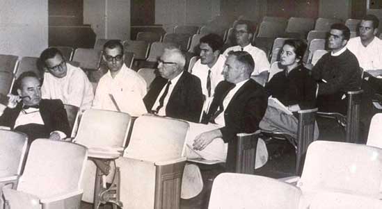
It
is only fitting to insert here a picture of Harlan Mossman, the greatest
comparative placentologist whose text is a "must". Here he is
in 1963 (fourth from the left) addressing Joseph Warkany (left) at a conference
held at Dartmouth Medical School, Hanover, NH. To the right of Mossman
is Vergil Ferm, then chairman of the department of anatomy at Dartmouth.
To the entire staff of the San Diego Zoo I am most grateful for your help. Most of the animal photographs are either the property of the Zoological Society of San Diego, or mine. Special thanks to Geoff Altshuler of Oklahoma City for his numerous suggestions. Most of all though I want to thank Dr. Mark Bogart for his support and Rand Uehara for making this such a fine internet presentation.
References
Amoroso, E.C.: Placentation. In, Marshall's Physiology of reproduction.
3rd. edition (ed. A.S. Parkes), Vol. 2, pp. 127-309, 1952. Longmans Green,
London.
Grosser, O.: Vergleichende Anatomie und Entwicklungsgeschichte der Eihäute und der Placenta. W. Braumüller, Vienna, 1909.
Grosser, O.: Frühentwicklung, Eihautbildung und Placentation des Menschen und der Säugetiere. J.F. Bergmann, München, 1927.
Ludwig, K.S.: Zur vergleichenden Histologie des Allantochorion. Rev. Suisse Zool. 75:819-831, 1968.
Mossman, H.W.: Vertebrate Fetal Membranes. Comparative Ontogeny and Morphology; Evolution; Phylogenetic Significance; Basic Functions; Research Opportunities. The MacMillan Press, Ltd. Houndmills, 1987.
Perry, J.S.: The mammalian fetal membranes. J. Reprod. Fert. 62:321-335, 1981.
Ramsey, E.M..: The Placenta of Laboratory Animals and Man. Holt, Rinehart and Winston, N.Y. 1975.
Ramsey, E.M.: The Placenta. Human and Animal. Praeger Publ., N.Y. 1982.
Soma, H.: Comparative Placentology. Modern Obstet. Gynecol. 123159, 1978, Nakayama Publ., Tokyo (in Japanese).
Starck,
D.: Vergleichende Anatomie und Evolution der Placenta. Verh. Anat. Gesellsch.
Zürich, 56. Versamml., Gustav Fischer, Jena, 1959.
Steven, D.H.: Comparative Placentation. Essays in Structure and Function.
Academic Press, London, 1975.
Strauss, F.: Probleme der Ovo-Implantation bei den Säugetieren. Z. Säugetierkunde 46:65-79, 1981.
Wimsatt, W.A.: Some aspects of the comparative anatomy of the mammalian placenta. Amer. J. Obstet. Gynecol. 84:1568-1594, 1962.
Addresse:
Kurt Benirschke, M.D.
8457 Prestwick Drive
La Jolla, CA 92037 USA
e-mail: kbenirsc@ucsd.edu

