
The study of the placenta is often difficult for pathologists. The terminology of the placental structures alone presents unusual terms, and its development and contribution to fetal life are difficult topics. They are not often taught and can best be understood by approaching the topic from a comparative understanding of the placental development. Mossman (1937) has aptly stated that " the placenta is probably more variable in structure than any other mammalian organ. And Kaufmann (1983) has similarly stated that there is much more variation in the structure of the placenta than is true of any other organ. How do placentas develop, how does their development influence fetal growth? Furthermore, are all placentas similar in their function? What is known of their transport and endocrine function? These and similar questions are best approached when one understands the evolution of placentas from their ultimate precursors of development from its egg ancestor, as the mammalian origin of the placenta finds its ancestry in the egg development of birds.
To the pathologist
This book is primarily directed to pathologists. Its content will be of marginal interest to placentologists who know many aspects much better than they can here be explained. Placentologists are more concerned with understanding the fine structure of the trophoblast/fetal-maternal interchange. That is not the reason for writing these pages. The reason for presenting these data and the many pictures I have added is to enable the pathologist, confronted with an unusual placenta, to undertake a proper examination of this strange structure. What are the features worth recording and what should be sectioned? In essence, is this a normal organ or what might be considered to be abnormal. Moreover, when there are problems with the neonate, is the placenta perhaps contributory to the problem? This is often true for human gestations and one would think that it holds for animal pregnancies as well.
While the pathologist is used to using formalin fixation for tissue processing, embryos and placentas are much better fixed in Bouin's solution as they are so delicate and water-laden. So, for optimal fixation, this is a better way to go for routine slides. That is not true for the electronmicroscopist, of course, and modern biologic questions are often best answered by the freezing of bits of placental tissue. Then, at some future date perhaps, it will be possible to get DNA or RNA for study.
We would like to know many aspects of the placenta that are not usually considered in texts, such as: the number of cotyledons, their sizes and distribution; how long is the umbilical cord and how many vessels does it contain? Are there ducts that lead to an allantoic sac? Is there inflammation (chorioamnionitis or abscesses)? And is the maternal organism infiltrated by trophoblast, something that can best be established when the placenta is still attached to the uterus. And are there infarcts or other areas of necrosis? Some of these features are essential for our understanding the cause of fetal demise, neonatal infection, and for an understanding of the genesis of fetal anomalies or premature birth. It must also be pointed out that the histologic details of placentas are further complicated by the fact that autolysis is often significant. For zoo pathologists, the placenta is frequently submitted many hours after it was passed by the dam. It may lie in the heat, be exposed to water and thus change its appearance considerably. It may then become difficult to compare the histology with published accounts that are generally based on optimal preservation. This is difficult to avoid, but one must take this into consideration when a decision is to be made as to whether the changes are artifact of preservation or represent true pathologic abnormalities.
General consideration
Birds
and other non-mammalian species possess membranes in their embryonic development
within the egg that, ultimately, eventuated in the placental structures,
as we now know them in mammals. Probably the first placental structures
can be inferred from the fossil evidence of Ichthyosaurus, more than 170
million years ago. Furthermore, a variety of placenta-like structures
are found in squamate (scale-possessing) reptiles. And other examples
can be cited for the earliest development of membranes that eventuated
into the mammalian placenta. Blackburn (1998) provided a useful overview
of these.
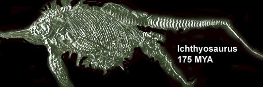
This photograph was shamelessly borrowed from Thenius, a specimen found
in Java.
The photograph of a Mesozoic reptilian fossil depicts the delivery of embryos that clearly must have had an intrauterine development and were liveborn. Whether it represents one of the first stages in placental evolution or whether the placenta of mammals was "invented" several times in the evolutionary history of mammals is unknown. But, from the analysis of placentas from a variety of mammals, it must be concluded that some of the diverse forms of placentas had separate origins. For instance, clearly the invasive simian placentas derived from prosimian ancestors with noninvasive placentas. The principal difference of simian and prosimian placentas is that, in the latter, the relationship to the maternal organism was appositional, in the former, invasive placentation evolved. Rather than reviewing the entire possible history from egg membranes to mammalian placentation, the reader is referred to Blackburn (1998) and Mossman (1987). Another excellent source is the book by Steven (1975) that presents in easily understood language the salient developmental features. Moreover, it is accompanied by excellent diagrams. Here we will discuss only the mammalian placenta.
This important change in placental development mentioned above, to become an invasive structure that infiltrates the maternal uterus, must have had major cosequences; for instance, the feature of non-rejection by the maternal organism of a genetically "foreign" genotype had to be accommodated. In a comprehensive essay, Haig (1993) reviewed this important topic that has not yet a final resolution. Did this invasion of the maternal organism happen only once? Probably not, as rodents for instance also have invasive placentas. Moreover, it is likely that the Afro-Asiatic simians had a separate origin from prosimians than that which occurred in South American platyrrhine primate species. And, these different origins of primates perhaps then explain not only their generally diverse phenotypes (e.g. platyrrhine vs. catarrhine, non-prehensile vs. prehensile, etc.) but also they may be the basis for the marked differences in placental structure and function of these simians. Other differences exist in the hormonal performance of the placenta, differences in routes and types of exchange performance, and so on.
Many other questions arise when patterns of placentation are questioned. Most recently, it has been learned that genetic "imprinting" of placental (trophoblastic) cells differs from that of the imprinting on the genome of embryonic cells. Paternal gene expression predominates in the placenta, while maternal expression dominates in embryo development. The presence or absence of certain enzymes, the presence of some structures (e.g. allantoic sac) and other aspects vary substantially in different animals' placentas, presumably signaling some totally different intrauterine physiologic behaviors of water exchange and its conservation. The regulation of the length of the umbilical cord varies widely, and so on. Thus, knowing the structure of one placenta (e.g. the sheep) is not always helpful for an understanding of the organ in another species, say the horse.
These considerations are the reason for writing this book, to enable to interested examiner of placentas to get a better understanding of the normality or the existence of possible abnormalities of a placenta from an animal with which he may be confronted for the first time. Ultimately, one can only hope that a more comprehensive understanding of this interesting organ evolves. Many questions will still confront the interested student.
This documentation is meant to be a guide for the study of any given placenta that comes your way for study. It should also provide a better understanding of the special nomenclature that has evolved to denominate the "fetal membranes", as the placenta is often referred to. This term alone, often also used to merely describe the "sac" of membranes (the so-called chorion laeve of human placentas), in contrast to the chorion frondosum (the actual placental disc), has presented problems. This is especially so for the complex rodent placenta, as its apparent homologue of "free" membranes has major exchange function and differs substantially from the "membranes" of primates. The references that are appended give access to the major contributions of students with a principal interest in comparative placentation.
Classification
Placentas are variously classified. It can be done by their macroscopic appearance, as for instance as being discoid (human), cotyledonary (ruminant), diffuse (whales), and zonary (carnivores). Another way is according to the intimacy of fetal-maternal contact. For the latter distinction, the "Grosser classification" is conventionally employed. Grosser (1927) distinguished the epitheliochorial, syndesmochorial, endotheliochorial, and hemochorial types. These denominate the layers between fetal trophoblast and maternal endometrial surface. But, some placentas (ruminants) may have some combination of these types of "invasion" and strict classification has often demanded electronmicroscopy for verification. In addition, King and Mossman (1974) have distinguished in primate placentas hemo-monochorial, -dichorial, and -trichorial implantation, as their electronmicroscopic studies showed that, frequently, more than one layer of trophoblastic cells makes up the villous surface. But this is less frequently an issue in understanding the placental barrier.
Then
there is the aspect of the development of the structure of villi that
has demanded separate classification such as being of the folded (pig),
lamellar (carnivores), trabecular (some primates), labyrinthine (rodents,
lagomorphs, insectivores) and villous types (human). Kaufmann & Burton
(1994) provided excellent diagrams to illustrate these features. Kaufmann
(1983) also showed a series of useful diagrams and a table that is here
placed for the purpose of orientation.
|
Order
|
Species
|
Placental
form
|
Maternal-fetal
Interdigitation
|
Maternal-fetal
separating membrane
|
| Insectivora |
Europ.
Mole |
Discoid Diffuse |
Labyrinth Labyrinth |
Hemochorial Hemochorial |
| Chiroptera (Bats) | Discoid | Labyrinth | Endotheliochorial Occas. Hemochorial |
|
| Primates | Tupaia | Bidiscoid | Labyrinth | Endotheliochorial |
| Galago | Diffuse | Folded | Epitheliochorial | |
| Rhesus | Bidiscoid | Villi | Hemochorial | |
| Ape, Human | Discoid | Villi | Hemochorial | |
| Lagomorpha | Rabbit, hare | Discoid | Labyrinth | Hemochorial |
| Rodentia | Guinea pig | Discoid | Labyrinth | Hemomonochorial |
| Rat, mouse | Discoid | Labyrinth | Hemotrichorial | |
| Beaver | Discoid | Labyrinth | Hemodichorial | |
| Carnivora | Dog, cat | Zonary | Labyrinth | Endotheliochorial |
| Sirenia | Manatee | Zonary | Labyrinth | Hemochorial |
| Artiodactyla |
Pig |
Diffuse Cotyledonary Cotyledonary |
Folded Villi Villi |
Epitheliochorial Epithelio- & Syndesmochorial Epitheliochorial |
| Perissodactyla | Horse | Diffuse, spec. | Villi, "cups" | Epitheliochorial |
| Cetacea | Whale, dolphin | Diffuse | Villi | Epitheliochorial |
This table indicates, incompletely though, the amazing variety of placental forms and types.
Evolution
The origin and evolution of the placenta are complex topics. Torpin (1971) emphasized in his account of the evolutionary relations between mammals that similarity of placentation must indicate evolutionary relationship between mammals. In other words, very unlike placentation of two given species would rule against their close relation. A good example would be the peculiar peripheral aspects of the villous structures seen in the Xenarthra (Edentata). The anteater and armadillos have a very specific and otherwise exceptional villous trabecular network of primitive villi that must relate to their evolutionary proximity. Such information may also be useful for future attempts at interspecies embryo transfers, as was discussed by Hradecky et al. (1987) with the examples of eland, giraffe, okapi exchanges.
There are numerous accounts of philosophical, anatomic and evolutionary kind that discuss the placental evolution. For instance, Mossman (1987), in his extensive classic on comparative placentation, devotes much time to explain the possible derivation of various placental structures. Ramsey (1989) traces the "History" of the human placenta and Soma (1978) provided a review in Japanese with many color illustrations of unusual species. Kaufmann & Burton (1994) have also published an excellent review that is replete with literature citation. It is primarily directed to the understanding of the human placenta and, i.a., it traces the origin of "chorion" to Aristotle and "placenta" (cake) to Realdo Columbus.
References
Amoroso, E.C.: Placentation. In, Marshall's Physiology of reproduction.
3rd. edition (ed. A.S. Parkes), Vol. 2, pp. 127-309, 1952. Longmans Green,
London.
Benirschke, K. and Kaufmann, P.: The Pathology of the Human Placenta, 4th ed. Springer-Verlag, NY, 2000.
Blackburn, D.G.: Placenta and placental analogs in reptiles and amphibians. In, Encyclopedia of Reproduction. E. Knobil and J.D. Neill, eds. 1998. Vol. 3. pp.840-847.
Enders, A.C.: A comparative study of the fine structure of the trophoblast in several hemochorial placentas. Amer. J. Anat. 116:29-68. 1965.
Grosser, O.: Vergleichende Anatomie und Entwicklungsgeschichte der Eihäute und der Placenta. W. Braumüller, Vienna, 1909.
Grosser, O.: Frühentwicklung, Eihautbildung und Placentation des Menschen und der Säugetiere. J.F. Bergmann, München, 1927.
Haig, D.: Genetic conflicts in human pregnancy. Quart. Rev. Biol. 68:495-532, 1993.
Hradecky, P., Benirschke, K. and Stott, G.G.: Implications of the placental structure compatibility for interspecies embryo transfer. Theriogenology 28:737-746, 1987.
Kaufmann, P.: Vergleichend-anatomische und funktionelle Aspekte des Placenta-Baues, Funkt. Biol. Med. 2:71-79, 1983.
Kaufmann, P. and Burton, G.: Anatomy and Genesis of the Placenta. Chapter 8 in, The Physiology of Reproduction, 2nd ed. E. Knobil and J.D. Neill, eds. Raven Press, NY, 1994. PP 441-484.
King, B.F. and Mossman, H.W.: The fetal membranes and unusual giant cell placenta of the jerboa (Jaculus) and jumping mouse (Zapus). Amer. J. Anat. 140:405-432, 1974
Ludwig, K.S.: Zur vergleichenden Histologie des Allantochorion. Rev. Suisse Zool. 75:819-831, 1968.
Mossman, H.W.: Comparative morphogenesis of the fetal membranes and accessory uterine structures. Carnegie Inst. Washington Publ. 479. Contrib. Embryol. 26:129-246, 1937.
Mossman, H.W.: Vertebrate Fetal Membranes. MacMillan, Houndmills, 1987.
Perry,
J.S.: The mammalian fetal membranes. J. Reprod. Fertil. 62:321-335, 1981.
Ramsey, E. M.: The Placenta of Laboratory Animals and Man. Holt, Rinehart
and Winston, N.Y., 1975.
Ramsey, E.M.: The Placenta. Human and Animal. Praeger Publishers, NY, 1982.
Ramsey,
E.M.: History. In, Biology of the Uterus. 2nd ed. R.M. Wynn, ed. Plenum
Publishing Corp. 1989. Pp. 1-17.
Soma, H.: Comparative placentology. Modern Obstetr. Gynecol. Series, Chapter
VII, Nakayama Publ., Tokyo. 1978, pp. 123-159.
Torpin, R.: Placentation and mammalian phylogeny. Obstetr. Gynecol. 37:942-948, 1971.
Spatz, W.B.: Nabelschnur-Längen bei Insektivoren und Primaten. Z. Säugetierk. 33:226-239, 1968.
Starck,
D.: Lehrbuch der Speziellen Zoologie. Vol. II, Part 5/2. Gustav Fischer
Verlag, Jena, 1995.
Steven, D.H.: Comparative Placentation. Essays in Structure and Function.
Academic Press, London, 1975.
Strauss, F.: Probleme der Ovo-Implantation bei den Säugetieren. Z. Säugetierkunde 46:65-79, 1981.
Wimsatt, W.A.: Some aspects of the comparative anatomy of the mammalian placenta. Amer. J. Obstet. Gynecol. 84:1568-1594, 1962.