(Clicking on the thumbnail images below will launch a new window and a larger version of the thumbnail.)


Zebra Species
Equus grevyi

Order: Perissodactyla
Family: Equidae

1) General Zoological Data

There are basically three species of zebra: The largest, and the most Northern African species, is Grevy's zebra (Equus grevyi). It is classified as being endangered. The most numerous zebra species is the Planes zebra with its several subspecies (Grant, Burchell, Damara - Equus quagga boehmi, etc.). The most Southern zebra species is the Mountain zebra (Equus zebra zebra), with Mrs. Hartmann's mountain zebra (Equus zebra hartmannae) a subspecies. Nowak (1999) provided an extensive consideration of the evolution of equidae, from their origin in North America, their ultimate increase in size and then their dispersion over Eurasia and Africa. So far as is known, their placental development and structure are essentially similar and also close to those of the better-studied horses.

Adult Grevy's zebras weigh around 400 kg, and newborns are around 40 kg. Gestation lasts from 358-438 days (average 409 days) according to Nowak. Their longevity is over 28 years.

Mountain zebras weigh around 300 kg, with neonates weighing 25 kg. Both subspecies are endangered, but Hartmann's mountain zebras may be seen in numerous zoological parks. Their longevity is over 29 years.

Planes' zebras have many more different phenotypes. This is most apparent in their differing striping patterns. They are probably conspecific with the now extinct quagga (Equus quagga) and are the most widely exhibited in zoos. Their weights are between 175 and 385 kg, neonates are 32 kg and are born after a gestational period of 360 to 396 days. Their longevity is as much as 40 years.

Most zebras produce a single young, but twins of opposite sex have been observed. We saw the placenta of such a pair of Hartmann's mountain zebra neonates, with one of the twins surviving. Twins and their placentas were significantly smaller than singletons and their placentas.

   
  Adult Equus grevyi at San Diego Zoo.
     
  Grevy's zebras at San Diego Wild Animal Park.
     
  Hartmann's mountain zebra in San Diego.
     
  Note the typical "double chin" ("Wamme") of the mountain zebra, parent of the "zebronkey" in the background. (Manila zoo).
     
  Burchell zebra in San Diego.


2) General Gestational Data

The gestational length and some other pregnancy data are summarized above. Generally speaking, the pregnancies of zebras last longer than that of the horse and twinning is as uncommonly successful as it is in the domestic horse. Whether monozygotic twinning occurs is unknown.

3) Implantation

Early stages of zebra implantation have not been described but they are well known from horse gestations (Enders & Liu, 1991; see also review by Wooding et al., 2001). I can only presume that great similarities exist in the implantation of animals that capably hybridize. The equine blastocyst is moved back and forth for the first two weeks between the two uterine horns by uterine contractions (Ginther, 1983). It lodges on day 16 for implantation. Allantochorionic villi become first evident on day 50. Enders & Liu (1991) further described the orientation and the attachment of the blastocyst to the endometrium. In superb electronmicrographs, they showed the interdigitation of trophoblastic microvilli with those of the endometrial epithelium, and the formation of the "girdle" with the subsequent endometrial cup development. There developed an extensive endometrial lymphatic system at this location that had already been alluded to by Amoroso (1952). The girdle was readily identified grossly as an opaque band. Unfortunately, no similar stages are known of zebra gestations. The cup and girdle disintegrate after 100 days of gestation and a diffuse epithelio-chorial placenta develops.

Near-term Hartmann's mountain zebra and placenta. Note spiraling of cord.
   
  Implanted immature placenta of Grevy's zebra. Note endometrial glands (purple) and the thin endometrial septa that separate villous lobules.
     
  Higher magnification of implantation site. The fetal villi are within the white spaces of endometrial glands.
     
  Surface of zebra placenta with villous ramifications and interspersed maternal tissue. The chorionic membrane is on top. Note the space in the top center with cylindrical trophoblast and questionable red blood cell mass - the probable origin of the ensuing brown pigment collection.
     
  4) General Characterization of the Placenta

The placenta of all equidae is similar. It is a thin organ whose external surface is diffusely covered by villi. The thickness is actually difficult to measure precisely; it is maximally 0.5 cm, usually thinner. The outside has a velvety-red appearance. It is an epithelio-chorial placenta. There is a large allantoic sac which normally contains hippomanes and which is connected to the fetal bladder by the allantoic duct of the umbilical cord. The umbilical cord is relatively long and, in horses, entangling of excessively long cords has occasionally caused abortion. I have had the opportunity of studying several zebra placentas and pregnant uteri of all three species that are summarized here.

The uterus of a nearly full term pregnant female Grevy's zebra was the most recently available specimen. The mare had died from volvulus, a serious condition of equidae. The female fetus weighed 15,750 g and had a crown-rump length of 80 cm. The placenta weighed 2,980 g and was uniformly thin and partially detached from the uterus. The fetus lay in the left horn, but the allantoic sac extended into the right uterine horn.

The term, delivered placenta of the Damara zebra (Equus quagga antiquorum) shown below (blue background) weighed 2,350 g. It is so thin that it is nearly translucent.

The placenta of the male stillborn Hartmann zebra shown above did not have a cervical star. The stillborn fetus weighed 32.5 kg and had an 82 cm CR length.

List of placental weights of different species of zebra:

   
Grevy's zebra Term 1,580 g
  Grevy's zebra Term 1,560 g
  Burchell's zebra Term 1,200 g
  Damara zebra Term 2,980 g
  Damara zebra Term 1,550 g +750 g membranes
  Hartmann's mountain zebra Term 1,400 g
  Hartmann's mountain zebra Term 1,700 g
  Hartmann's mountain zebra Term 2.350 g
       
       
  Grevy's zebra uterus with foal. The right uterine horn is lying on top.
     
  Near-term Grevy's zebra uterus with foal.
     
  Placenta of near-term Grevy's zebra.
     
  Placenta of Damara zebra. Hippomanes at arrows.
     
  Chorionic plate of delivered zebra placenta with villi below. Note the cylindrical epithelium beneath the vascularized chorion.
     
  This picture exhibits a chorionic surface of placenta with a much more cylindrical trophoblast between the villous stems and accumulation of deep brown pigment.
     
  This picture also exhibits a chorionic surface of placenta with a much more cylindrical trophoblast between the villous stems and accumulation of deep brown pigment.
     
  Villous ramification of zebra placenta. A cuboidal epithelium covers the villi which are dominated by fetal capillaries. The endometrial surface is single-layered and cuboidal.
     
  Higher magnification of the villi of a delivered zebra placenta. The fetal capillaries dominate the villous structure and are located directly beneath the single-layered trophoblast.


5) Details of fetal/maternal barrier

In general, zebras have an epithelio-chorial relationship to the uterus. The single-layered trophoblast apposes closely the maternal endometrial epithelium with which it interdigitates by microvilli. Only at the site of the "girdle", with the development of the endometrial cups, is there trophoblastic invasion of the endometrium. This occurs by specialized, binucleated trophoblast cells with endometrial epithelial destruction ensuing. An intense inflammatory cell reaction subsequently destroys the invading trophoblastic cells commencing early and terminating around day 100 in the horse. It is presumed, but unknown, that the zebras have similar features in early gestation, especially since hybridization is possible.

6) Umbilical cord

The umbilical cord of the Grevy's zebra placenta that is shown above within its placenta was 45 cm long and minimally twisted. It contained three large blood vessels, two arteries and a vein, as is typical of equidae. The amnionic surface was finely granular due to areas of squamous metaplasia. This cord was not spiraled at all. It had a large allantoic duct and numerous small blood vessels surrounded the duct in the allantoic portion of the cord (see Whitwell, 1975 for details in horse cords).
The cord of the stillborn Hartmann zebra had 4 large blood vessels, heavily spiraled and possibly the cause of fetal demise as it is considerably longer than other zebra species' cords as well.

The frayed umbilical cord of the delivered Damara zebra placenta shown above measured 58 cm in length and was 5 cm in diameter. In addition, it had significant spiraling, as is occasionally seen in horses.

In addition to these two cases, I have examined the following additional zebra placental umbilical cords:

List of umbilical cord lengths of different species of zebra

   
Grevy's zebra Term 68 cm
  Grevy's zebra Term 68 cm
  Burchell's zebra Term 62 cm
  Damara zebra Term 58 cm
  Damara zebra Term 65 cm allant.+17 cm amn.portion
  Hartmann's mountain zebra Term 68 cm
  Hartmann's mountain zebra Term 55 cm
  Hartmann's mountain zebra Term 20 cm (?complete)
  Hartmann's mountain zebra Term Twins 60 cm each
  Hartmann's mountain zebra Term stillborn 84 cmx2.5 cm


The length of the umbilical cord has been studied in a large sample of thoroughbred horses (Whitwell, 1975). It varies from 36 to 83 cm (55 cm mean), with complications often ensuing from excessively long and, less often, short cords. Horses also have two arteries, one vein and an allantoic duct that may become constricted by fetal motions. The long cord allows the fetus to reside in the uterine horn in which the placenta is not implanted because of the large quantity of amnionic fluid. As a result of tactic stimulation, the fetus moves actively and, when it has a long cord, it may strangulate.

7) Uteroplacental circulation

I know of no detailed descriptions in zebras.

8) Extraplacental membranes

A 5 x 3 x 0.5 cm flattened piece of dark brown hippomanes was present in the allantoic sac of the Grevy's zebra placenta shown above within its uterus. The amnionic sac contained recently discharged flakes of meconium. Likewise, the Damara zebra placenta had substantial hippomanes associated with the allantois. An additional Damara zebra placenta contained in addition to hippomanes, the remains of a shed fetal hoof.

   
  Photograph of the undersurface of the chorionic plate.
     
  At left is the amnionic membrane, at left is the vascularized allantoic membrane with its cylindrical epithelium.


9) Trophoblast external to barrier

There is no trophoblastic infiltration of the endometrium in equidae, with the exception of the trophoblastic cells in the endometrial cups of the girdle. While this has been studied extensively in horses, no direct information on zebras can be found in the literature.

10) Endometrium

The endometrium of horses has been studied in detail by biopsies, including during early pregnancy (Keenan et al., 1987; others). The surface has complex microvillous extensions with which the trophoblast interacts (Enders & Liu, 1991). True decidua is not formed, but extensive alterations occur at the site of the girdle, with ultimate sloughing and necrosis by what appears to be an immunologic rejection of the allogeneic trophoblast (Enders et al., 2000). Large lymphatic sinuses develop in the endometrial cup regions (Amoroso, 1952) through which the eCG reaches the maternal vascular system. The FSH- and LH-like eCG secretions take place from the binucleated trophoblastic girdle cells that invade the endometrium and partially phagocytoze it.

11) Various features

The fetal gonads of all equids are characteristically large and stimulated (Mossman & Duke, 1973). There is massive interstitial cell development in testes and yellow pigmentation of these cells is striking. Similarly, the ovarian stroma has a massive, pigmented, interstitial cell development; the oocytes are localized in a thin rim at the ovarian periphery. The precise origin of the stimulus for this development is unknown (see below).

   
  Uterus and large ovaries of the Grevy's zebra fetus
     
  Cross-sectioned ovary of Grevy's zebra fetal ovary.
     
  Ovarian cortex of the ovary of the Grevy's zebra foal. At arrows are a few oocytes within the fibrous cortex; the majority of the ovary is replaced by pigmented interstitial cells.
     
  Higher magnification of pigmented interstitial cells in the neonatal ovary.
     
  The fetal testis has a similar degree of interstitial cell stimulation. A germ cell is indicated by the arrow.
     
  In very young fetuses, the interstitial cell stimulation is extensive, but the pigmentation is less apparent.
     
  12) Endocrinology

A detailed study of fecal estrogen and progesterone secretion in Grevy's zebra was undertaken by Asa et al. (2001) as well as Kirkpatrick et al. (1990) Asa et al. also studied the urinary gonadotropin (eCG) excretion. Progesterone levels were found to be relatively low when compared with those of numerous other species. Urinary estrone levels became elevated and were then diagnostic of pregnancy at about 8 months before the end of pregnancy (Czekala et al., 1990). In horses, it showed a rise in serum levels as early as the 40th day of gestation. Higher levels were found in horses than in Hartmann's zebras. Asa et al. (2001) found eCG (equine chorionic gonadotropin) to be present after 35-40 days, as is the case in the domestic horse. It completely disappeared at 195 days of their 425 day gestation. The steroid profiles were similar to those of the horse.
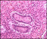
Horses 
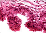
and, presumably all equidae, have special sites of eCG secretion in their fetally-derived "endometrial cups". These unique structures have been studied by numerous investigators, both structurally and functionally, but they have not been studied in zebras, and their remains are not recognizable in term placentas. In horses, eCG secretion from these cups begins on day 32; some time after day 100 the cups are destroyed by an intense maternal lymphocyte reaction (reviewed in detail by Wooding et al., 2001). While it is easy to speculate that this hormone may also be the cause of the fetal gonadal stimulation, its rapid decline after day 100 in maternal serum argues against this possibility, as the gonads remain large and apparently stimulated until term. Nevertheless, the unique fetal gonadal development and the uniqueness of endometrial cups suggest a relationship.
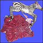
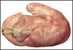
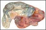
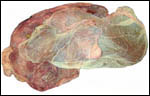
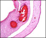
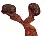
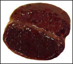
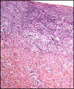
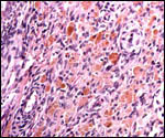
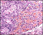
As is usual for all equidae, the fetal gonads of my Grevy's zebra gestation were much enlarged and dark brown. They were autolyzed and somewhat diffluent. They weighed 42 and 39 g.

13) Genetics

The chromosome number of Grevy's zebra is 2n=46; the Planes' zebras with all their subspecies have 2n=44, and the Mountain zebras have 2n=32 (Benirschke & Malouf, 1967). Numerous other cytogenetic studies on the equidae have since appeared, e.g. Gadi & Ryder (1983) who described the distribution of the nucleolus-organizing regions.

A wide variety of zebra hybrids have been recorded (Gray, 1972). This includes hybrids among zebra species, as well as with other equidae. Depending on their chromosome number, most are sterile offspring (Benirschke et al., 1964; 1967).

Numerous genetic studies, other than those of the cytogenetic descriptions, have been done in equidae. For instance, Ryder & Hansen (1979) studied (G+C)-rich satellites in horses in order to investigate the C-banded regions of chromosomes. Microsatellite loci of the SINE families were studied by Gallagher et al. (1999). The mitochondrial DNA evolution of the genus Equus was investigated by George & Ryder (1986). That study showed that zebra species diverged most recently from the equid line.
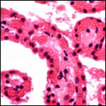
14) Immunology

The most remarkable aspect of an immunologic interaction between fetus and mare occurs at the implantation site of the chorionic girdle. Here, the binucleated trophoblast invades the endometrium, partially destroys it, and thus elicits an immunological maternal response that ultimately leads to the destruction of the invading trophoblast with cessation of its eCG secretion. This process appears to be genotypically controlled, as horse-into-donkey embryo transplants survive, while those of donkey-into-horse are usually rejected (Enders et al., 1996). This failed pregnancy of the latter transfers (abortion) appears to be due to the absence of an appropriate endometrial cup development In part this occurs because of a reduced degree of trophoblastic invasion, and also because of a more intense early immunological response. The suggestion that this is genotypically regulated is made. Perhaps imprinting is another part of this spectrum, as phenotypic sequelae of imprinting have been described in the phenotypes of hybrids. More complex transfers are described in this paper, but they are beyond the scope of this chapter, as zebra studies are still forthcoming.
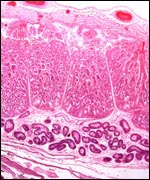
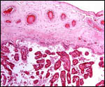
15) Pathological features

In horses, fetal death has been described as occasionally being due to excessively twisted, long cords. They have also caused indentations of the fetal skin. Short cords may rupture and cause demise. One case of a single umbilical artery was associated with renal anomalies, not unlike that seen commonly in human gestations (Whitwell, 1975; Benirschke & Kaufmann, 2000).

Griner (1983) listed as principal causes of zebra mortality the following: Stress and trauma, especially after immobilization; interspecific aggression; enteritis; volvulus; ascaridiasis; occasional milk aspiration in foals, and rare intestinal perforation. Neoplastic lesions were not found.

16) Physiologic data

I know of no relevant zebra studies.

17) Other resources

Cell lines of these two species, as well as most other equidae are available from CRES by contacting Dr. Oliver Ryder at: oryder@ucsd.edu.

18) Other remarks - What additional Information is needed?

It is highly desirable to obtain early implantational stages of the zebra placentas with special attention to the development of endometrial cups. Likewise, studies of eCG secretion the comparison to other equids are desirable. The secretory output of fetal gonads should be studied and the stimulus for their development should be investigated.

Acknowledgement

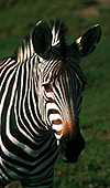
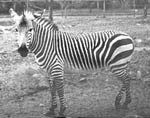
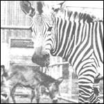
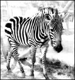
Most of the animal photographs in this chapter come from the Zoological Society of San Diego. I appreciate also very much the help of the pathologists at the San Diego Zoo.

References

Amoroso, E.C.: Placentation. In, Marshall's Physiology of Reproduction, 3rd ed.. A.S. Parkes, ed. V. 2:127-311, 1952, Longmans Green, London.

Asa, C.S., Baumann, J.E., Houston, E.W., Fischer, M.T., Read, B., Brownfield, C.M. and Roser, J.F.: Patterns of excretion of fecal estradiol and progesterone and urinary chorionic Gonadotropin in Grevy's zebras (Equus grevyi): Ovulatory cycles and pregnancy. Zoo Biol. 20:185-195, 2001.

Benirschke, K. and Kaufmann, P.: The Pathology of the Human Placenta. Springer-Verlag, NY, 2000.

Benirschke, K., Low, R.J., Brownhill, L.E., Caday, L.B. and de Venecia?Fernandez, J.: Chromosome studies of a donkey/Grevy zebra hybrid. Chromosoma 15:1?13, 1964. (Benirschke, K.: Corrigendum. Chromosoma 15:300, 1964.)

Benirschke, K. and Malouf, N.: Chromosome studies of Equidae. In: Equus, Vol. 1 & 2. H. Dathe, ed, pp. 253?284, 1967.

Czekala, N.M., Kasman, L.H., Allen, J., Oosterhuis, J. and Lasley, B.L.: Urinary steroid evaluations to monitor ovarian function in exotic ungulates: VI. Pregnancy detection in exotic equidae. Zoo Biol. 9:43-48, 1990.

Enders, A.C. and Liu, I.K.M.: Lodgement of the equine blastocyst in the uterus from fixation through endometrial cup formation. J. Reprod. Fertil. Suppl. 44:427-438, 1991.

Enders, A.C. and Liu, I.K.M.: Trophoblast-uterine interactions during equine chorionic girdle cell maturation, migration, and transformation. Amer. J. Anat. 192:366-381, 1991.

Enders, A.C., Jones, C.J., Lantz, K.C., Schlafke, S. and Liu, I.K.M.: Simultaneous exocrine and endocrine secretions: trophoblast and glands of the endometrial cups. J. Reprod. Fertil. Suppl. 56:615-625, 2000.

Enders, A.C., Meadows, S., Stewart, F. and Allen, W.R.: Failure of endometrial cup development in the donkey-in-horse model of equine abortion. J. Anat. 188:575-589, 1996.

Gadi, I.K. and Ryder, O.A.: Distribution of silver-stained nucleolus-organizing regions in the chromosomes of the Equidae. Genetica 62:109-116, 1983.

Gallagher, P.C., Lear, T.L., Coogle, L.D. and Bailey, E.: Two SINE families associated with equine microsatellite loci. Mammalian Genome 10:140-144, 1999.

George, M. and Ryder, O.A.: Mitochondrial DNA evolution in the genus Equus. Mol. Biol. Evol. 3:535-546, 1986.

Ginther, O.J.: Mobility of the early equine conceptus. Theriogenology 19:603-511, 1983.

Gray, A.P.: Mammalian Hybrids. A Check-list with Bibliography. 2nd edition. Commonwealth Agricultural Bureaux Farnham Royal, Slough, England, 1972.

Griner, L.A.: Pathology of Zoo Animals. Zoological Society of San Diego, 1983.

Keenan, I.R., Forde, D., McGeady, T., Wande, J. and Roche, J.F.: Endometrial histology of early pregnant and nonpregnant mares. J. Reprod. Fertil. Suppl. 35:499-504, 1987.

Kirkpatrick, J.F., Lasley, B.L. and Shideler, S.E.: Urinary steroid evaluations to monitor ovarian function in exotic ungulates: VII. Urinary progesterone metabolites in the equidae assessed by immunoassay. Zoo Biol. 9:341-348, 1990.

Mossman, H.W. and Duke, K.L.: Comparative Morphology of the Mammalian Ovary. University of Wisconsin Press, Madison, 1973.

Nowak, R.M.: Walker's Mammals of the World. 6th ed. The Johns Hopkins Press, Baltimore, 1999.

Ryder, O.A. and Hansen, S.K.: Molecular cytogenetics of the equidae. I. Purification and cytological localization of a (G+C)-rich satellite DNA from Equus przewalskii. Chromosoma 72:115-129, 1979.

Whitwell, K.E.: Morphology and pathology of the equine umbilical cord. J. Reprod. Fertil. Suppl. 23:599-603, 1975.

Wooding, F.B.P., Morgan, G., Fowden, A.L. and Allen, W.R.: A structural and immunological study of chorionic gonadotrophin production by equine trophoblast girdle and cup cells. Placenta 22:749-767, 2001.

   
back to top 

© 2005. All rights reserved.
welcome | home | index | intro | placentation | glossary | author | contact