| |
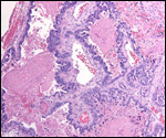
9) Trophoblast external to barrier
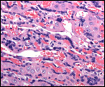
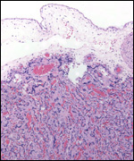
No pregnant uterus has become available to ascertain the depth of trophoblastic
invasion; if one assumes it to be similar to cats, then the myometrium is
not infiltrated and only the endometrium has trophoblast, eroding the glands.
No vascular invasion is demonstrable.
10)
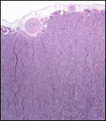
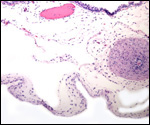
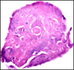
Endometrium
There is minimal decidualization.
11)
Various features
None are described.
12)
Endocrinology
Gonadotropins of pregnancy have not been described for tigers, and no
information on other endocrine secretions has been published. There is
much information on domestic cats, however (Please see the chapter on
cat).
13)
Genetics
Tigers have 38 chromosomes, as do the majority of Felids, except some
of the South American species with 2n=36.
We have seen 39 chromosomes with XXY sex chromosomes ("Klinefelter
Syndrome" - in humans) in an infertile but otherwise normal tiger.
This (39,XXY) is a well-recognized error in domestic cats, where triploidy
and chimerism are also common. We have also seen an autosomal trisomy
in a fetus of a domestic cat.
Fertile hybridization among subspecies, and with lions occurs. Leopard
x Tiger hybrids have aborted (Gray, 1972).
Genetic studies are restricted to the cat chromosomes. Murphy et al. (1999a)
reported virtual complete conservation of the X-chromosome, including
complete synteny) between cat and man. The Y chromosome was also exceedingly
similar. Other genetic studies compare chromosome 12 and 22 (Murphy et
al., 1999b). O'Brien et al. (1997) reviewed the evolution and comparative
genomics of cats, but the tiger was not included. Other karyological studies
can be found in the publications by Hsu & Rearden (1965), Roubin et
al. (1973) and Wurster-Hill & Gray (1973). Newman et al. (1985) described
electrophoretic biochemical variation among various felids, including
tigers.
14)
Immunology
MHC molecules, NK cells, and other immune cell populations have not been
described.
15)
Pathological features
Tigers are susceptible to a large variety of infectious organisms, much
as cats are. We have seen cataracts, renal failure ("Tiger-Krankheit")
and they have normally lipuria.
Habitually aborting domestic cats were studied by Huxtable et al. (1979)
who found multifocal placental necrosis without identifying the cause
of this lesion. The postpartum uterine involution of the domestic cat
was described by Dawson (1946), and McEntee (1990) described other features
of uterine and ovarian pathology. He also summarized the occasional uterine
torsion in pregnancy and infections. In domestic cats, herpes virus infection
leads to necrotizing placentitis and abortion (Hoover & Griesemer,
1971). Numerous genetic diseases have been described in cats (Migaki,
1982), but few (other than albinism) have been assigned to the tiger (Berrier
et al., 1975).
16)
Physiological data
There are no data on uterine blood flow, blood volume, or blood pressure.
It is known, however, that tigers have lipuria (Hewer et al., 1948), a
feature we have been able to confirm.
17) Other resources
Cell lines are available from the "Frozen zoo" of CRES at the
Zoological Society of San Diego. A sweeping review of the phylogeny of
felids comes from Thenius (1967).
18)
Additional needs for data to be collected
Virtually no pathology has been recorded of pregnancies. But, the accidental
finding of a 39,XXY tiger ("Klinefelter syndrome-equivalent")
suggests that more studies of placentas and neonates could be useful.
References
For cell lines write to: www.frozenzoo@sandiegozoo.org
Benirschke,
K., Edwards, R. and Low, R.J.: Trisomy in a feline fetus. Amer. J. Vet.
Res. 35:257-259, 1974.
Berrier,
H.H., Robinson, F.R., Reed, T.H. and Gray, C.W.: The white tiger enigma.
Vet. Med./Small Anim. Clin. 70:467-472, 1975.
Centerwall,
W.R. and Benirschke, K.: An animal model for the XXY Klinefelter syndrome
in man: Tortoiseshell and calico male cats. Amer. J. Vet. Res. 36:1275-1280,
1975.
Dawson, A.B.: The effects of lactation on the postpartum involution of
the uterus of the cat. Amer. J. Anat. 79:241-265, 1946.
Gray,
A. P.: Mammalian Hybrids. A Check-list with Bibliography. 2nd ed. Commonwealth
Agricultural Bureaux, Farnham Royal, 1972.
Hoover,E.A.
and Griesemer, R.A.: Experimental feline herpesvirus infection in the
pregnant cat. Amer. J. Pathol. 65:173-188, 1971.
Hsu,
T.C. and Rearden, H.H.: Further karyological studies in felidae. Chromosoma
16:365-371, 1965.
Huxtable,
C.R., Duff, B.C., Bennett, A.M., Love, D.N. and Butcher, D.R.: Placental
lesions in habitually aborting cats. Vet. Pathol. 16:283-291, 1979.
Leiser,
R.: Blastocystenimplantation bei der Hauskatze. Licht- und elektronenmikroskopische
Untersuchung. Zbl. Vet. Med. C, Anat. Histol. Embryol. 8:79-96, 1979.
Ludwig, K.S.: Zur vergleichenden Histologie des
Allantochorion. Rev. Suisse Zool. 75:819-831, 1968.
Malouf,
N., Benirschke, K. and Hoefnagel, D.: XX/XY chimerism in a tri-colored
male cat. Cytogenetics 6:228-241, 1967.
McEntee,
K.: Reproductive Pathology of Domestic Mammals. Academic Press, San Diego,
1990.
Migaki,
G.: Section VIII, compendium of inherited metabolic diseases in animals.
In, Animal Models of Inherited Metabolic Disease, pp. 473-501. R. Desnick,
D. Patterson and D.G. Scarpelli, eds., 1982.
Mossman,
H.W.: Vertebrate Fetal Membranes. MacMillan, Houndmills, 1987.
Murphy,
W.J., Sun, S., Chen, Z.-C., Pecon-Slattery, J. and O'Brien, S.J.: Extensive
conservation of sex chromosome organization between cat and human revealed
by parallel radiation hybrid mapping. Genome Res. 9:1223-1230, 1999a.
Murphy,
W.J., Menotti-Raymond, M., Lyons, L.A., Thompson, M.A. and O.Brien, S.J.:
Development of a feline whole genome radiation hybrid panel and comparative
mapping of human chromosome 12 and 22 loci. Genomics 57:1-8, 1999b.
Newman,
A., Bush, M., Wildt, D.E., v.Dam, D., Frankenhuis M.Th., Simmons, L.,
Phillips, L. and O'Brien, S.J.: Biochemical genetic variation in eight
endangered or threatened felid species. J. Mamm. 66:256-267, 1985.
Nowak, R.M. and Paradiso, J.L., eds.: Walker's Mammals of the World, 4th.
ed. Vol. II. The Johns Hopkins Press, Baltimore, 1983.
O'Brien,
S.J., Wienberg, J. and Lyons, L.A.: Comparative lessons from cats.TIG
13:393-399, 1997.
Perry,
J.S.: The mammalian fetal membranes. J. Reprod. Fert. 62:321-335, 1981.
Ramsey,
E. M.: The Placenta of Laboratory Animals and Man. Holt, Rinehart and
Winston, N.Y., 1975.
Roubin,
M., deGrouchy, J. and Klein, M.: Les félidés: Évolution
chromosomique. Ann. Génét. 16:233-245, 1973.
Schaller,
G.B.: The Deer and the Tiger. Univ. Chicago Press, 1967.
Srivastava,
S.C.: Contribution to our knowledge of the structure of the placenta of
Panthera tigris tigris (Lin.) Agra Univ. J. Res. 1:37-47, 1952.
Thenius,
E.: Zur Phylogenie der Feliden (Carnivora, Mamm.). Z. zool. Syst. Evolutionsforschung
5:129-143, 1967.
Wimsatt,
W.A.: Some aspects of the comparative anatomy of the mammalian placenta.
Amer. J. Obstet. Gynecol. 84:1568-1594, 1962.
Wislocki,
G.B.: Experimental studies on fetal absorption, II. Behaviour of the fetal
membranes and placenta of the cat toward colloidal dyes injected into
the maternal blood stream. Carnegie Contrib. to Embryol. 11: 1920
Wurster-Hill,
D.H. and Gray, C.W.: Giemsa banding patterns in the chromosomes of twelve
species of cats (Felidae). Cytogenet. Cell Genet. 12:377-397, 1973.
Hewer, T.F., Matthews, L.H. and Malkin, T.: Lipuria in tigers. Proc. Zool.
Soc. London 118:924-928, 1948.
Wynn,
R.M. and Björkman, N.: Ultrastructure of the feline placental membrane.
Amer. J. Obstet. Gynecol. 102:34-43, 1968.
Zhemkova,
Z.P.: The use of sex chromatin in identifying embryonic and maternal tissues
in the placenta: New observations on the haemochorial nature of the cat
placenta. J. Embryol. Exp. Med. 10:127-139, 1962.
|