(Clicking on the thumbnail images below will launch a new window and a larger version of the thumbnail.)
Thomson's Gazelle
Gazella thomsonii

Order: Artiodactyla
Family: Bovidae

1) General Zoological Data


The Thomson's gazelle comes from the arid areas of eastern Africa. It still occurs there in sizeable numbers (Nowak, 1999). These gazelles aggregate in herds of 10-30 animals, occasionally there are many more in a group. Tommies are a grazing species, and a male dominates a group of females and their territory (Walther, 1972). Estes (1967) did a more recent analysis of the territorial behavior of these gazelles. Numerous colonies exist in various zoological gardens. The longevity is more than 9 years according to Mentis (1972), but 15 years have been recorded in captivity, according to Jones (1993). Although the Thomson's gazelle was not specifically studied in the phylogenetic analysis of bovidae by Matthee & Robinson (1999), they related the Dama gazelle to a precursor of the Oribi and, in turn, related it to springbok, saiga and blackbuck.
   
  Male Thomson's gazelle at San Diego Wild Animal Park.
     
  Pair of Thomson's gazelle with typical lateral stripe.
     
  2) General Gestational Data

Females may conceive twice annually because of the post partum estrus experienced by these animals. The gestation lasts 160-180 days with usually single offspring resulting that weigh between 2.2 and 3 kg.

The female animals whose specimens are here described weighed 7.5 kg. The younger animal died from pneumonia. The placental weight of this gestation, or of other specimens, is as yet unknown.

3) Implantation

In these two immature specimens, the implantation occurred in the right horn. The left horn and the endocervical canal were filled with thick mucus in one, in the other it was occupied by cotyledons. There was no extension of the membranes into the other horn in the first specimen, but was found in the second. This is also described by Kellas (1966) for the young oribi specimens that he examined. The fetuses of both gestations were male embryos, measuring 4 cm in length in the first and 19 cm in the second. The younger umbilical cord was 0.8 cm long, had four vessels and an allantoic duct in the first and 7 cm long in the second. They were not twisted. The approximately 40 and 55 cotyledons were arranged in four rows and implantation was mesometrial. The caruncles of the empty horn are well displayed in the photographs. They appear as yellowish protrusions from the endometrial surface.

A second specimen received was a pregnant uterus of an animal that died in February. It contained a 280 g male fetus with a crown-rump length of 19 cm. The placenta was implanted into both uterine horns, although more cotyledons were in the right horn, where the fetus lay. This placenta had 55 cotyledons, although very many were diminutive. They were concave on their surface. Uterus and placenta together weighed 218 gm.

   
  Immature gestation (February) with 19 cm fetus, short umbilical cord, large allantoic sac (right), and 55 cotyledons in four rows.
     
  Uterus of the younger Thomson's gazelle with embryonic implantation in the right horn.
     
  Opened uterus with 4 cm embryo in the bicornuate uterus. Note the four rows of caruncles in the unoccupied left horn.
     
  Embryonic sac in opened uterine horn. Note the four rows of pale caruncles in the other horn.
     
  Pregnant uterus with implanted cotyledons of second specimen, also present in left horn (top right).
     
  4) General Characterization of the Placenta

Krölling (1931) reported on the structure of the antelope placentome, especially on Kobus kob and Gazella rufifrons. He found the placentomes of gazelles to be much flatter than those of most other ruminants. The Thomson's gazelle has an epithelio-chorial placenta without invasion of trophoblast into the endometrium or into deeper structures. Despite the fact that this species is not immediately related to the Oribi, its placentation is similar to that species. For the oribi (Ourebia ourebi), Kellas (1966) described the placental development in great detail. It includes excellent illustrations. Unlike the sheep, the oribi and Thomson's gazelle lack the hematomas adjacent to the placentomes that are found in some ruminants, as in the sheep. At the lateral portion of the cotyledons, one observes that the endometrium has a few glands that are largely absent below the implanted cotyledon and, as in oribi, the endometrium consists mostly of the trabecular endometrial stroma.

Numerous binucleate cells (presumed to produce relaxin) are found in the otherwise cuboidal trophoblastic surfaces of villi. The villi interdigitate with the endometrial caruncular lacunae that, in immature uteri, are apparently less well developed than are those of the slender-horned gazelle. Over the areolar regions, between the cotyledons, the trophoblast is taller, and cellular and proteinaceous debris is found below this layer. There is no trophoblastic infiltration of the endometrium.
   
  Enlarged view of second placenta's concave cotyledons.
     
  Entire concave cotyledon of young implantation with myometrium below.
     
  Edge of cotyledon. A few endometrial glands extend between the caruncles to form the areolae. The "membranes" between the cotyledons are clad by tall trophoblast for absorption of glandular secretions.
     
  Fetal surface with bluish villi extending between broad maternal septa.
     
  Base of young placentation of Thomson's gazelle. Infiltration/or modification of endometrium is apparent here. The pink maternal septa are interdigitated with fetal villi.
     
  Higher magnification of implantation site with the pinker maternal septa and interdigitating fetal villi (V). Binucleate trophoblast (B.N.).
     
  Implanted placenta of the immature gestation in Thomson's gazelle. Huge maternal blood vessels of endometrium.
     
  Villous branches extending from the very immature chorionic membrane into the caruncle. The congested vessels are in the maternal trabeculae.
     
  "Floor" of the youngest specimen's implanting cotyledon. The interdigitations of villi with caruncles are obviously without invasion. Large maternal vessels below.
     
  5) Details of fetal/maternal barrier

This epithelio-chorial placenta shows no direct contact between the trophoblast and endometrial epithelium, other than appositional. Although a space appears between the trophoblast and the endometrial surface of the trabeculae, this is surely the result of shrinkage from fixation. As Kellas (1966) has pointed out for oribi placentas, it is most likely that the two surfaces are closely applied in vivo. Thus, the microvilli would interlock for nutrient exchange. While the trophoblast is cuboidal and columnar, with a moderate number of binucleate cells and occasional giant nuclei, the endometrial epithelium is thin and often has hyperchromatic nuclei. There are numerous microvilli on the trophoblastic surface, none are apparent on the endometrium. Many trophoblastic cells have vacuoles, presumably signs of phagocytosis. Binucleate cells are present in the placentomes as well as in the intercotyledonary membrane. These observations, however, are restricted to H&E preparations and more extensive microvilli may be apparent on the endometrium electronmicroscopically. The fetal villous capillaries are relatively sparse and do not indent the trophoblast, as is the case in camelidae, for instance. The same absence of interdigitation with trophoblast was observed in the oribi placenta.

The endometrial septa (trabeculae) that separate the villi are markedly congested in this very young gestation. The endometrial epithelium is flat and frequently seemingly absent. Kellas (1966) described it as having a syncytial character in oribi. He also referred to degenerative regions as having a "holocrine" nature, with lysis of cells, especially between the cotyledons.
   
  Villi interdigitating with maternal endometrial caruncular crypts. The maternal tissue is well vascularized. Binucleated trophoblast (Bin.) at arrows.
     
  This is the edge of the cotyledon with the more cylindrical trophoblast over the areolae in the younger specimen. Fetal chorioallantoic vessel is seen above.
     
  6) Umbilical cord

The umbilical cord contains two arteries and two veins. In addition, there is an allantoic duct. At these early gestations, the epithelium is cylindrical. Very small blood vessels accompany the duct and few additional vessels are found in the remainder of the cord's substance. Hippomanes have not been found. The amnionic cover is very delicate, and has a thin, flat epithelium. There are also no metaplastic nodules at the early stage of development, but they become pronounced later, as shown next.
   
  Fetal umbilical cord with "callosities" (squamous metaplasia) at arrows. The allantoic duct in the center is surrounded by small vessels.
     
  One of the small regions of squamous metaplasia on cord surface.
     
  Allantoic duct surrounded by small blood vessels.
     
  Allantoic duct of this very immature gestation's umbilical cord. Note the small number of accompanying small blood vessels.
     
  7) Uteroplacental circulation

The vasculature beneath the implanted cotyledons is strikingly enlarged. The remainder of the circulation has not been studied in this species. Kellas (1966), however, gave a detailed description of the extensive maternal vasculature, which he studied in serial sections. Large venous sinuses were observed at the cotyledonary base, similar to what is seen in the present species.

Kellas (1966) observed a set of male twin gestations in oribi with two amnions. The two chorions had vascular anastomoses between the fetal circulations. Perhaps they were monozygotic twins, but the ovaries were not described as to their number of corpora lutea.

8) Extraplacental membranes

The general architecture of the membranes is much the same as in other ruminants. The amnion is thin and avascular, the allantois has virtually no epithelium at this stage, and the chorioallantoic membrane has a mucoid appearance. Kellas (1966) remarked that, near term, the allantoic fluid was virtually absent in oribi. The same finding was true for the slender-horned gazelle (see that chapter). It cannot be judged in the current very immature specimen but this presents a challenging observation, as it is usually the receptacle of fetal urine. It was thus no surprise that in our older specimen with the 19 cm fetus, a large quantity of fluid was found.

   
  This section is of intercotyledonary membranes above the areolae. There is no decidua capsularis.
     
  9) Trophoblast external to barrier

There is no infiltration of trophoblast into the endometrium or uterine muscle.

10) Endometrium

There is no such decidual transformation as is found in primate gestations, but the endometrium changes markedly in pregnancy. Kellas (1966) preferred to denominate the large, hyperchromatic and basophilic stromal cells as decidua. They are also sudanophilic and often granular. They are not shed at birth, as is the case in primates, for instance. But little more than the histology is known of these cells, as was amply discussed in Kellas' two publications (1961, 1966). The same can be said of the nature and purpose of the degenerated debris found in the areolae. Kellas has discussed the various interpretations in detail and more is said in this book in the chapter on sheep. Glands remain in the intercaruncular regions to term, and they appear to secrete a proteinaceous material into the areolae. The tissue below the cotyledons is more fibrous and its epithelium adjacent to the trophoblast is flat and the nuclei are hyperchromatic.

The caruncles of the nonpregnant horn were poorly formed. They stood out as being located between regions of endometrial glands and generally lacked glands almost altogether. There was little epithelium and few cisternae were seen, as are so obvious in the nonpregnant horn's caruncles of the slender-horned gazelle. In this respect, these species have a remarkably different structure of the endometrium. Their early formation can be seen in the illustration of an uterus from a neonate, shown below.

   
  Caruncle of nonpregnant uterine horn. There are virtually no crevices, as seen in slender-horned gazelles. Intercaruncular debris is also present.
     
  This is from a 2 day old newborn's uterus showing the earliest suggestion of caruncle formation.
     
  11) Various features

There are no other unusual features, like a subplacenta or metrial glands.

12) Endocrinology

The finding of interstitial cell stimulation in even this tiny embryo suggests that LH or some other hormone must circulate in the fetus. Its origin may be placental, but there has been no evidence produced to affirm this and this seems to be unlikely at this point. No endocrine studies have been reported in this species. It is likely, however, that relaxin is produced by the binucleate cells, as in other ruminants.

   
  Testis (T) of this 4 cm embryo with stomach (S), pancreas (P), and mesonephros (M) adjacent.
     
  Higher magnification of embryonic testis with stimulated interstitial (Leydig) cells (IC) at arrows.
     
  13) Genetics

Thomson's gazelles have 58 chromosomes in both sexes (Nelson-Rees et al., 1967; Wurster & Benirschke, 1968). They lack the X/autosome translocation found in so many other African antilopinae (Effron et al., 1976).

A male hybrid with Gazella rufifrons was born in London but died the next day (Gray, 1972)

14) Immunology

I know of no immunological studies.

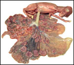
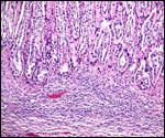
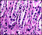
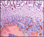
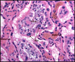
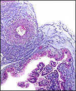
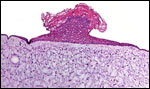
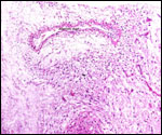
15) Pathological features

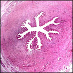
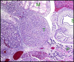
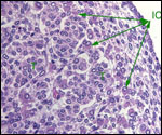
Trauma was found to be the commonest cause of mortality (Griner, 1983).

16) Physiologic data

No data are known to me.

17) Other resources

Cell strains of this gazelle species are available from CRES at San Diego zoo by contacting Dr. Oliver Ryder at: oryder@ucsd.edu.

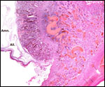
18) Other remarks - What additional Information is needed?

No weights of mature placentas, nor have the lengths of their term gestation umbilical cords been recorded. Endocrine studies of gestation are needed.

Acknowledgement

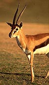
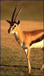
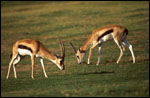
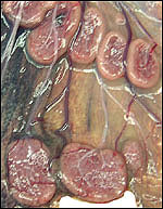
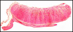
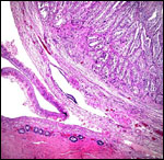
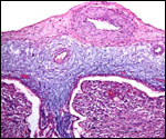
The animal photographs in this chapter come from the Zoological Society of San Diego. I appreciate also very much the help of the pathologists at the San Diego Zoo.

References

Effron, M., Bogart, M.H., Kumamoto, A.T. and Benirschke, K.: Chromosome studies in the mammalian subfamily Antilopinae. Genetica 46:419-444, 1976.

Estes, R.D.: The comparative behavior of Grant's and Thomson's gazelles. J. Mammal. 48:189-209, 1967.

Gray, A.P.: Mammalian Hybrids. A Check-list with Bibliography. 2nd edition. Commonwealth Agricultural Bureaux Farnham Royal, Slough, England, 1972.

Griner, L.A.: Pathology of Zoo Animals. Zoological Society of San Diego, San Diego, California, 1983.

Jones, M.L.: Longevity of ungulates in captivity. Int. Zoo Ybk. 32:159-169, 1993.

Kellas, L.M.: An intra-epithelial granular cell in the uterine epithelium of some ruminant species during the pregnancy cycle. Acta Anat. 44:109-xxx, 1961.

Kellas, L.M.: The placenta and foetal membranes of the antelope Ourebia ourebi (Zimmermann). Acta Anat. 64:390-445, 1966.

Krolling, O.: Über den Bau der Antilopenplazentome. Z. Mikrosk. Anat. Forsch. 27:216-232, 1931.

Matthee, C.R. and Robinson, T.J.: Cytochrome b phylogeny of the family bovidae: Resolution within the alcelaphini, antilopini, neotragini, and tragelaphini. Molec. Phylogenet. Evol. 12:31-46, 1999.

Mentis, M.T.: A review of some life history features of the large herbivores of Africa. The Lammergeyer 1-89, 1972.

Nelson-Rees, W.A., Kniazeff, A.J., Darby, N.B. and Malley, R.I.: Chromosomes of a male gazelle. Gazella thomsoni and a female tapir. Tapirus terrestris columbianus. Mamm. Chromosomes Newsl. 8:229-230, 1967.

Nowak, R.M.: Walker's Mammals of the World. 6th ed. The Johns Hopkins Press, Baltimore, 1999.

Walther, F.R.: Territorial behaviour in certain horned ungulates, with special reference to the examples of Thomson's and Grant's gazelles. Zoologica Africana 7:303-307, 1972.

Wurster, D.H. and Benirschke, K.: Chromosome studies in the superfamily Bovoidea. Chromosoma 25:152-171, 1968.


back to top

© 2002. All rights reserved.
welcome | home | index | intro | placentation | glossary | author | contact