Malayan Tapir
Tapirus indicus

Order: Perissodactyla
Family: Tapiridae


1) General Zoological Data


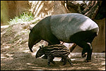
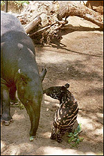
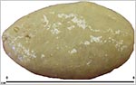
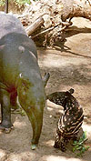
Of the four species of tapirs, Tapirus indicus is the only Asiatic form; the other three species are South American tapirs. The Malayan tapir is characterized by its white saddle and larger size. The saddle is also the reason for its name "saddle backed tapir" or "Schabrackentapir", in German. The young of all species have fine white stripes on brown skin. Malayan tapirs are generally solitary animals. They are the largest of the four species, weighing in excess of 350 kg. The females are usually somewhat larger than males. Tapirs need warm environments in order to thrive in captivity, and they must ideally have access to a pool. Tapirs are excellent swimmers. The longevity of this species is over 30 years in captivity. Many animals are kept and bred in various zoological gardens. All tapirs are endangered species.

The name "tapir" derives from a South American Indian tribe, the Tupi (Gotch, 1979). The designation "indicus", however, is misleading, as these animals do not come from India. Rather, they are "East Indian" from Sumatra and the Malayan peninsula. Some taxonomists have preferred to assign the genus name "Acrocodia" to the Malayan tapir because of its being somewhat distinct from the South American forms. There is a detailed description of morphology and phylogeny in Starck (1995). Barongi (1993) presented an inclusive report on the management and conservation of tapirs in 1993.

The tapiridae were once distributed world-wide and the East Indian and American tapirs are believed to have diverged from one another about 20-30 MYA (Ashley et al., 1996). The South American tapirs immigrated about 3 MYA from North America where they became extinct. Between 1000-3000 Malayan tapirs may still be in the wild, according to Ashley et al. (1996).
   
  Female Malayan tapir with neonate at San Diego Zoo.
     
  Female Malayan tapir with neonate at San Diego Zoo.
     
  Pair of Malayan tapirs at San Diego Zoo to show their aquatic ability and long snouts.


2) General Gestational Data

Sexual maturity is attained at the age of 3-4 years [30 months according to Barongi, 1993], and the estrous cycle is 30 days. The neonate of the placentation described here weighed 8.5 kg, and it died neonatally from cold exposure. It was otherwise normal. The usual weight of the normally one neonate is between 6 and 13 kg (Puschmann, 1989; Barongi, 1993). Twins are not recorded. Gestation lasts 390-405 days (Nowak, 1999).


3) Implantation

Implantation is mesometrial with the yolk sac, but more early stages need to be described. The cord also attaches mesometrially. Starck (1995) infers that implantation is as delayed as is that of horses.


4) General Characterization of the Placenta

This species has an epithelio-chorial, diffuse placentation with small multiply-branched villi. The villous stems are considered to be "microcotyledons" by several authors. Very few placentas of any of the tapiridae have been described, and no really early stages are on record (Mossman, 1987). Thus, it is not known whether tapirs possess endometrial "cups" as the equidae do. The placentas of tapirs are confined to one uterine horn of their bicornuate uterus, whose body is relatively small. Portions of the placental surface are "smooth", with trophoblastic cover but very few villi.

I have had the opportunity to study two full-term placentas from a Malayan tapirs. Both were diffuse organs, weighing 240 and 1,400 g and one measuring 45 cm in greatest diameter, the other 140 x 80 cm. Another was 80 cm in length and up to 0.3 cm in thickness. Much of the chorionic surface was studded with small villi, but many had been rubbed off during the process of delivery for which reason the weight may be too low. Some surface areas were smooth and may represent the yolk sac region described by Dolinar (1967). The remarkable feature of these placentas is their very thinness.

   
  Full-term placenta of Malayan tapir. It is so thin that one can see the green floor through most of the membranes. Thus, to identify the "smooth" areas is difficult.
     
  Site of cord insertion with some (reddish) villus tissue seen adjacent. Tiny yellowish amnionic "callosities" seen at left.
     
  Second specimen spread out to suggest lay-out in uterus. It is very translucent.
     
  One of the "microcotyledons" of this delivered placenta from a Malayan tapir. The arborized villi are obvious. Naturally, there is no maternal tissue. The amnion is seen above the chorion, somewhat detached.


5) Details of fetal/maternal barrier

No implanted placentas have been described in any detail. Thus, what is described here is inferred from the similarity of all perissodactyl placentations, and their morphology, development, and phylogeny. If this is correct, then the tapirs have a diffuse epithelio-chorial relation between the branched villous trophoblastic cover and the endometrial epithelium. The villi infiltrate the endometrial glandular lumens and are closely applied to the endometrium, without invasion.

   
  Final arborization of a villus with single-layer trophoblast. It has a very fine microvillous brush border.
     
  Marked edematous blebs in the membranes of the second tapir placenta.


6) Umbilical cord

The umbilical cord of this specimen was only 5 cm long and had four large vessels as well as a large allantoic duct. There were no spirals. Numerous small blood vessels were also present within the cord, many of them concentrating around the allantoic duct. The allantoic duct was lined by a tall urothelial epithelium. Small smooth muscle bundles were also present. The amnionic surface epithelium of the cord was squamous with keratinization.

   
  Surface of the umbilical cord with a keratinized squamous epithelium and some detached keratin.
     
  This is the allantoic duct within the umbilical cord. Its lining is a multi-layered transitional epithelium, similar to that of the urinary bladder. Small muscle bundles and blood vessels are seen in the wall.
     
  Surface of amnion. Much of the epithelium has degenerated, but the "callosities" are seen as small protrusions.


7) Uteroplacental circulation

This has not been studied.


8) Extraplacental membranes

The allantoic sac is much larger than the amnionic sac. This is typical of equidae as well. Indeed, the amnion does not contact the chorionic sac at term; it is surrounded by the allantoic sac. The amnion has a flat epithelium that was much degenerated in my specimen. Many small "callosities", however, were present and consisted of connective tissue protrusions. The allantoic sac had a single-layered cylindrical epithelium. Small vacuoles were found frequently in this epithelium. There were no hippomanes. There was no decidua capsularis, and areolar regions were not appreciated.

Dolinar (1967) described two delivered placentas of Malayan tapirs and drew attention to the small remains of the yolk sac placenta ("Omphaloplazenta") in this species and the other two families of perissodactyla. He observed a small sac near the insertion of the cord in all these species with a persisting omphalomesenteric vasculature and a small patent duct.

   
  The amnionic surface with "callosities. Some have balls of squamous epithelium.
     
  The allantoic sac is above, with single-layered epithelium and numerous blood vessels. The amnion (below) has degenerated (autolyzed) epithelium and moderate round cell infiltration.
     
  Hippomanes (6x4x0.5 cm) of second tapir placenta.


9) Trophoblast external to barrier

No implanted specimen has been described. Thus, it cannot be verified that there is no trophoblast infiltration into the endometrium. Judging by the similarities to equine and rhinoceros placentas, however, trophoblastic infiltration is not expected. Whether "cups" exist must be explored in the future when implanted specimens become available, and also by endocrine studies.


10) Endometrium

The nonpregnant endometrium has few glands and is otherwise "usual". Whether it allows cup formation remains to be explored. In a neonatal uterus it is readily apparent how short the uterine corpus is, as compared to the horns.

   
  Neonatal uterus at the site of the extensions of the two uterine horns.


11) Various features

None.


12) Endocrinology

The first endocrine study of tapir pregnancies was undertaken by Kasman et al. (1985). It indicated that urinary estrone sulfate levels rose in the latter part of gestation and provided a means of making the diagnosis of pregnancy. A study of serum steroids during pregnancy in Baird's tapir (!) was reported by Brown et al. (1994). It showed elevating levels of progesterone and, late in gestation, of estrogens. Finally, in an endocrine study of equids, rhinos, and four tapir species, Ramsay et al. (1994) showed that urinary equine chorionic gonadotropin secretion was very low in tapirs. Nevertheless, the neonatal gonads are remarkable and very similar to those found in horses. The ovary consists practically entirely of luteinized stromal cells in which the ova are difficult to identify in the thin peripheral rim of cortex. The neonatal testis also has large numbers of stimulated interstitial (Leydig) cells.

   
  Neonatal ovary of Malayan tapir showing massive luteinization of stromal cells.
     
  Neonatal ovary with oocyte mantle in the top one-half; below is the luteinized layer.
     
  Testis of neonatal Malayan tapir with stimulated interstitial cells (I.C.).


13) Genetics

Houck et al. (2000) described the chromosomes of all four species of tapir in some detail. Tapirus indicus has the lowest number of chromosomes (2n=52, vs. 76 and 80 of the South American species). No hybrids have been described.

Phylogenetic relationships were sought by protein analysis (Graur et al., 1993); closer kinship to carnivores was thus uncovered. Ashley et al. (1996) studied mtDNA sequences and established that the South American species are all very similar, but quite distinct from the Malayan tapir.

Karyotype of female Malayan tapir 2n=52.


14) Immunology

I know of no studies.


15) Pathological features

Trauma was the principal cause of death of the animals autopsied by Griner (1983). He also observed pneumonia and, more frequently, rectal prolapse. Barongi (1993) who provided a general survey of tapir husbandry emphasized the foot problems, helminths and rectal prolapse.


16) Physiologic data

I know of no studies.


17) Other resources

Cell strains of this species are available from CRES at the San Diego Zoo by contacting Dr. Oliver Ryder at: oryder@ucsd.edu.


18) Other remarks - What additional Information is needed?

It is imperative that more placentas be studied, especially implanted placentas and early implantations. In addition, endocrine studies during pregnancy and of the neonates are needed.


Acknowledgement

The animal photographs in this chapter come from the Zoological Society of San Diego. I appreciate also very much the help of the pathologists at the San Diego Zoo.


References

Ashley, M.V., Norman, J.E. and Stross, L.: Phylogenetic analysis of the perissodactylan family tapiridae using mitochondrial cytochrome c oxidase (COII) sequences. J. Mammal. Evol. 3:315-326, 1996.

Barongi, R.A.: Husbandry and conservation of tapirs. Int. Zoo. Ybk. 32:7-13, 1993.

Brown, J.L., Citino, S.B., Shaw, J. and Miller, C.: Endocrine profiles during the estrous cycle and pregnancy in the Baird's tapir (Tapirus bairdii). Zoo Biol. 13:107-117, 1994.

Dolinar, Z.J.: Uber die Omphaloplazenta der Perissodactyla. Anat. Anaz. (Suppl.) 120:637-640, 1967.

Gotch, A.F.: Mammals - Their Latin Names Explained. Blandford Press, Poole, Dorset, 1979.

Graur, D., Gouy, M. and Duret, L.: Evolutionary affinities of the order perissodactyla and the phylogenetic status of the superordinal taxa ungulate and altungulata. Mol. Phylogen. Evol. 7:195-200, 1997.

Griner, L.A.: Pathology of Zoo Animals. Zoological Society of San Diego, San Diego, California, 1983.

Houck, M.L., Kingswood, S.C. and Kumamoto, A.T.: Comparative cytogenetics of tapirs, genus Tapirus (Perissodactyla, Tapiridae). Cytogenet. Cell Genet. 89:110-115, 2000.

Kasman, L.H., McCowan, B. and Lasley, B.L.: Pregnancy detection in tapirs by direct urinary estrone sulfate analysis. Zoo Biol. 4:301-306, 1985.

Mossman, H.W.: Vertebrate Fetal Membranes. MacMillan, Houndmills, 1987.

Nowak, R.M.: Walker's Mammals of the World. 6th ed. The Johns Hopkins Press, Baltimore, 1999.

Puschmann, W.: Zootierhaltung. Vol. 2, Säugetiere. VEB Deutscher Landwirtschaftsverlag Berlin, 1989.

Ramsay, E.C., Moran, F., Roser, J.F. and Lasley, B.L.: Urinary steroid evaluations to monitor ovarian function in exotic ungulates: X. Pregnancy diagnosis in perissodactyla. Zoo Biol. 13:129-147, 1994.

Starck, D.: Säugetiere. In, Lehrbuch der Speziellen Zoologie (A. Kaestner, Founder). Vol. II Wirbeltiere. Teil 5/2. Gustav Fischer Verlag, Jena, 1995.


back to top

© 2010. All rights reserved.
welcome | home |index |intro | placentation | glossary | author | contact