(Clicking on the thumbnail images below will launch a new window and a larger version of the thumbnail.)
Golden Lion Tamarin
Leontocebus rosalia rosalia
(Leontopithecus rosalia, Leontideus r.)

Order: Primates
Family: Callitrichidae (Callithricidae)

1) General zoological data of species


Marmosets and tamarins are Brazilian, Colombian, Peruvian arboreal monkeys. The South American monkeys in general have presumably developed independently from Prosimian precursor species and, perhaps for that reason, they have very different modes of placentation and reproductive physiology than other Simiae. There are many breeding colonies in zoos and, especially, at the New England Primate Research Center of Harvard University. Other marmoset colonies are kept at the Rio de Janeiro Primate Center. Golden lion tamarins had nearly become extinct, were bred in sufficient numbers at the National Zoo, Washington DC, and subsequently also in other institutions. They were reintroduced to Brazil in the 1980s-1990s with hundreds again living in the wild. This species has at least three subspecies. New species of tamarins and marmosets are still being discovered in Brazil. This large group of small, arboreal New World primates presents a very similar placental morphology that has been studied in only a few representative species. But it is reasonable to infer from those studies and from their general similarity, their physiology and genetics, that the general structure of their placentas is similar, if not identical, except for their sizes and weights.

Because of these close relationships, the placenta of tamarins and marmosets is treated in only this one representative species.

When differences have been observed, special attention will be drawn to these and the species in question. These animals have served in a large number of biomedical research studies, especially the common marmoset, Callithrix jacchus.

2) General gestational data

The general litter size of Callithricidae is composed of fraternal twins, but triplets are also common (Windle et al., 1999). Occasional singletons occur but they may also be the result of prenatal demise of a twin.

Weights of neonates and of the organs of neonates vary widely with species. It was recorded for the common marmoset (Callithrix jacchus) by Wadsworth et al. (1981). Gestational lengths are around 145 to 150 days, and puberty is reached around 18-24 months. Newborns weigh from 30-50 g, depending on the species. Adult weights vary from 360 to 710 g (Nowak & Paradiso, 1983).

   
  Golden lion tamarin (Leontopithecus rosalia).
     
  Twin golden lion tamarin fetuses and placenta from Ryan et al., 1961.
     
  Uterus of pregnancy in previous figure before section.
     
 
3) Implantation

Implantation is antimesometrial with one lobe (placenta) anterior, the other posterior, but having extensive vascular connections. It is superficial and amnion forms by cavitation. Wislocki (1939) described occasional side-by-side implantation. The villous tissue is trabecular, without typical cotyledons. It is a hemochorial placenta and moderate decidua formation of the uterus simplex occurs. There is no allantoic sac. The remarkable features of marmoset and tamarin placentas are the inter-twin vascular connections between fraternal twins, and the villous hematopoiesis of the placentas. An early implantation has been described by Benirschke & Layton (1969). This case and two other early gestations described by Wislocki (1939) show the chorionic membranes to be fused prior to the development of fetal blood vessels. The exact time of implantation was unknown until the descriptions of Moore et al. (1985). They examined pregnant uteri from timed matings in Callithrix jacchus, the common marmoset and determined that implantation takes place around day 12 with invading syncytium. Later studies (Chambers & Hearn, 1985) examined the uterus every 10 days during pregnancy and provided an excellent guide of this species' placentation and fetal growth. Much of their work was done in thin sections and correlated with progesterone levels. Implantation appeared to occur over the mouth of an endometrial gland and was achieved by trophoblastic infiltration, rather than by such destruction as seen in other primates. Although some syncytial invasion into maternal vessels was observed (later, Smith et al., 1987, refuted this assumption in an electron microscopic study), most were free of trophoblast. There was early and marked trophoblastic proliferation, cytotrophoblast was very sparse but present.

A detailed study of seven marmoset implantation sites between days 12 and 15 in Callithrix jacchus, accompanied by superb electronmicrographs was published by Enders & Lopata (1999). This paper also provides references to the endocrine means for precise timing of gestation in marmosets. This study confirmed the unusual growth of the early blastocyst/embryo that grows within the uterine lumen, rather than implanting deeply into the endometrium, as is the case in humans, for instance. This may relate to the delay of embryonic disk formation and the easier ability to culture their blastocysts. Fusion of syncytium with endometrial epithelial cells was observed. True invasion of the endometrium by the syncytiotrophoblast began on day 15. Cytotrophoblast always was the precursor of syncytium. In one of their cases the two blastocysts implanted on the same uterine surface, rather than on opposite sides as is the norm.

The callithricid placenta contains maternal blood vessels, which were studied in detail by Merker et al. (1987). By day 48, the endometrial vessels around these vessels had disappeared and a prominent collagen-rich (types IV & V) layer (which also had laminin plus proteoglycan MB-1) developed, with gaps to allow perfusion. The basement membrane material appeared to have been produced by the endothelium.
Electronmicroscopic studies were undertaken by several authors. Wynn et al. (1975) studied term placentas of two species and found a poorly developed decidual reaction, a "pseudolabyrinthine" (trabecular) organ, with an acellular junctional zone. The villous surface was similar to other primates with syncytium (endowed with much microvillous surface), and cytotrophoblast. There was little villous connective tissue. The "junctional zone" with the endometrium is relatively acellular with a small amount of Nitabuch fibrinoid and some mononuclear elements. Their study of amnion showed it is like that of other primates. They also described retention to term of a yolk sac, not seen by other students. Broad columns of trophoblast are rare. They do exist, however, in broad columns at early implantation.
   
  Implantation zone of tamarin placenta. Endometrium below
(E = endometrial gland; F = Nitabuch fibrinoid; V = villi;
C = broad sheets of cytotrophoblast anchoring the villi to endometrium).
     
  Early twin blastocysts in golden lion tamarin (Benirschke & Layton, 1969). [E1 and E2 embryos, C=Chorion, A=amnion]. Note broad zone of invasion and large endometrial glands.
     
  Same as previous, actual photograph.
     
  Wall of large intraplacental maternal blood vessel with calcification and abundant basement membrane deposits.
     
  Fetal surface with amnion and fetal blood vessels.
     
  Complete cross section of tamarin placenta, amnion above. Note the trabecular structure.
     
  Higher power of tamarin villous tissue. Note clumped syncytium and focal villous hematopoiesis.
     
  Occasional singletons are born, like this pregnancy with a single placenta and no remnant of a twin. This is Saguinus oedipus.
     
 
4) General characteristics of placenta

The two disks (one for each twin) are monochorionic and usually closely approximated. Wislocki reported (1939) implantation of blastocysts in Saguinus geoffroyi to have occurred on opposite sides of the uterus or, in others, side-by-side. This author also reported on presomite embryos with early implantation and found equally early chorionic fusion. But the timing of this early implantation (or size/age of the embryos) was not stated.

A detailed study of the hematopoiesis in placental trabeculae, first observed by Wislocki (1943), was provided by Jollie et al. (1975). They also gave fine-structural details. Hematopoiesis in the placenta occurred at all stages of gestation; it took place in the sinuses but it was primarily of the erythropoietic type.

5) Details of barrier structure

The surface is made up of typical syncytium with abundant microvilli, underlain by cytotrophoblast. There is little villous connective tissue, thus the barrier between maternal and fetal blood is small.

   
  Higher power view of villous structures with fetal vessels, pronounced syncytial knots on villous surface.
     
 
6) Umbilical cord

There are three vessels in the umbilical cord and no ducts. The length of cords has been described by Spatz (1968) for pygmy and common marmosets as being 20-40 cm. It is eccentrically inserted and has few spirals. It is covered by thin amnionic epithelium.

7) Uteroplacental circulation

Maternal vessels enter the villous tissue and are surrounded by a dense collagenous membrane. Trophoblast does not appear to enter the maternal vessels significantly.

   
  Maternal floor of tamarin placenta showing large maternal vessel surrounded but not invaded by trophoblast. Areas of necrosis and Nitabuch fibrin layer are apparent.
     
 
8) Extraplacental membranes

Amnion is like that of other primates; there are in atrophied villi in the chorion laeve, but it contains bridging vessels.

9) Trophoblast external to barrier

There is very early and broad endometrial infiltration by syncytium but it is not very deep. There appears to be no maternal vascular invasion by trophoblast.

10) Endometrium

There is only scant decidua formation.

11) Various features

Metrial glands do not exist.

12) Endocrinology

Ryan et al. (1961) showed that placental tissue aromatizes androgens to estrogens and assumed that this mechanism prevents freemartinism of similarly chimeric female bovine fetuses. Hearn et al. (1978) summarized much of reproductive physiology of these species. Other steroid and gonadotropic hormones are presented in numerous papers listed here. A remarkable feature of callithricid gestations is the extensive luteinization of the ovaries, making it essentially impossible to identify the corpora lutea of pregnancy.

Hobson and Wide (1981) showed great similarity among various primates' gonadotropins (including the common marmoset) and of the chorionic gonadotropin subunits.

13) Genetics

Marmosets and tamarins normally produce twins, occasionally triplets; when singletons are born, they are often chimerae, indicating that in utero demise of a twin had occurred (Gengozian & Merritt, 1970). Unilateral oophorectomy does not lead to the production of singletons. We have seen one such fetus papyraceus, apparently an acardiac twin.

Because of regularly present inter-twin placental vascular anastomoses, the fraternal twins are all blood (marrow) chimerae; their solid tissues are not involved. They are thus also immunologically tolerant of one another.

The chromosome number of most species is 2n=46, except for the pygmy marmoset (Cebuella pygmaea), and possibly a few other species, with 2n=44. A Robertsonian mechanism of fusion is presumably the cause of this difference. A variety of hybrids have been described (Coimbra-Filho & Mittermeier, 1973; Gray, 1971).

14) Immunology

Because of the congenital hematopoietic chimerism, tolerance among fraternal twins is secured. This has been tested by Cooper (1968) with skin graft exchange.
Transplantation MHC molecules have been studied by Cadavid et al. (1997). Callithricidae "exhibit limited variability of their MHC class I genes", and have genus-specific clustering of MHC c-DNA sequences.

15) Pathological features

Tamarins and marmosets have a long-known tendency of suffering from rickets. This led to the recognition that their demand is for vitamin D3, not D2 (Hunt et al., 1967). Supplementation with this vitamin now has essentially eliminated the osteodystrophy fibrosa in these species. This vitamin is now incorporated into commercial diets. Other skeletal defects of presumably hereditary nature were described by Chalifoux and Elliott (1986). Golden lion tamarins have suffered from a hereditary form of congenital diaphragmatic hernia (Bush et al., 1980). Toxoplasmosis has been fatal to callithricid monkeys, and intestinal infection with Acanthocephalans worms (Prosthenorchis elegans), transmitted by cockroaches, is a frequent problem in captive marmoset colonies. Microfilariasis (Acanthocheilonema perstans) infection is also common in Callithricidae.

A hamartomatous uterine tumor was described by Moreland and Woodard, 1968), and Lunn (1980) described a case of placenta previa in a common marmoset pregnancy. Abortions are common in marmosets and tamarins. Single fetuses may be accompanied by a macerated or an acardiac co-twin fetus papyraceus. Wislocki (1939) also reported one twin to be macerated.

Intrauterine demise of a singleton was reported by Pauly & Strauss (2002) in a Leontopithecus chrysomelas pregnancy. The mummified fetus was delivered by Cesarean section, but the placenta could not be retrieved and was left behind. The female recovered but the cause of fetal death remained unresolved.

Erythroblastosis (hemolytic disease) has been described by Gengozian et al. (1966), and a complete placenta previa was seen by Lunn (1980) in a term twin pregnancy, attended by much bleeding in a common marmoset, and dystocia was also described there.

   
  Term infant and macerated embryo with placenta of pygmy marmoset (Pygmaea cebuella).
     
  Occasional triplets occur, here one with maceration (right), but not with a separate placenta, Saguinus labiatus.
     
  Spontaneous abortus in Leontopithecus rosalia. One fetus (left) had died much earlier.
     
 
16) Physiological data

No relevant data are reported

17) Other resources

A number of fibroblast cell lines are available through CRES, the research arm of the Zoological Society of San Diego (see: http://www.sandiegozoo.org/conservation/cres_home.html. Please direct your inquiries to Dr. Oliver Ryder (oryder@ucsd.edu).

18) Additional needs for study

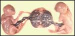
We know too little about the frequency of demise of one of the twins. In human gestation, this can be due to the so-called twin-to-twin-transfusion syndrome, or it may be due to the conversion into acardiac fetus because of the presence of vascular anastomoses. How often does this happen in marmosets and tamarins? Also, the real physiologic reason why the female twin, who is chimeric with the male co-twin, is spared the freemartin syndrome of artiodactyls (cattle especially) is not understood.

References

There is a very extensive bibliography on golden lion tamarins. An especially important reference is:

Kleiman, D.: Social and sexual behaviour of Leontopithecus rosalia during the reproductive cycle. In, Kleiman, D., The Biology and Conservation of the Callithricidae . Smithsonian Press, Washington, 1976.

Barnhart, D.D. and Gengozian, N.: An evaluation of the mixed lymphocyte culture reaction in marmosets. Transplantation 20:107-115, 1975.

Bedard, M.T., Ma, S.F. and Jones, T.C.: Chromosomal banding patterns and nucleolar organizing regions in three species of Callithricidae (Saguinus oedipus, Saguinus fuscicollis, and Callithrix jacchus). J. Med. primatol. 7:82-97, 1978.

Bender, M.A. and Mettler, L.E.: Chromosome studies of primates. II. Callithrix, Leontocebus and Callimico. Cytologia (Tokyo) 25:400-404, 1960.

Benirschke, K., Anderson, J.M. and Brownhill, L.E.: Marrow chimerism in marmosets. Science 138:513-515, 1962.

Benirschke, K. and Brownhill, L.E.: Further observations on marrow chimerism in marmosets. Cytogenetics 2:331-341, 1963.

Benirschke, K. and Layton, W.: An early twin blastocyst of the golden lion marmoset, Leontocebus rosalia L. Fol. Primatol. 10:131-138, 1969.

Cadavid, L.F., Shufflebotham, C., Ruiz, F.J., Yeager, M., Hughes, A.L. and Watkins, D.I.: Evolutionary instability of the major histocompatibility complex class I loci in New World primates. Proc. Natl. Acad. Sci. USA 94:14536-14541, 1997.

Cell lines inquiries: Dr. Oliver Ryder (oryder@ucsd.edu).

Chalifoux, L.V. and Elliott, M.W.:Congenital anomalies in two neonatal tamarins (Saguinus oedipus and Saguinus fuscicollis). J. Med. Primatol. 15:329-337, 1986.

Chambers, P.L. and Hearn, J.P.: Embryonic, foetal and placental development in the common marmoset monkey (Callithrix jacchus). J. Zool. London 207:545-561, 1985.

Coimbra-Filho, A.F. and Mittermeier, R.A.: New data on the taxonomy of Brazilian marmosets of the genus Callithrix Erxleben, 1777. Folia primatol. 20:241-264, 1973.

Coimbra-Filho, A.F. and Mittermeier, R.A., eds.: Ecology and Behavior of Neotropical Primates. Academia Brasileira de Ciências. Rio de Janeiro, 1981.

Cooper, R.W.: Small species of primates in biomedical research. Lab. Anim. Care 18:267-279, 1968.

Cronin, J.E. and Sarich, V.M.: Molecular systematics of the New World monkeys. J. Human Evol. 4:357-375, 1975.

Dunn, F.L.: Acanthocephalans and cestodes os South American monkeys and marmosets. J. Parasitol. 49:717-722, 1963.

Enders, A.C. and Lopata, A.: Implantation in the marmoset monkey: Expansion of the early implantation site. Anat. Rec. 256279-299, 1999.

Flurer, C.I. and Zucker, H.: Evaluation of serum parameters relevant to vitamin D status in tamarins. J. Med. Primatol. 16:175-184, 1987.

Egozcue, J., Perkins, E.M. and Hagemenas, F.: Chromosomal evolution in marmosets, tamarins, and pinchés. Folia Primatol. 9:81-94, 1968.

Gengozian, N.: Marmosets: their potential in experimental medicine. Ann. N.Y. Acad. Sci. 162:336-362, 1969.

Gengozian, N. and Merritt, C.B.: Effect of unilateral ovariectomy on twinning frequency in the marmoset. J. Reprod. Fert. 23:509-512, 1970.

Gengozian, N., Batson, J.S. and Nelson, B.M.: Bone-marrow grafting attempts in marmosets after whole-body irradiation. Int. J. Rad. Biol. 11:553-561, 1966.

Gengozian, N., Lushbaugh, C.C., Humason, G.L. and Kniseley, R.M.: "Erythroblastosis foetalis" in the primate, Tamarinus nigricollis. Nature 209:731-732, 1966.

Gray, A.P.: Mammalian Hybrids. Commonwealth Agricultural Bureaux, Farnham Royal, Slough SL2 3BN, 1971.

Hearn, J.P., Abbott, D.H., Chambers, P.C., Hodges, J.K. and Lunn, S.F.: Use of the common marmoset, Callithrix jacchus, in reproductive research. Prim. Med. 10:40-49, 1978.

Hershkovitz, P.: Notes on tertiary platyrrhine monkeys and description of a new genus from the late miocene of Colombia. Folia Primatol. 12:1-37, 1970.

Hershkovitz, P.: Comments on the taxonomy of Brazilian marmosets (Callithrix, Callitrichidae). Folia Primatol. 24:137-172, 1975.

Hampton, J.K., Hampton, S.H. and Levy, B.M.: Reproductive physiology and pregnancy in marmosets. pp. 527-535 in Medical Primatology, Karger, Basel 1970.

Hearn, J.P., Abbott, D.H., Chambers, P.C., Hodges, J.K. and Lunn, S.F.: Use of the common marmoset, Callithrix jacchus, in reproductive research. Prim. Med. 10:40-49, 1978. Karger, Basel.

Hobson, B.M., Hearn, J.P., Lunn, S.F. and Flockhart, J.H.: Urinary excretion of biologically active chorionic gonadotrophin by the pregnant marmoset (Callithrix jacchus jacchus). Fol. Primatol. 28:251-258, 1977.

Hobson, B.M. and Wide, L.: The similarity of chorionic gonadotrophin and its subunits in term placentae from man, apes, old and new world monkeys and a prosimian. Fol. Primatol. 35:51-64, 1981.

Hunt, R.D. and Garcia, F.G.: Hypervitaminosis D in New World monkeys. Am. J. Clin. Nutrition 22:358-366, 1969.
86:221-223, 1963.

Hunt, R.D., Garcia, F.G. and Hegsted, D.M.: A comparison of vitamin D2 and D3 in New World primates. I. Production and regression of osteodystrophia fibrosa. Lab. Anim. Care 17:222-234, 1967.

Jollie, W.P., Haar, J.L. and Craig, S.S.: Fine structural observations on hematopoiesis in the chorioallantoic placenta of the marmoset. Amer. J. Anat. 144:9-38, 1975.

Lasley, B.L., Monfort, S.L., Hodges, J.K. and Czekala, N.M.: Comparison of urinary estrogens during pregnancy in diverse species. In, Fetal Endocrinology, pp. 111-126, Academic Press, 1981.

Merker, H.-J., Bremer, D., Barrach, H.-J. and Gossrau, R.: The basement membrane of the persisting maternal blood vessels in the placenta of Callithrix jacchus. Anat. Embryol. 176:87-97, 1987.

Nowak, R.M. and Paradiso, J.L.: Walker's Mammals of the World, 4th ed. Vol. 1.Johns Hopkins Univ. Press, Baltimore, 1983.

Pauly, A. and Strauss, G.: Kaiserschnit bei einem Goldkopflowenaffchen (Leontopithecus chrysomelas) mit Entwicklung einer mummifizierten Frucht. Milu 10:583-587, 2002.

Preslock, J.P., Hampton, S.H. and Hampton, J.K.: Cyclic variations of serum progestins and immunoreactive estrogens in marmosets. Endocrinology 92:1096-1101, 1973.

Ryan, K.J., Benirschke, K. and Smith, O.W.: conversion of andro-stenedione-4-C14 to estrone by the marmoset placenta. Endocrinology 69:613-618, 1961

Sarich, V.M. and Cronin, J.E.: South American mamma molecular systematics, evolutionary clocks, and continental drift. In, Evolutionary Biology of the New World Monkeys and Continental Drift. R. Ciochon & A.B. Chiarelli, eds. Plenum Press, N.Y. 1980. Chapter 20.

Seuanez, H.N., L. Forman, L. and Alves, G.: Comparative chromosome morphology in three Callithrichid genera: Cebuella, Callithrix, and Leontopithecus. J. Hered. 79:418-424, 1988.

Smith, C.A., Moore, H.D.M. and Hearn, J.P.: The ultrastructure of early implantation in the marmoset monkey (Callithrix jacchus). 175:399-410, 1987.

Spatz, W.B.: Nabelschnur-Länge bei Insektivoren und Primaten. Z. Säugetierk. 33:226-239, 1968.

Turton, J.A., Ford, D.J., Bleby, J., Hall, B.M. and Whiting, R.: Composition of the milk of the common marmoset (Callithrix jacchus) and milk substitutes used in hand-rearing programmes, with special reference to fatty acids. Fol. Primatol. 29:64-79, 1978.

Windle, C.P., Baker, H.F., Ridley, R.M., Oerke, A-K. and Martin, R.D.: Unrearable litters and prenatal reduction of litter size in the common marmoset (Callithrix jacchus). J. Med. Primatol. 28:73-83, 1999.

Wislocki, G.B.: Hematopoiesis in the chorionic villi of the placenta of platyrrhine monkeys. Anat. Rec. 85:349-363, 1932.

Wislocki, G.B.: Observations on twinning in marmosets. Amer. J. Anat. 64:445-483, 1939.

Wislocki, G.B.: Placentation in the marmoset (Oedipomidas geoffroyi) with remarks on twinning in monkeys. Anat. Rec. 52:381-399, 1932.

Wynn, R.M., Richards, S.C. and Harris, J.A.: Electron microscopy of the placenta and related structures of the marmoset. Amer. J. Obstet. Gynecol. 122:60-69, 1975.

Wynn, R.M., Richards, S.C. and Harris, J.A.: Electron microscopy of the placenta and related structures of the marmoset. Amer. J. Obstet. Gynecol. 122:60-69, 1975.
Moore, H.D.M., Gems, A. and Hearn, J.P.: Early implantation stages in the marmoset monkey (Callithrix jacchus). Amer. J. Anat. 172:265-278, 1985.


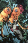
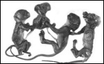
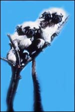
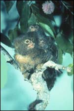
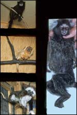
Various species of marmosets/tamarins

   
  Cottontops (Oedipomidas [Saguinus] oedipus).
     
  Pygmy marmoset (Cebuella pygmaea). Moustached tamarin (Tamarinus nigricollis).
     
  Pygmy marmoset (Cebuella pygmaea). Middle left: Golden lion tamarin (Leontopithecus rosalia). Bottom left: Cotton tops (Oedipomidas [Saguinus] oedipus) Right: Common marmoset (Callithrix jacchus).


back to top

© 2002. All rights reserved.
welcome | home | intro | placentation | glossary | author contact me: kbenirsc@ucsd.edu