 |
|
(Clicking
on the thumbnail images will launch a new window and a larger version
of the thumbnail.)
|
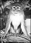
Miopithecus talapoin
Order: Primates
Family: Cercopithecidae
1) General Zoological Data
This single species of the genus Miopithecus is of west African equatorial regions. It is the smallest African monkey, males weighing 1,230 g and females between 745 and 820 g (Nowak, 1999). In German, this species is referred to as "Zwergmeerkatze". Another species of Miopithecus (M. ogouensis) of regions north of the Congo River has been delineated (see Groves, 2001). This animal has a shorter tail and minor facial differences. This opinion is further supported by the identification of different endogenous viral sequences identified in these two species/subspecies by v.d. Kuyl et al. (2000). This publication also suggested that the probability exists now that talapoins split from colobine stock 9-14 MYA. Excellent web sites exist of all possible features and publications on this animal and can be accessed by "Google". Some authorities have considered that this species should not receive separate genus rank (and that it be listed as Cercopithecus talapoin), others have disagreed with that view. The current status is as listed above. Jones (1982) gives 27 years 8 months as maximal recorded life span. A few zoos and several Primate Research Centers carry this species.
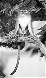 |
Talapoin monkey at San Diego Zoo, many years ago. |
The length of gestation was given as 158-166 days by Rowell (1977). A single, large newborn is commonly born that weighs around 230 g. I am not aware that twins have been reported.
3)
Implantation
No data are available on implantation of this species but it is likely
to be similar to other closely related cercopithecidae. The animals have
a typical unicornuate single uterus and, like the other species, one lobe
must implant anterior, the other posterior with connecting vessels coursing
laterally.
4)
General Characterization of the Placenta
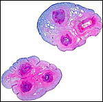
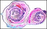
The placenta available from this single animal was a bilobed placenta
weighing 65 g, the smaller lobe weighing 25 g. The lobes measured 7.5
x 0.5 and 8 x 0.5 cm. The umbilical cord inserted at the large lobe eccentrically,
measured 16 cm in length and had a marked right spiral. A retroplacental
hematoma was found behind the larger disk and it also had a large marginal
infarct.
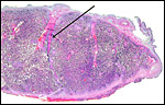 |
Complete cross section of disk. The arrow points to the connective tissue band that connects the chorion (above) and decidua (below). |
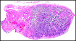 |
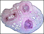
Complete section of the marginal portion of placental disk with marginal infarct (left). Amnion is on top. |
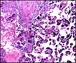
This is a hemochorial placenta, similar to all other cercopithecidae. Relatively sparsely branched villi are covered by cytotrophoblast and, peripheral to this, syncytiotrophoblast. The trophoblast is bathed in maternal blood. Focal detachment of syncytial "knots" are free in the intervillous space and may be swept away into the maternal lung. The amount of syncytiotrophoblast is particularly pronounced. In addition, there are numerous small islands of cytotrophoblast ("extravillous trophoblast, X-cells") with attached fibrinoid throughout the placenta. Also characteristic is the presence of several straight connective tissue fibers that span from the chorion's under-surface to the decidua basalis. These are covered by trophoblast. The villi contain small numbers of fetal capillaries, a small quantity of connective tissue and very few Hofbauer cells (macrophages). In between the villi one often finds small foci of calcifications. These are normal deposits within fibrin and fibrinoid and they occur in greater numbers with advancing gestational age and have no pathological significance.
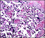 |
Subchorionic region of villous tissue. Dark pink/purple regions are fibrinoid and focal calcifications. The chorion is at top right. |
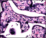 |
Terminal villi with numerous capillaries in scant stroma and surface of syncytiotrophoblast. One large syncytial "knot" is suspended in the intervillous blood and is presumably traveling to maternal lung. |
The umbilical cord contains two arteries and one vein. There are no areas of squamous metaplasia and there are no ducts. The cord was 16 cm in length and had a marked right twist. It is unknown at this point whether there is a communicating artery, known as "Hyrtl's anastomosis" at the placental base of the cord. This aspect has been addressed for baboon placentas by Houston & Hendrickx (1968) and should be studied in other species.
 |
Cross sections of umbilical cord. The vein in the top picture is at right. The bluish background is Wharton's jelly. |
 |
Higher magnification of umbilical cord. Vein at left. |
No specific studies are known to me but the probability of similarities to other cercopithecidae is great. Thus, reference to the chapter by Ramsey (1975) may be helpful.
8)
Extraplacental membranes
The membranes are composed of amnion on the inside, beneath which is the
chorion that carries fetal blood vessels to the villous tissue. A thin
layer of trophoblast and extremely little decidua capsularis are even
more peripheral. The membranes carry several large fetal blood vessels
from one lobe to the other, vessels that course laterally in the uterus.
As in the other cercopithecids described, there are no atrophied villi
in the free membranes, as is the case in humans. This is especially surprising
as this must be a shallow implantation and one might have expected such
atrophic villi. No allantoic sac or vitelline cavity remains at term.
 |
Roll of membranes with trophoblast (purple) at outside. The large pink vessels represent the connecting blood vessels between the two disks. |
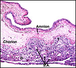 |
Free membranes with D= decidua, S.A. = spiral arteriole. |
Trophoblast invades the decidua basalis, but whether it infiltrates the uterus or deeper into the spiral arterioles awaits the description of an implanted placenta.
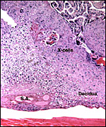 |
Implantation site of mature placenta. S.A. = spiral arteriole; X-cells = extravillous trophoblast. |
The endometrium undergoes typical decidualization as seen in other primates. The delivered placenta, however, has very little attached decidua basalis, and the amount of decidua covering the membranes is diminutive. The decidua basalis is infiltrated by cytotrophoblast ("extravillous trophoblast, X-cells") and these cells are also found in the walls of the spiral arterioles. They are rather purpler in staining quality than the lighter-stained decidual cells. They also produce the "fibrinoid" that makes up the thin layer of Nitabuch.
11)
Various features
None.
12)
Endocrinology
Eberhart et al. (1983) studied the cortisol and prolactin levels of socialized
talapoins grouped with estrogen-treated or non-treated females. Genital
swelling during estrus is pronounced.
13) Genetics
Talapoin monkeys have 54 chromosomes, as shown below (Hsu & Benirschke,
1971). In the references just cited, previous chromosome studies of talapoins
are listed. More recently, Dutrillaux et al. (1978) and Ponsa et al. (1980)
have published various banding studies on talapoin monkeys. I am not aware
that hybrids have been described.
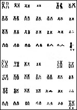 |
Karyotypes of male and female talapoin monkeys. |
I am not aware of any specific immunological studies in this species.
15)
Pathological features
The villous tissue of talapoins has infarcts, as is so typical of most
cercopithecidae. While such infarcts are most commonly observed in preeclampsia
of human gestations and are due to occlusion of maternal spiral arterioles
with atherosis and thrombosis, no such lesions have been observed in talapoins,
and there is also no evidence that they suffer preeclampsia.
 |
At left is the edge of the old infarct, at right is normal villous tissue. |
Hunt et al. (1995) studied the DNA sequences of the blue cone photopigments in talapoin and marmoset and found great similarities. They suggested separation of Old and New World monkeys some 43 MYA. These animals are frugivorous and insectivorous.
17)
Other resources
There are no cell strains available at the San Diego Zoo, but may well
be available from some Primate Research Center.
18)
Other remarks - What additional Information is needed?
Early stages of implantation and endocrine data are needed.
Acknowledgement
The animal photograph in this chapter comes from the Zoological Society
of San Diego.
References
Dutrillaux, B., Viegas-Pequignot, E., Couturier, J. and Chauvier, G.:
Identity of euchromatic bands from man to cercopithecidae (Cercopithecus
aethiops, Cercopithecus sabaeus, Erythrocebus patas, and Miopithecus talapoin).
Hum. Genet. 45:283-296, 1978.
Eberhart, J.A., Keverne, E.B. and Meller, R.E.: Social influences on circulating levels of cortisol and prolactin in male talapoin monkeys. Physiol. Behav. 30:361-369, 1983.
Groves, C.: Primate Taxonomy. Smithsonian Institution Press, Washington, 2001.
Houston, M.L. and Hendrickx, A.G.: Observations on the vasculature of the baboon placenta (Papio sp,) with special reference to the transverse communicating artery. Folia primatol. 9:68-77, 1968.
Hsu, T.C. and Benirschke, K.: An Atlas of Mammalian Chromosome. Vol. 5, Folio 248, 1971. Springer-Verlag, N.Y.
Hunt, D.M., Cowing, J.A., Patel, R., Appukuttan, B., Bowmaker, J.K. and Mollon, J.D.: Sequence and evolution of the blue cone pigment gene in Old and New World primates. Genomics 27:535-538, 1995.
Jones, M.L.: Longevity of captive mammals. Zool. Garten 52:113-128, 1982.
Kuyl, A.V.v.d., Dekker, J.T. and Goudsmit, J.: Primate genus Miopithecus: evidence for the existence of species and subspecies of dwarf guenons based on cellular and endogenous viral sequences. Mol. Phylogenet. Evol. 14:403-413, 2000.
Nowak, R.M.: Walker's Mammals of the World. 6th ed. The Johns Hopkins Press, Baltimore, 1999.
Ponsa, M., Estop, A.M., Miro, R., Rubio, A. and Egozcue, J.: Banding patterns of the chromosomes of Miopithecus talapoin compared with Macaca mulatta and Cercopithecus aethiops. Cytogenet. Cell Genet. 28:41-46, 1980.
Ramsey, E.M.: The Placenta of Laboratory Animals and Man. Holt, Rinehart and Winston, New York, 1975.
Rowell, T.E.: Reproductive cycle of the talapoin monkey (Miopithecus talapoin) Folia Primatol. 28:188-202, 1977.