(Clicking on the thumbnail images below will launch a new window and a larger version of the thumbnail.)
 
Last updated:
April 17, 2004.
Springhaas (Springhare)
Pedetes capensis

Order: Rodentia
Family: Pedetidae

1) General zoological data

The name Pedetes derives from the Greek word for dancer or leaper. It is most appropriate for this charming small nocturnal rodent (Gotch, 1979). This is the sole member of the family. It lives in Africa and extends from Kenya to the Cape . Some authors have considered that a second species ( Pedetes surdaster ) exists, while most authorities believe it to be the same animal (see, however, the discussion by Matthee & Robinson, 1997b) . In addition, regional subspecies have been considered (see Otiang'a-Owiti et al., 1992).

The taxonomic position of Pedetidae is uncertain. Indeed, Simpson (1945) referred to this genus as "incertae sedis". That position has not improved since those considerations, because Nowak (1999) still needed to state that: "The family is of highly uncertain systematic position". The springhaas is characterized by having extremely long legs, which give the animal the jumping ability and that also remind of kangaroos. It is thus also referred to as being bipedal. Also striking are a relatively large head, very large (nocturnal-adapted) eyes, and the long, bushy tail.

Adults weigh around 3-4 kg and neonates are 250-300 g. Fischer & Mossman (1969) found one newborn to weigh 500 g. Springhaas are now less common because of habitat destruction and extensive hunting for meat. They were once plentiful. They prefer living singly in several burrows, which they routinely construct and which are also used at night. Foraging for vegetable matter begins at dusk and extends through most of the night. Longevity in captivity is at least 13 years.

   
  A characteristic springhaas in the San Diego Zoo.
     
  A characteristic springhaas in the San Diego Zoo.
     
 
2) General gestational data

Reproduction occurs throughout the year and was studied extensively by Merwe et al. (1980). These authors also studied some endocrine parameters. A single young is born after an estimated 78-82 days of gestation. The exact length of pregnancy, however, had not been determined decisively and was disputed (Merwe et al., 1980). Now it is thought to be 60-77 days (see below). Puschmann (1989) stated that there might be 3-4 pregnancies per year in captive animals. Twins are rare but they have been reported (Nowak, 1999; Rosenthal & Meritt, 1973). Butynski (1979) who made an extensive ecological study estimated that twins occur in less than 1% of pregnancies. One authors' young weighed 300 g, another was 278 g. Butynski (1979) found the average neonatal weight to be 250 g. He received a pregnant female and described the neonate in some detail. The neonate weighed 250 g, the mother weighed 1,785 g and a male weighed 2,665 g. He provided detailed information on the growth of that male young.

The young leave the burrows when they are 1.5 kg in size and spermatogenesis begins at 2.5 kg size and is continuous throughout life. Most males have inguinal testes, rarely were they found to be scrotal; they can then be readily retracted into abdominal pouches (Coe, 1969). The reproductive tracts of both sexes were described in great detail by Coe (1969), and also by Fischer & Mossman (1969). The latter authors stated that the uterus is "duplex" and only occasionally did the cervix assume a "V-shape". In those cases, there is only one os while, normally, there are two cervical ostia. The ovaries are contained in a bursa. There are no cervical glands, but the clitoris has two glands. The cervix is lined by squamous epithelium and the penile glans has spines. Implantation occurred on the side of the corpus luteum, mostly on the left. But, ovulation does occur on both sides and fetuses were five times on the right, and six times on the left side, according to Coe (1969). Likewise, Otiang'a-Owiti et al. (1992) found an equal number of gestations on the right as on the left side of the uterine horns.

It should be stated here that the publication by Coe (1969) is on Pedetes surdaster, the Kenyan "species". Since that publication, however, this species has been joined with P. capensis and probably there is no further use here to separate them. All anatomical aspects appear to be similar, as are weight, breeding, and length of gestation (estimated to be 2 months by Coe, others state 77 days). An earlier publication on reproduction by vs. Horst (1935) was not accessible to me.

3) Implantation

The implantation of the blastocysts occurs antimesometrially, with the inner cell mass being mesometrial in location according to Fischer & Mossman (1969). Otiang'a-Owiti et al. (1992), who did a comprehensive study disagree with this conclusion. They found a mesometrial location as implantation site. Prior to the establishment of the relatively small definitive chorioallantoic placenta, a large "preplacenta" is formed within which large maternal blood channels develop. The preplacenta consists of mostly trophectoderm. It is analogous to the "Träger" of other rodents and has also been called the "ectoplacenta". Its periphery becomes syncytial and erodes maternal capillaries. Then the blastocyst expands within the endometrial slit into which it has implanted and contacts the decidual lining at the opposite pole. Fischer & Mossman (1969) depicted these early stages of placentation in excellent diagrams that make the complexity better understood. In fact, rather than recounting the extensive data their detailed study has yielded, the reader must review their publication to fully understand the complexity of this organ. Nevertheless, although that publication appears to be authoritative, a newer study by Otiang'a-Owiti et al. (1992) on large and well-prepared material takes exception with many of their findings.

In considering all aspects of placentation in this rodent and comparing it with other genera, Fischer & Mossman (1969) came to the conclusion that it is most similar to that of Ctenodactylidae (the "Gundis" of North Africa). These authors offered much evidence that specific aspects of placentation may be useful for establishing a taxonomic position for disputed species. But one must read this paper as well as the newer study from Kenya (Otiang'a-Owiti et al., 1992) before passing judgment.

4) General Characterization of the Placenta

I have had only one slide of a placental disc available for study, although the animals have bred in San Diego, as they have also in many other institutions. This slide was made available by Dr. Dennis Meritt, then at Lincoln Park Zoo. Therefore, the description of this animal's placenta must follow primarily the report of the extensive study on 37 specimens undertaken on the placentation of Pedetes by Fischer & Mossman (1969) and, importantly, the newer study by Otiang'a-Owiti et al. (1992).

In addition to following the stages of placental development, the former authors found two preimplantational blastocysts with trophoblastic surfaces and inner cells masses. An implanting blastocyst had no zona pellucida and was allegedly attached antimesometrially.

The final placenta consists of a disk with large maternal blood channels entering from the basal decidua. The blood gets reflected and sent back through the trophoblastic channels. According to Fischer & Mossman (1969), there is a late-developing inverted yolk sac that persists to term and whose epithelial cells appose the uterine endometrium. It becomes very thin. That is contrary to the detailed observations by Otiang'a-Otiwi et al. (1992) who did not find the yolk sac to invert. Instead the external epithelium joins the uterine endometrium, the endodermal membrane's epithelium remaining internal. The allantoic sac is small.

Fischer and Mossman (1969) considered the preplacenta to be a most striking aspect of springhaas placentation. This area is composed of three principal regions and within it the maternal blood channels develop. There is a central proliferative zone which is covered by thin syncytium, and which has a lateral extension of cytotrophoblast. As with other rodents, the placental disc is composed of labyrinth and trophospongium (see the chapter on mouse). Another unusual aspect of springhaas placentation delineated by these students is the occurrence of blood islands in the villi. They do not occur in other rodents.

The final disc of the mature placenta is mainly composed of labyrinth (90%) and trophospongium, the remainder. The base is made up largely of degenerating cells and trophoblast intermixed.

5) Details of fetal/maternal barrier

It had initially been believed that the springhaas placenta was endotheliochorial in its composition. Fischer & Mossman's (1969) detailed study, however, then suggested that its relationship is really hemodichorial. This was reexamined by two groups: Otiang'a-Owiti et al. (1992) who did the light microscopy and by Owiti et al. (1985) with electronmicroscopy. They worked with exceptionally well-fixed material and found that the placenta has principally an endotheliochorial relationship. To be sure, near term, the endothelium and is wispy connective tissue base becomes very attenuated and may be difficult to identify. In addition, there is the syndesmochorial relationship of the membranous dome of the placenta, not the inverted yolk sac placental portion described by Fischer & Mossman (1969).

   
  At left is the maternal floor with the numerous giant cells and endometrial debris.
     
  At left is a large fetal blood vessel (bottom) that distributes fetal blood into the villi.
     
  The large maternal vessel at right borders on trophoblast but the endothelium is not visible in this slightly autolyzed specimen.
     
 
6) Umbilical cord

The umbilical cord was not available to me and is not even mentioned in the exhaustive study of Fischer & Mossman (1969). The only probable features are the presence of yolk sac- and allantoic ducts. Otiang'a-Otiwi et al. (1992) found the cord to have three allantoic vessels (2A, 1V), and two yolk sac vessels (1A, 1V). They did not mention ducts and did not measure the lengths.

7) Uteroplacental circulation

The circulation of springhaas placentas was alluded to and diagrammatically shown in Fischer & Mossman's study (1969). A large maternal vessel develops in the preplacenta, divides and send its blood to the placental surface whence it is returned in fine channels. Here it borders trophoblast and allows exchange. These authors considered this to be a typical counter-current setup of the labyrinth. The stem villi of lobules are perfused by a thin-walled fetal arteriole.
   
  Fetal surface of disc with fetal vessel, small allantoic cavity, and large (central) maternal channel. In a few areas one may see some endothelial cells.
     
 
8) Extraplacental membranes

The "free membranes" are the bilaminar omphalopleure with thick Reichert membrane. The external epithelium is not endodermal, but it is the inner epithelium. Thus, this is not an inverted yolk sac placentation. Instead the ectodermal epithelium fuses with the endometrium and here forms, in essence, a syndesmochorial region. This membrane and the uterine wall are very thin.

9) Trophoblast external to barrier

Only the decidual and vascular infiltration of trophoblast are seen external to the placenta. This does not involve the uterine muscle.

10) Endometrium

The decidua undergoes considerable changes during placental development. This has been described in great detail by Fischer & Mossman (1969) but does no seem to be important for this consideration of the placenta.

11) Various features

No true "sub-placenta" is present, but the early stages of development produce a very large "pre-placenta", akin to former designations "ectoplacenta". This consists then of pure trophoblastic epithelial cells and leads to the development of maternal blood channels.

12) Endocrinology

Merwe et al. (1980) found that the progesterone levels in pregnancy correlated with the size of the corpus luteum (from 61 to 92 ng/ml) and were relatively similar throughout gestation. Luteinizing hormone (LH) concentrations were high in early pregnancy and declined later. The LH was measured by a species-cross-reacting sheep antibody, but values were not given in the publication. Estrogens were not reported.
Fischer & Mossman (1969) assumed that gonadotropins and steroids are produced by trophoblast, as in other rodents. But they provided no evidence for it and merely referred to an old paper by Deane et al. (1962).

13) Genetics

Only one chromosomal study has been reported on these animals. We have studied a single male and female specimen and found its chromosome number to be 2n=38. The karyotype that was then published was done with an early Giemsa banding method (Bogart et al., 1976; Hsu & Benirschke, 1977) . It needs to be repeated. Hybrids with other species are unknown. Matthee & Robinson (1997 a,b ) studied mtDNA in an attempt to better place the species phylogenetically. Although their results are also not definitive, they placed the species with Heteromyidae, Geomyidae and Muridae in one clade, separating it from a clade containing the Hystricidae, Thryonomidae and Sciuridae. They suggested that this is consistent with the placental findings. In addition, however, they were able to delineate the karyotypes of these two distinct subspecies of springhare more extensively (Matthee & Robinson, 1997b). They found that the East African population ( P. c. surdaster ) had 2n=40 while the South African population ( P. c. capensis ) had 2n=38 with a single Robertsonian translocation (between acrocentrics ## 16 and 17) being the mechanism of this difference. The authors provide a map of the distribution of these two groups and suggested that: “Although the pronounced mitochondrial phylogeographical break, which characterizes the southern and east African springhare populations, is indicative of long-term historical population separation, there is little doubt that at some stage in their evolutionary past the species was continuously distributed between the two geographic isolates”. Finally they consider older taxonomic studies that separated Pedetes into separate species.

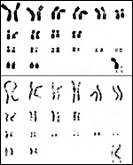
Karyotypes of Pedetes c. capensis from Hsu & Benirschke, 1977.

14) Immunology

No studies are known to me.

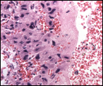
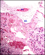
15) Pathological features

A newly imported springhaas with widespread filariasis was described by Anderson et al. (1998). The organism (Filaria versterae) is apparently normally confined to subcutaneous tissue but in this animal, it invaded into thoracic and peritoneal cavities. Because of pulmonary involvement, the animal had to be euthanized. Butynski (1979) found many animals to be infected with a stomach nematode (Physaloptera capensis) that did not have any apparent deleterious impact.

16) Physiologic data

An interesting study on osmoregulation was done by Peinke & Brown (1999). Since the animal is exposed to very arid conditions, they deprived animals of water for up to seven days to test osmotic regulation. They lost up to 30% body weight during that period and regulated their hematocrits and electrolytes appropriately. They thus maintained their plasma volume despite the water deprivation by producing concentrated urine and reducing fecal water loss.

17) Other resources

Many animals exist in zoological gardens, but breeding colonies are not known to exist. We have no cell lines for study in our collection of cell strains at CRES.

18) Other remarks - What additional Information is needed?

Because of the great complexity of the organ, additional studies are needed. It will be of interest to know the length of the umbilical cord, the number of cord blood vessels, and actual data on gonadotropin and steroid production with cellular localization. Chromosomal studies with comparing the results to other rodents are desirable.

Acknowledgement

Most of the animal photographs in these chapters come from the Zoological Society of San Diego. I appreciate also very much the help of the pathologists at the San Diego Zoo. I am appreciative for the material obtained from Dennis Meritt, Ph.D.

References

Anderson, R.C., Gustafson, B.W. and Williams, E.S.: Acute filariasis in a springhaas. J. Wildl. Dis. 34:145-149, 1998.

Bogart, M.H., Scollay, P.A., Cooper, R.W. and Benirschke, K.: Springhaas (Pedetes capensis). Chromos. Inform. Serv. 30:14-15, 1976.

Butynski, T.M.: Reproductive ecology of the springhaas Pedetes capensis in Botswana. J. Zool. (London) 189:221-232, 1979.

Coe, M.J.: The anatomy of the reproductive tract and breeding in the spring haas, Pedetes surdaster larvalis Hollister. J. Reprod. Fertil. Suppl. 6, 159-174, 1969.

Deane, H.W., Rubin, B.L., Driks, E.C., Lobel, B.L. and Leipsner, G.: Trophoblast giant cells in placenta of rats and mice and their probable role in steroid hormone production. Endocrinology 70:407-419, 1962.

Fischer, T.V. and Mossman, H.W.: The fetal membranes of Pedetes capensis, and their taxonomic relevance. Amer. J. Anat. 124:89-116, 1969.

Gotch, A.F.: Mammals - Their Latin Names Explained. Blandford Press, Poole, Dorset, 1979.

Hediger, H. Gefangenschaftsgeburt eines afrikanischen Springahasen, Pedetes caffer. Zool. Garten 17:166-169, 1950.

Horst, C.J. v.d.: On the reproduction of the springhare, Pedetes caffer. Pamph. S. Afr. Boil. Soc. 8:47, 1935.

Hsu, T.C. and Benirschke, K.: An Atlas of Mammalian Chromosomes. Vol. 10, Folio 456, 1977.

Matthee, C.A. and Robinson, T.J.: Molecular phylogeny of the springhare, Pedetes capensis , based on mitochondrial DNA sequences. Mol. Biol. Evol. 14:20-29, 1997a.

Matthee , C.A. and Robinson, T.J.: Mitochondrial DNA phylogeography and comparative cytogenetics of the springhare, Pedetes capensis (Mammalia: Rodentia). J. Mammalian Evol. 4:53-73, 1997b.

Merwe, M.v.d.. Skinner, J.D. and Millar, R.P.: Annual reproductive pattern in the springhaas, Pedetes capensis. J. Reprod. Fertil. 58:259-266, 1980.

Nowak, R.M.: Walker's Mammals of the World. 6th ed. The Johns Hopkins Press, Baltimore, 1999.

Otiang'a-Owiti, G.E., Oduor-Okelo, D. and Gombe, S.G.: Foetal membranes and placenta of the springhare (Pedetes capensis larvalis Hollister). Afr. J. Ecol. 30:74-86, 1992.

Owiti, G.E.O., Oduor-Okelo, D. and Gombe, S.: Ultrastructure of the chorioallantoic placenta of the springhare Pedetes capensis larvalis Hollister. Afr. J. Ecol. 23:145-152, 1985.

Peinke, D.M. and Brown, C.R.: Osmoregulation and water balance in the springhare (Pedetes capensis). J. Comp. Physiol. [B] 169:1-10, 1999.

Puschmann, W.: Zootierhaltung. Vol. 2, Säugetiere. VEB Deutscher Landwirtschaftsverlag Berlin, 1989.

Rosenthal, M.A. and Meritt, D.A.: Hand-rearing springhaas Pedetes capensis at Lincoln Park Zoo, Chicago. Intern. Zoo Yearb. 13:135-137, 1973.

Simpson. G.G.: The principles of classification and a classification of mammals. Bull. Amer. Museum Nat. Hist. 85:1-350, 1945.


back to top

© 2002. All rights reserved.
welcome | home | intro | placentation | glossary | author contact me: kbenirsc@ucsd.edu