(Clicking on the thumbnail images below will launch a new window and a larger version of the thumbnail.)


Three-toed Sloths
Bradypus tridactylus, B. torquatus, B. variegatus

Order: Xenarthra
Family: Bradypodidae

1) General Zoological Data


Three-toed sloths occur in most regions of South America. Three species have been delineated (Nowak, 1999). Wetzel & Kock (1973) provided a detailed consideration of the pelage and other distinguishing features, as well as the distribution of the various species (see also Wetzel & Avila-Pires, 1980). The most easily followed nomenclature and distribution map, however, is that of Wetzel (1985) which indicates that the brown-throated three-toed sloth (Bradypus variegatus) is the most widely distributed sloth species in South America. It, as the other species, has had many synonyms. A chromosomal difference was detected in some of these by Jorge et al. (1985). B. infuscatus (probably variegatus) had 2n=54, B. tridactylus had 2n=52, but a specimen of B. variegatus had 2n=55. Later, Jorge and Pinder (1990) studied three specimens of B. torquatus from Northern Brazil, perhaps Brazil's most endangered species. They found them to have only 50 chromosomes. Thus, the picture is still confusing, although it must be admitted that their species designation is not necessarily concordant with that supplied by Wetzel (1985). For a detailed discussion of the complex xenarthran phylogeny and many further specifics, the reader is directed to the inclusive monograph of Montgomery (1985). In addition, Wetzel (1982) has published a detailed review of xenarthran systematics.

Adult three-toed sloths weigh around 2.25-6.2 kg. Their shaggy hair is gray-brown, often greenish because of algal growth and much depends on the subspecific designation. Unlike Choloepus, the two-toed sloth, Bradypus, has a short tail and, of course, three toes on all extremities. Its forelegs are also longer. It is further distinguished by having 8-9 neck vertebrae, which allow their marked rotational ability of the head. They are nearly totally arboreal, can swim well, and are preyed upon, i.a., by harpy eagles. Their folivorous diet is mainly directed to consuming Cecropia, but some other leaves may also be eaten. Nowak (1999) wrote many fascinating details and described differences between the 2-toed and 3-toed sloths, and a charming, popular account on sloths was written by Hoke (1987).

The evolutionary relationship of Xenarthra, including the sloths, is poorly resolved as yet. This is a very diverse group of animals and they have a long history. Large ancestors to the sloths existed in their past. The descendency of extant sloths has recently been studied with molecular tools (Greenwood et al., 2001). These investigations have suggested two distinct lineages for the two genera of sloths: The two-toed sloths were related to the mylodontids, while the three-toed sloths appeared to be molecularly more similar to megatherid ancestors. A somewhat different interpretation was arrived at by the molecular studies of Delsuc et al. (2001). They found sloths and anteaters to group together and assigned a more recent origin of sloths than is commonly held to be the case.
   
  Three-toed sloth with young (Courtesy, G.G. Montgomery).
     
  2) General Gestational Data

There is usually a single offspring, after a gestation of 106 days. Wislocki (1927), who had the largest material, never saw more than one implanted embryo. Newborns weigh 200-250 g. The placental weight of term organs is around 60-80 g.

3) Implantation

Implantation occurs mainly at the fundus of the pear-shaped uterus. At first, the entire conceptus is covered with villous/lobular tissue. Later in development, however, in much of the lower portion (over the cervix and some anterior uterine parts) of the uterus, the villous tissue atrophies. The implantation occurs with invasion of the decidua.

Wislocki (1935) studied the uteri of four early gestations in B. griseus and remarked that the anterior portion of the uterus is larger, the posterior flatter and thinner in these early specimens. The decidua also had a different appearance in this material. The allantois is very rudimentary and was observed only in serial sections of the body stalk. The yolk sac, on the other hand, was prominent and was described in detail. No omphalo-placental circulation ever developed because the yolk sac detaches from the chorion very early. The entire blastocyst surface contacts and invades the endometrium, removing the epithelium in the process. As a result of the trophoblastic infiltration of the decidua, the maternal vessels and their endothelium hypertrophy significantly. While initially the trophoblast is of the cytotrophoblastic variety, it soon becomes primarily and then exclusively syncytial. The syncytium appears to be the invasive cell type, but it never breaches the maternal vascular endothelium.

4) General Characterization of the Placenta

The first comprehensive description comes from the pen of Becher (1921) who reviewed earlier contributions on two-toed sloths and highlighted the differences in structure. Becher reviewed the inaccessible contribution by Klinkowstrom (1895) who had three different developmental stages available. Remarkably, in the youngest stage, the chorionic lobules were distributed all over the chorion and only later did they concentrate into 1 or 2 connected lobes at the fundus. The fetus of Becher's specimen was 10 cm long. The placenta had the usual lobular projections on the fetal surface which he equated which cotyledons. He, as well as other students, was much impressed with the ease of placental separation and described in considerable detail the decidua vera and that which separates with the placenta at its floor. The lobules ("cotyledons") are separated one from another by decidual septa. Moreover, he then discussed numerous giant cells that he considered to originate from blood cells. This is probably incorrect. The chorion and villi are covered by a syncytium that lies directly adjacent to the maternal vascular endothelium. Thus, an endothelial-chorial relation was established for this placenta.

Wislocki (1925) made the next important observations (on a specimen of B. griseus) which was followed by several additional studies. The fetus of his specimen was 24.5 cm long. In that specimen, the lobulated cotyledonary tissue covered 3/4 of the uterine surface ("bell-shaped") and appeared to be constructed of two adjoining halves. The cord, centrally inserted between the two lobes, had a smooth surface. He preferred to speak of placental "lamellae" and considered the designation "villi" to be an inappropriate designation for the placental labyrinth. Like Becher, Wislocki found a trophoblastic syncytium that surrounds the rather larger-than-capillaries maternal blood vessels in the labyrinth and then considered this placenta to have a syndesmo-chorial relationship because of the thin layer of connective tissue that remained around many maternal vessels. In all of the descriptions thus far, much emphasis was placed on the coexistence of many regressive changes occurring, with atrophy of whole cotyledons.

In a subsequent publication, Wislocki (1926) described 14 pregnant uteri of various stages of gestation. Much like the "fourth membrane" of the camelids, the sloths have a thin "epitrichium", a delicate membrane that is attached to the nostrils, anus, toes, etc. Later (1927), he elaborated on these specimens and was finally convinced that the placenta is actually an endothelio-chorial organ, and he likened it to the barrier of carnivores. Later, in reply to de Lange's paper (1926), Wislocki (1928) emphasized that the trophoblast was syncytial everywhere, without any evidence of cytotrophoblast. He strongly disputed de Lange's assumption of sloths having a hemochorial placenta.

De Lange (1926), who had two uteri for examination, was of the opinion that the trophoblast relation to the uterus was mixed, between endothelio-chorial and hemochorial. But, King et al. (1982) laid all these arguments to rest when they studied uteri by electron microscopy. In young specimens they found a syndesmochorial relation but the more advanced specimens were definitely endothelio-chorial. They reminded these authors of the structure of the shrew's placenta. This conforms to our own observations (Benirschke & Powell, 1985). Two other aspects should be mentioned:

1) there is a large number of large macrophages within the villous connective tissue that were described as "epithelioid cells" by Wislocki (1927), and 2) the endothelium of the labyrinthine maternal vessels are unusually large and thus confusing on first observation.

Through the generosity of Drs. Z. Silva and W. Jorge in Brazil I have been able to examine one three-banded sloth conceptus that is depicted here (Benirschke & Powell, 1985). Other reports will be cited from the literature. The entire uterus was available and contained a fully-grown male fetus (27 cm long, 185 g) that was attached by a 15 cm umbilical cord to a multilobulated placenta that weighed 60 g and measured 11 x 7 cm. It had a marginal "infarct" (areas of probably normal atrophy). There were about 20 placental lobules that had fused to a single disk of placental tissue. The membranes made up approximately 50% of the whole specimen. The placenta was readily detached from the uterus. The uterus was single, unicornuate, and measured 12 x 5 cm. The placenta had a fundal implantation. The fallopian tubes were 7.5 cm long and the distance between their origins was 4 cm. The ovaries were large and located in a bursa (8 x 3 cm).

   
  Full term gestation of three-toed sloth, with placenta at left. Its "infarct" is at the top left, white. (Courtesy Drs. Silva & Jorge, Brazil).
     
  The same placenta at its fundal implantation site. Right ovary with corpus luteum at top right.
     
  Uterus with bladder and anterior wall removed to expose the fetal position.
     
  Opened uterus with fetus deflected to the right. It shows that the lower portion of the formerly cotyledonary tissue is undergoing atrophy (white areas at bottom of placenta, at arrows).
     
  Opened uterus with fetus deflected to the right. It shows that the lower portion of the formerly cotyledonary tissue is undergoing atrophy (white areas at bottom of placenta, at arrows).
     
  Portion of attached placenta with uterus below. The two protuberant "lobules" are evident. They are separated by the septum (S) composed of decidual tissue.
     
  5) Details of fetal/maternal barrier

The term placenta is an endothelio-chorial organ. Maternal blood vessels, lined by hypertrophic endothelial cells, are surrounded by trophoblastic syncytium. In younger stages of development, the syncytium is seen to destroy the decidua and a remnant of connective tissue surrounds the vessels thus, technically speaking, the placenta is a syndesmochorial organ at that time.
   
  One of the protuberant placental lobules, markedly congested and separated from the adjacent lobule by the septum on the right. Note the large number of small chorioallantoic blood vessels in the chorion.
     
  A septum of decidual tissue protrudes between two regressing lobules of labyrinth. Myometrium at bottom.
     
  The relation of a "villous/lamellar" fetal structure with its villous connective tissue (VCT) that harbors numerous "epithelioid" cells, corresponding to macrophages or "Hofbauer cells". The endothelium (E) of the maternal vessel is hypertrophic.
     
  Higher magnification of maternal-fetal relation. MV= maternal vessel. Note how closely the trophoblastic syncytium is attached to the endothelium.
     
  It is at this plane that the placenta separates in the decidua, leaving some decidua basalis and decidual septa with the placenta. Note the large maternal artery that separates with the placenta.
     
  Cross section through the lamellar labyrinth of a sloth placenta. The dark red areas are the maternal vasculature, the lighter areas are "villous" tissue.
     
  6) Umbilical cord

The cord of our specimen was 15.5 cm long, had very few spirals, and contained two arteries and one vein. No ducts were found to persist and there were no surface excrescences. Wislocki (1926) found the cord usually attached to the midsagittal posterior plane of the uterus. It has a Y-shaped bifurcation shortly before its placental insertion, with the vessels then running to the two nearly joined lobes.

7) Uteroplacental circulation

This has not been studied in any detail. The maternal blood vessels of the decidua, surrounded by trophoblastic syncytium, are large and do not really have much of a capillary structure. There is no ingrowth of trophoblast into the maternal vessels. Physiologic studies or perfusion experiments have not been performed.

8) Extraplacental membranes

Becher (1921) described numerous small amnionic nodules in his young specimen that were not present in our term placenta, nor in Wislocki's specimen (1925). Later, however, Wislocki (1926; 1927) observed amnionic "villi", small projections of connective tissue that bulge up the amnionic epithelium. The amnion consists of thin single-layered epithelium. Wislocki (1927) stated that, the portions of the membranes that lack cotyledons (where they had regressed, would be a better description), always contain blood vessels. We also found in addition to the large, "normal" fetal blood vessels a host of tiny vessels to persist in these membranes (Benirschke & Powell, 1985). We also found numerous pigmented macrophages in the amnionic connective tissue. Meconium, however, has never been observed in the sloths' uteri.

Becher also made extensive observations on the decidua formation in this species. He found large glands early in gestation which, by the stage Wislocki was able to examine this tissue (1925), had shrunken. Although the yolk sac is a prominent structure in early gestation, vitelline remnants have not been identified at term.

   
  This is the edge of the placenta with the membranes showing a decidua capsularis on the outside, much as is seen in human placentas.
     
  9) Trophoblast external to barrier

The invasion of syncytial trophoblast into the decidua has best been described in Wislocki's 1927 contribution. He was of the opinion that the trophoblast destroys decidual cells and the maternal vascular walls, up to their endothelium. He thus considered that the maternal tissue was gradually transformed into the lamellae of the labyrinthine tissue.

10) Endometrium

There is a large quantity of decidua capsularis and basalis that separates with the placenta (see Becher, 1921).

11) Various features

There is no true "subplacenta" but a decidua basalis with remnants of glands is present and the placenta separates in a decidual plane. We have not found trophoblast invasion of either spiral arterioles or the myometrium.

12) Endocrinology

I know of no endocrine studies done in sloths. It is remarkable, however, to witness the size of the fetal adrenal gland. It possesses a wide "fetal zone", much like the nine-banded armadillo and human fetus has (Moser & Benirschke, 1962). In the human fetus, this region produces dehydroepiandrosterone, as precursor for estriol. Similarly, the interstitial cell component of the testis is markedly stimulated.

   
  The adrenal gland of the sloth has a striking "fetal zone" that disintegrates after birth.
     
  The fetal testis has a large component of stimulated interstitial cells in between the tubules.
     
  13) Genetics

Personal communications from Dr. W. Jorge in Brazil indicates that all five specimens studied by him had 54 chromosomes, with an XX/XY sex determining mechanism. The same author, however, provided additional details in his later contribution (Jorge et al., 1985). B. tridactylus had 52 elements, B. infuscatus (probably variegatus) had 54 chromosomes, and a specimen of B. variegatus had 55 chromosomes. In a later contribution, Jorge & Pinder (1990) studied the very much endangered Bradypus torquatus from Northern Brazil and found them to have 2n=50, with XX/XY sex determining mechanism.

No hybrids have been described.

14) Immunology

I know of no immunological studies done in sloths.

15) Pathological features

Diniz and Oliveira (1999) described the clinical and pathological conditions of sloths in captivity. They found digestive and respiratory problems to be the most notable. A variety of parasites were found in fecal examinations and they stated that the first 6 months in captivity were the most critical. Thereafter, management is less problematic. Seymour et al. (1983) isolated a large number of poorly studied viruses from sloths in Panama. Further details on parasites infecting the sloths may be found in contributions to the monograph by Montgomery (1985).

16) Physiologic data

A variety of physiologic studies have been undertaken in Bradypus. For instance, da Mota et al. (1992) have studied pancreatic endocrine cells and found that glucagon-producing cells are much commoner than found in the pancreas of human or laboratory animals. They related this to their folivorous diet and possibly to their phylogeny. A comprehensive review of sloths' physiology is found in the contribution by Gilmore et al. (2000). In their additional paper of 2001, these authors specifically elaborated on the hair structure of sloths with its algal growth, as well as their ecology of carrying specific disease agents (viruses, parasites, and leishmania). The cardiovascular adaptation to postural change was studied by Duarte et al. (1982). Many other fascinating details of their physiology are highlighted in an editorial (1974), such as the abdominal testes, the conservation of temperature, the infrequent urination and defecation.

17) Other resources

Some cell strains are available from CRES at the San Diego Zoo by contacting Dr. Oliver Ryder at: oryder@ucsd.edu.

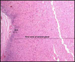
18) Other remarks - What additional Information is needed?

Endocrine studies of placentas and gestation are needed to identify the possible presence or the absence of placental gonadotropins. The presence of so large a fetal adrenal gland demands the study of fetal endocrine aspects.

Acknowledgement

I appreciate very much the help of the pathologists at the San Diego Zoo.

References

Becher, H.: Zur Kenntnis der Placenta vom Bradypus tridactylus. Z. Anat. Entwicklungsgesch. 61:114-136, 1921.

Benirschke, K. and Powell, H.C.: On the placentation of sloths. Pp. 237-241, In, Montgomery, G.G., ed.: The Evolution and Ecology of Armadillos, Sloths, and Vermilinguas. Smithsonian Institution, Washington, 1985.

Delsuc, F., Catzeflis, F.M., Stanhope, M.J. and Douzery, E.J.: The evolution of armadillos, anteaters and sloths depicted by nuclear and mitochondrial phylogenies: implications for the status of the enigmatic fossil Eurotamandua. Proc. Roy. Soc. Lond. B Biol. 268:1605-1615, 2001.

Diniz, L.S. and Oliveira, P.M.: Clinical problems of sloths (Bradypus sp. and Choloepus sp.) in captivity. J. Zoo Wildl. Med. 30:76-80, 1999.

Duarte, D.P., da Costa, C.P. and Huggins, S.E.: The effects of posture on blood pressure and heart rate in the three-toed sloth, Bradypus tridactylus. Comp. Biochem. Physiol. A 73:697-702, 1982.

Editorial: Sloth's compensations. The Lancet ii:1054, 1974.

Gilmore, D.P., da-Costa, C.P. and Duarte, D.P.: An update on the physiology of two- and three-toed sloths. Braz. J. Med. Biol. Res. 33:129-146, 2000.

Gilmore, D.P., da Costa, C.P. and Duarte, D.P.: Sloth biology: an update on their physiological ecology, behavior and role as vectors of arthropods and arboviruses. Braz. J. Med. Biol. Res. 34:9-25, 2001.

Greenwood, A.D., Castresana, J., Feldmaier-Fuchs, G. and Paabö, S.: A molecular phylogeny of two extinct sloths. Mol. Phylogenet. Evol. 18:94-103, 2001.

Heuser, C.H. and Wislocki, G.B.: Early development of the sloth (Bradypus griseus) and its similarity to that of man. Contrib. Embryol. Carnegie Inst. Washington 25:1-13, 1935.

Hoke, J.: Oh, it's so nice to have a sloth around the house. Smithsonian 18:88-98, 1987.

Jorge, W., Orsi-Souza, A.T. and Best, R.: The somatic chromosomes of Xenarthra. Pp.121 in, The Evolution and Ecology of Armadillos, Sloths, and Vermilinguas. G.G. Montgomery, ed. Smithsonian Institution, Washington, 1985.

Jorge, W. and Pinder, L.: Chromosome study on the maned sloth Bradypus torquatus (Bradypodidae, Xenarthra). Cytobios 62:21-25, 1990.

King, B.F., Pinheiro, P.B.N. and Hunter, R.L.: The fine structure of the placental labyrinth in the sloth, Bradypus tridactylus. Anat. Rec. 202:15-22, 1982.

Lange, D.de: Quelques remarques sur la placentation de "Bradypus". Compt. Rend. Assoc. Anat. Liège 21:321-333, 1926.

Meritt, D.A. and Meritt, G.F.: Sex ratios of Hoffmann's sloth, Choloepus hoffmanni Peters, and three-toed sloth, Bradypus infuscatus Wagler, in Panama. Amer. Midl. Nat. 96:472-473, 1976.

Montgomery, G.G., ed.: The Evolution and Ecology of Armadillos, Sloths, and Vermilinguas. Smithsonian Institution, Washington, 1985.

Moser, H.G. and Benirschke, K.: The fetal zone of the adrenal gland in the nine-banded armadillo, Dasypus novemcinctus. Anat. Rec. 143:47-60, 1962.

Da Mota, D.L., Yamada, J., Gerge, L.L. and Pinheiro, P.B.: An immunohistochemical study on the pancreatic endocrine cells of the three-toed sloth, Bradypus variegates. Arch. Histol. Cytol. 55:203-209, 1992.

Nowak, R.M.: Walker's Mammals of the World. 6th ed. The Johns Hopkins Press, Baltimore, 1999.

Seymour, C., Peralta, P.H. and Montgomery, G.G.: Viruses isolated from Panamanian sloths. Amer. J. Trop. Med. Hyg. 32:1435-1444, 1983.

Wetzel, R.M. and Avila-Pires, D.D. de: Identification and distribution of the recent sloths of Brazil (Edentata). Rev. Bras. Biol. 40:831-836, 1980.

Wetzel, R.M. and Kock, D.: The identity of Bradypus variegatus Schinz (Mammalia, Edentata) Proc. Biol. Soc. Wash. 86:25-34, 1973.

Wetzel, R.M.: Systematics, distribution, ecology, and conservation of South American edentates. Pp. 345-375, in "Mammalian Biology in South America. Special Publication Series of the Pymatuning Laboratory of Ecology, University of Pittsburg, Vol. 6, 1982.

Wetzel, R.M.: The identification and distribution of recent Xenarthra (=Edentata). Pp. 5-21, In, Montgomery, G.G., ed.: The Evolution and Ecology of Armadillos, Sloths, and Vermilinguas. Smithsonian Institution, Washington, 1985.

Wislocki, G.B.: On the placentation of the sloth (Bradypus griseus). Contrib. Embryol. Carnegie Inst. Washington 16:7-19, 1925.

Wislocki, G.B.: Further observations upon the placentation of the sloth (Bradypus griseus). Anat. Rec. 32:45-51, 1926.

Wislocki, G.B.: On the placentation of the tridactyl sloth (Bradypus griseus) with a description of some characters of the fetus. Contrib. Embryol. Carnegie Inst. Washington 19:211-228, 1927.

Wislocki, G.B.: Further observations upon the minute structure of the labyrinth in the placenta of the sloths. Anat. Rec. 40:385-395, 1928.


back to top

© 2002. All rights reserved.
welcome | home | index | intro | placentation | glossary | author | contact