 |
| (Clicking
on the thumbnail images will launch a new window and a larger version
of the thumbnail.) |
| Last updated: June 28, 2011. |
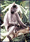
Silvered Leaf Monkey
Trachypithecus cristatus
Order: Primates
Family: Cercipothecidae
Subfamily: Colobinae
1. General Zoological Data
According to Nowak (1999) they are also listed as Presbytis or Semnipithecus . Thus, Hayssen et al. (1993) list them as P. cristatus. According to Jones (personal communication) they may reach an age of 31 years in captivity. Gray (1972) quotes several authors who described hybrids with T. phayrei. These animals come from Indonesia (Wilson & Reeder, 1992) and the remarkable newborns are yellow for a period of at least one year. They make loud calls and live in the canopy of large trees. They eat leaves, fruit and seeds but their maintenance in many Zoological Gardens is difficult, as their diet requires largely leaves and fruit (Puschman, 1989, 2004).
 |
Adult animal from pictures in Sleeper & Wolfe (1997). |
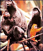 |
Adult animals with one yellow young from Sleeper & Wolfe (1997). |
General Gestational Data
Hayssen et al. (1993) suggest that their gestation is around 140 days.
Implantation
No data are available on early implantational stages.
General Characterization of the placenta
As is true of many other primates, the placenta is composed of two separate lobes. The one with the cord was 37.3 g; the secondary lobe was 38.8 g. Small marginal infarcts may be seen at the left and right of the accessory lobe. Large vessels traversed from one lobe to the other. A detailed description of Colobine placentas was published by Benirschke (2008).
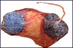 |
Fetal side of the placenta with its two disks. The large vessels between the lobes are clearly evident in the connecting membranes. The lateral infarcts are seen in the secondary disk. |
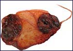 |
Maternal side of the placenta with the connecting vessels in the membranes. |
Details of fetal/maternal barrier
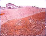 |
Low power view of the fetal surface with amnion partially detached and fetal vessel in the chorion. |
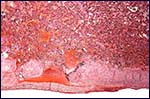 |
Maternal surface of the placenta with decidua basalis at base. |
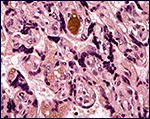 |
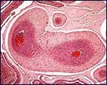
Villi of this hemochorial placenta with syncytial knots and large (brownish) fetal blood vessel. |
Umbilical cord
The umbilical cord inserted eccentrically on one disk and large membranous blood vessels traversed to the other disk. The cord was 18 cm long and contained three blood vessels.
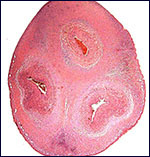 |
Cross section of the normal placenta. |
Uteroplacental circulation
The intervillous circulation is fed by typical decidual spiral arterioles.
Extraplacental membranes
 |
Large blood vessels in the connecting membranes between the two lobes. |
Trophoblast external to barrier
No trophoblast was encountered but then the uterus was not available.
Endometrium
There is typical decidua basalis, as seen in human placentas.
Various Features
The stomachs have several partitions as shown by Scott (1992), as are commonly seen in most langur species.
Endocrinology
Endocrine features are listed by Hayssen et al. (1993) but they refer to P. entellus only.
Genetics
This species, as most langurs, has 44 chromosomes. Their details were described in a contribution by Bigoni et al. (1997). The karyotype shown next is from O’Brien et al. (2006).
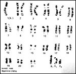 |
Karyotype of Trachypithecus from O’Brien et al. (2006). |
Immunology
No details are described.
Pathological features
Infarcts were seen in the secondary lobe. Griner (1983) described the autopsy following euthanasia of a senile animal dying from Salmonella infection. Scott (1992) lists a few other pathological features but he is not specific as to which species he refers to.
Physiologic data
The sacculations of the langur stomach are depicted by Scott (1992).
Other resources
The cells of this species are frozen in the “Frozen Zoo” of the San Diego Zoo and can be made available by requesting them from Dr. Oliver Ryder
(oryder@ucsd.edu).
Other remarks – What additional information is needed?
Early implantational stages are needed.
Acknowledgement:
The colored photos were taken from: “Primates” (Subtitle: ‘The amazing world of lemurs, monkeys, and apes’) by B. Sleeper & A. Wolfe; Chronicle Books, San Francisco 1997.
References
Benirschke, K.: The placenta of the colobinae. Vietnamese J. Primatol. 2:393-339, 2008.
Bigoni, F., Koehler, U., Stanyon, R. Ishada, T. and Wienberg, J.: Fluorescence in situ hybridization establishes homology between humans and silvered leaf monkey chromosomes, reveals reciprocal translocations between chromosomes homologous to human Y/5, 1/9, and 6/16, and delineates an X1X2Y1Y2/X1X1X2X2 sex-chromosome system. Amer. J. Primatol. 102:315-327, 1997.
Gray, A.P.: Mammalian Hybrids. A Check-list with Bibliography. 2nd edition.
Commonwealth Agricultural Bureaux Farnham Royal, Slough, England, 1972.
Griner, L.A.: Pathology of Zoo Animals. Zoological Society of San Diego, San Diego, California, 1983.
Hayssen, V., van Tienhoven, A. and van Tienhoven, A.: Asdell’s Patterns of Mammalian Reproduction: a Compendium of Species-specific Data. Comstock/Cornell University Press, Ithaca, 1993.
Nowak, R.M.: Walker’s Mammals of the World. 6th ed. The Johns Hopkins Press, Baltimore, 1999.
O’Brien, S.J., Menninger, J.C. and Nash, W.G., eds.: Atlas of Mammalian Chromosomes. Wiley-Liss; A. John Wiley & Sons, Inc. Hoboken, N.J., 2006.
Puschmann, W.: Zootierhaltung. Tiere in menschlicher Obhut. Säugetiere. Verlag Harri Deutsch, Frankfurt, 2004.
Puschmann, W.: Zootierhaltung. Vol. 2, Säugetiere. VEB Deutscher Landwirtschaftsverlag Berlin, 1989.
Sleeper, B., with pictures by A. Wolfe: Primates (The amazing world of lemurs, monkeys, and apes). Chronicle Books, San Francisco, 1997.
Wilson, D.E. and Reeder, D.A.M.: Mammal Species of the World. A Taxonomic and Geographic Reference. 2nd ed. Smithsonian Institution Press, Washington, DC, 1992.