(Clicking on the thumbnail images below will launch a new window and a larger version of the thumbnail.)
 

Last updated:
May 20, 2004.

Domestic Sheep
Ovis aries


Order: Artiodactyla
Family: Bovidae


1) General zoological data of species

Domestic sheep have presumably derived from Ovis musimon, the mouflon, a species that is distributed across Mediterranean islands. Domestication is thought to have begun approximately 10,000 years ago. There are six species and numerous subspecies of sheep distributed throughout Asia and North America, but the domestic sheep now occurs worldwide. Because some sheep species and/or subspecies are significantly endangered, they now require protection and have been listed as CITES I species (Nowak, 1999). Longevity varies with species and race. For mouflons, the maximal life span is given as 19-20 years (Puschmann, 1989).

   
  Bighorn sheep.
     
  Domestic ewe with mouflon offspring from embryo transfer (Courtesy Dr. T.D. Bunch).
     
  2) General gestational data

Keisler (1999) has provided a detailed review of the reproductive parameters of domestic sheep and goats. The reader is also referred to the Chapter on Cretan Goat. That species' reproductive aspects have many similarities. There are about 800 different "breeds" of sheep with many different physical characteristics. Depending on the breed, puberty occurs between 5 and 12 months. Breeding occurs throughout the year in domestic sheep and is somewhat dependent on photoperiods. The duration of estrus is in between 3 and 72 hrs. (average 29 hrs.); it is influenced by the presence of a male. The estrous cycle averages 16 days.

While the length of gestation in wild sheep species varies between 150 and 180 days, the domestic sheep has an average length of gestation of 148 days. Most sheep produce singletons, but some races regularly have twins, even triplets. Singletons weigh about 1.5 kg (Alexander, 1964). The placental weight is difficult to ascertain as the membranes are voluminous and of little importance to fetal growth as such. Kleemann et al. (2001) provided weights around 600 g. They were interested in the relation of placental and fetal sizes after progesterone treatment in early gestation. Whether these weights reflect a direct relation to the provision of nutrients to the fetus can be questioned. Therefore, investigators have weighed what they considered to be the most meaningful weight - that of blotted cotyledons. This is discussed below in detail (Alexander, 1964).

3) Implantation

Within three to four days, the fertilized ovum reaches the uterus as a morula. The blastocyst develops by day 6 and sheds its zona pellucida by day 8 or 9. The blastocyst enlarges rapidly after the tenth day (Assheton, 1906). It becomes "sticky" and, on about day 17-18, it attaches with cytoplasmic protrusions to the endometrial surface. Naturally, this attachment is principally at the caruncles of the endometrium. The placenta remains diffuse for the first month or so after its mesometrial attachment (King et al., 1982). Lawn et al. (1969) did an electronmicroscopic study of goat and sheep placentas. Their youngest sheep gestation was at 42 days of pregnancy; the cotyledons were then just beginning to develop a villous structure. The trophoblast was apposed to an intact endometrium that had the appearance of protein secretion. Microvilli were abundant and occasional binucleated trophoblastic cells were present. Interdigitation of the cells progressed thereafter and the endometrial epithelium began to transform. Moreover, multinucleated giant cells became apparent. The syncytial cytoplasm then also acquires characteristic cytoplasmic granules.
   
  Uterine caruncles of immature common waterbuck (Kobus kob) with normal endometrium in between. (Courtesy, Dr. P. Hradecky).
     
  4) General characteristics of placenta

The sheep placenta is a polycotyledonary placenta with 70 to 100 cotyledons. Number and size of cotyledons varies widely with maternal age, strain of sheep and perhaps it depends on yet some other factors. The cotyledons are the fetal portions of the placentomes. The latter term, placentome, additionally encompasses the maternal caruncle.

In between the cotyledons is the intercotyledonary chorion, which has an additional and different absorptive function. It is covered by simple trophoblast and modified only over the mouths of endometrial glands. These regions are referred to as the "areolae". In between the areolae there are the "arcades" with focal hematomas and absorption of blood by trophoblast.

After the initial expansion of the blastocyst at about day 10, the elongated chorionic sac fills both of the uterine horns. It is entirely covered with trophoblast. This large sac contains a large, elongated allantoic cavity that apposes the chorion to the end of the outer chorionic sac and apposes the amnion directly over its central portions. The yolk sac is short-lived and plays no major role in ovine placentation and it cannot be found at term.

A bighorn sheep placenta received in May, 2004 weighed 300g, measured 83 cm in longest dimension, had 51 small concave cotyledons and a 27 cm long umbilical cord with four vessels.
   
  Diagram of opened sheep placenta. The largest compartment is the allantoic sac. Inside is the smaller amnionic cavity with the fetus. Dark spots are the placentomes - cotyledons.
     
  Immature sheep placentome. The pale areas are villous tissue; in between villi are the maternal sheaths.


Mossman (1987) differentiated between three types of cotyledons: the "flat" kind of certain deer, the "convex" kind of other deer and giraffes, and the "concave" kind of sheep and caprines. The concave cotyledonary sheep placenta implants primarily upon the uterine caruncles. In between the cotyledons, the chorion is simple and lacks villi. The trophoblast intertwines with the uterine endometrium in the caruncular regions, to form the so-called "placentome". Following death of a pregnant animal it is easy to peel the placenta away from the endometrium, without significant damage to either structure. There is a large allantoic sac filled with urine. Urine is led to it through the allantoic duct and that passes from the dome of the bladder through the umbilical cord. Some urine must also pass through the urethra into the amnionic cavity. Sheep possess multiply branched villi, in contrast to some other ruminants. In reconstructions they appear as folds, rather than the multiply branched thin villi that are seen in primate placentas.

Alexander (1964) sought to explore the relation between fetal weight and cotyledonary weights in Merino sheep gestations; Corredale sheep had slight differences and were not described in detail. He extensively discussed the differences between different breeds, and emphasized the need to be mindful of the existence of wide differences. He suggested that it is necessary to collect normative data for breeds and altitude when studies are undertaken to ascertain influences of altitude etc. on fetal development. The number of cotyledons was poorly related to birth weight, in contrast to their weights. Not all caruncles are used for cotyledonary development and this apparently varies widely. Moreover, it appears that some caruncles disappear with aging. Male fetuses occupy more caruncles than do female fetuses. Alexander separated the cotyledons (without intervening membranes) from the caruncles by traction; after blotting them dry, he weighed them. There was an excellent correlation between fetal weight and cotyledonary mass at different stages of gestation. Male fetuses and cotyledons were slightly heavier. Those of twins were slightly smaller. The average number of uterine caruncles was 73 in Merino sheep (from 64 to 145). Also, the number of caruncles was generally similar in both uterine horns. Older ewes had a slightly heavier cotyledonary mass which varied from 115 to 130 g. Individual cotyledons weighed about 1.6-1.8 g. There were, however, enormous differences in their weights - 0.1 -12 g of fetal cotyledons and 0.1-45 g of complete placentomes. The fetal part of a complete cotyledon was 40% and was darker than the maternal part. Alexander made another important observation. He found that whereas in immature cotyledons the maternal tissue (caruncle) tissue surrounds the cotyledon (which is concave, according to Mossman), towards term the opposite is true. The components then become convex. It is further interesting to contemplate that most of these investigators considered only the cotyledonary weight although it had been long appreciated that the intercotyledonary region serves also as an important exchange (nutritive) region. Perhaps it should be included in the weights when an association with fetal growth is sought.
Osgerby et al. (2003) studied the effects of maternal nutrition on the condition of neonates and placenta. They showed convincingly that the type of nutrition significantly influences placental cotyledonary structure as well as fetal condition.

5) Details of barrier structure

The nature of the interface between maternal epithelium and trophoblast was described in the electronmicroscopic study of Lawn et al. (1969). This interface comprises also an area in which maternal blood is extravasated "to supply the fetus with iron". Wimsatt (1950) had studied this region in great detail and it will be discussed more fully below.

The endometrial crypt is lined with epithelium that has a striking microvillous surface. This, in turn, is closely adherent to the trophoblast with which its microvilli interdigitate. The maternal epithelium is continuous, is not interrupted, and it is often multinucleated. The microvilli of trophoblast and endometrium interdigitate diffusely and thus, this placenta is here epitheliochorial (-bichorial to be correct), as was first clearly defined by Ludwig (1962). Because of degeneration of some endometrial epithelium during the course of gestation, areas of syndesmochorial relation exist. Mossman (1987), however, correctly pointed out that there is little vascular supply in those areas and, from a functional viewpoint, they are probably of little importance. Thus, one should consider that the sheep placenta is principally an epitheliochorial one.

Lawn et al. (1969) paid special attention to the development of the well-known binucleate cells of the ruminant placenta. This cell has spawned an enormous amount of literature. These investigators traced the cells' development from early nuclear division (without cytoplasmic division) to their accumulation of numerous cell organelles. Among these the Golgi complex appeared first and was then succeeded by a large number of granules. In the mature cell, the granules fill the cellular cytoplasm, nearly crowding out all other components. It was also shown that these cells never completely bordered the endometrial epithelium - a rim of cytoplasm always separated the two cell types. Contrary to earlier investigators then, Lawn et al. (1969) correctly identified the binucleate cells as having derived from trophoblast. The organelles, as well as some of the endometrial crypt lining cells, stain intensely with the PAS method. They are now known to contain placental lactogen.

The original classification of the sheep placenta as being an epitheliochorial organ, however, is no longer considered being entirely correct. (Please see also the chapter on Cretan Goat for additional considerations.) Autoradiographic studies by Wooding et al. (1981) probably now supersedes the older information. Relative to its cotyledons, the ovine/caprine placenta must now be considered as a focally syndesmochorial organ. The intercotyledonary regions remain epitheliochorial in the mature placenta, as had been previously determined.

The most controversial aspect perhaps of ungulate placentation has been the origin of the syncytium that lines the crypts of the cotyledons. Similarly, the nature of, or the relation of the syncytium to the binucleated cells has been interpreted differently. Binucleate cells are clearly trophoblastic in origin and they produce the placental lactogen. The autoradiographic study by Wooding et al. (1981) investigated the origin of syncytial cells. It appeared to me that it convincingly showed that the syncytium has a trophoblastic origin and that it is not derived by fusion with endometrial epithelium, as was once thought. Presumably, the binucleate cells initiate the process of fusion and subsequent migration within the trophoblastic layer, and they continue to do so during the entire pregnancy. Moreover, the fact that there are no mitoses in the syncytium, while they do occur in the cellular trophoblast, supports this notion. Thus, the syncytium was considered to develop much like other fusion-induced cells. Wooding (1984) later showed that some fusion with maternal epithelium also produces trinucleate hybrid cells. These cells are quite specific, and secretion of the lactogen-containing granules (perhaps to maintain gestation) was described in his paper. Wooding emphasized that there is no immune reaction as the result of maternal epithelial erosion and this fusion ("hybridization" of cells). This may be an erroneous conclusion, as will be seen shortly.

In addition to the binucleate cells, numerous giant cells are found deep in the placentome. According to Lawn et al. (1969) they originate from the endometrial epithelium in later gestation; more likely they are part of the "syncytium". In addition to these developments, the endothelium of the maternal capillaries becomes markedly hypertrophied.

   
  Immature sheep placenta with loosely constructed villi and thin region of maternal tissue, "plates".
     
  Immature sheep placenta with loosely constructed villi and thin region of maternal tissue, "plates".
     
  Implantation site of immature sheep placenta. Note the pink maternal tissue tongues with blood vessels penetrate between the villi.
     
  Branching villus with trophoblast cover (T) adjoining the maternal epithelium.
     
  Branching fetal villus with fetal blood vessel covered by trophoblast (see binucleate cell at right). The maternal epithelium seems to be focally broken.
     
  Mature villous tissue with binucleate cells at arrowheads. The maternal tissue is adjacent but very difficult to identify. Many giant cells.
     
 
For many years, the nature and origin of these giant cells has been disputed in the literature. Wimsatt (1950) considered them to be trophoblastic in origin. A later electronmicroscopic study by Davis and Wimsatt (1966) reaffirmed the trophoblast derivation of the syncytial (giant) cells. These authors then suggested movement of binucleate cells into the crypts where the giant cells develop. Wooding (1983) stated that the binucleate cells comprise as many as 1/5th of the trophoblastic population of ruminant placentomes. Another 1/5th of the binucleate cells was "migrating" and, in sheep, they were closer to the maternal tissues than the remainder of trophoblast. They arose early in gestation and decreased significantly towards term. A syncytium with uterine epithelium develops it is later replaced by cellular endometrial epithelium. One or two mitoses per 100 cells are present in the trophectoderm, but none in the syncytial giant cells.

Lee et al. (1986) studied binucleate cells with an antibody (SBU-3) that was found to interact commonly with most ruminants (deer, sheep, and cow). Binucleate cell cytoplasm stained with this antibody, both in the placentomes and in the intercotyledonary regions. These investigators, however, did not find this immunological marker to be expressed in the syncytial giant cells. This has to make one reconsider the true origin and nature of the syncytial giant cells. I believe that their origin from trophoblastic binucleate cells is not totally verified. Additional studies are needed.

Another area of special study by Wimsatt (1950) was the subchorial hematoma and pigmentation. At the top of the villous tree (and to a lesser extent in the "arcades" of the intercotyledonary membrane) significant amounts of maternal blood extravasate. The liberated red cells are here lysed and phagocytosed by trophoblast. As a result, large quantities of yellow ("nonferruginous") pigment accumulate. These hematomas are well developed in late pregnancy, and bleeding may occur intermittently throughout gestation. The precise vascular location of the origin of these hemorrhages is also uncertain. The blood may derive from peripheral capillaries of the septal tips in which Wimsatt observed degenerative changes. Others have suggested that the hematomas are the result of trophoblastic invasion which is, however, less evident at these sites than elsewhere in the cotyledons.
   
  Surface of cotyledon with large maternal hematoma and adjacent pigment-laden trophoblast (mouflon).
     
  Surface of placenta with massive pigment accumulation in trophoblast. This is derived from former maternal hemorrhages.
     
  6) Umbilical cord

The umbilical cord attaches mesometrially. It has four large allantoic blood vessels (2A, 2V) and a large, patent allantoic duct. In addition to the four large blood vessels, there are numerous smaller allantoic vessels. They are especially congregated around the large allantoic duct.
The cord of one bighorn sheep measured 27 cm in length and had no spirals.

The surface of the umbilical cord has numerous plaques (verrucae) that are composed of foci of squamous metaplasia with much keratin. There are few descriptions of the length of the ovine umbilical cord. Arvy & Pillery (1976) gave the length of ovine cords as being 7 cm, while Reynolds (1952) found it to be 10 cm. The umbilical cord of the sheep has no spirals. Reynolds studied the compression of Wharton's jelly by forcefully injecting the cord vessels and found that Wharton's jelly was only slightly compressible in sheep and goats, much less so than was found for human cords.
   
  The umbilical cord surface has many plaques of squamous metaplasia (verrucae), next to its thin amnionic epithelium.


7) Uteroplacental circulation

In his study of sheep placentation, Wimsatt (1950) injected the fetal artery with colored dyes and thus characterized the villous vascular tree. A single artery injects blood centrally into the cylindrical main stem villus; it then divides, sending a single vessel into the flattened villous ramifications, where a plexiform capillary network lines the surface of the villi with protrusions into the trophoblast. A complex network of venules drains the villous structures, the veins having larger caliber and being less muscular.

Makowski (1968) illustrated the injected maternal/fetal vasculature of individual cotyledons. He found that there was no evidence of a true counter-current blood flow. The maternal arterioles apparently have a sphincter-like arrangement at their bases. This is presumed to dictate the blood flow through the maternal plates. There was no evidence of a neural supply to the vessels.

8) Extraplacental membranes

   
  Diagram of the intercotyledonary membranes attached to the uterus.
     
  "Internal" membranes of sheep placenta with the amnion above. Beneath it is the allantoic membrane. Note that it is the allantoic chorion that contains the blood vessels. The amnionic connective tissue is avascular.


There are no "free" membranes in the sheep placenta. Between the cotyledons, however, the intercotyledonary chorion is located, which is a relatively simple membrane. It is avillous and covered on the outside with a simple layer of cylindrical trophoblastic epithelium that has a microvillous surface. The trophoblastic epithelial cells have a varied morphology, as is evident in the next photograph. They are pressed against the endometrium by pressure from within the allantoic cavity. In late stages, the exocoelomic space is completely obliterated. The allantoic sac, which is of endodermal origin, fuses with the chorionic sac as early as on the 22nd day of development (Davies & Wimsatt, 1962). It is very large and elongate. Its columnar epithelium possesses only very short microvilli and, in the mature placenta, it is relatively flat. Davies (1952) was perhaps the first to draw attention to the accumulation of large amounts of fructose in the allantoic fluid.

Wimsatt (1964) paid special attention to this region and observed that, soon after implantation, the endometrium of these areas is lost and the trophoblast apposes the connective tissue of the uterus. The placental relation then is temporarily of a syndesmochorial kind. In later stages (after 100 days) the epithelium of the endometrium has regenerated and then the trophoblast has the usual epitheliochorial relation again.

A small amount of acellular debris separates the trophoblast from the endometrial epithelium. The trophoblastic epithelium of these areas also accumulates various electron-dense inclusions in advanced gestation. They are of uncertain nature and not comparable to the lactogen granules. They probably relate to the small extravasations of maternal blood in the "arcades". The intercotyledonary membrane region is also characterized by the "areolae", an old description dating back to Eschricht (reviewed by Ludwig, 1968). Numerous studies have suggested that the areolae are situated above the mouths of several endometrial glands and that uterine "milk" secreted here serves a nutritive function for the fetus. The areolae increase in size with gestation, up to a 3mm diameter, and were beautifully studied by Wimsatt (1964). He showed PAS positive material to increase in the course of gestation at these locations, and found several modifications of the histologic appearance of the trophoblastic epithelium over time. With the secretion of the glands, a "pit" forms and the trophoblastic epithelium becomes focally elevated. Wimsatt also segregated three "epochs" in the development of the areolae, which are not usually discussed further. These are presumably related to an increased nutritive uptake or the production of uterine milk. Ludwig (1968) suggested the term "enteroid" for these areolae, while he called the cotyledonary regions as subserving a "nephropneumoid" function.

Wimsatt (1964) was also the first author to study the vasculature of the intercotyledonary membranes in some detail. Interestingly, here the arteries are located in such a manner that they are situated above the veins, which are closer to the trophoblast. That is the same arrangement as is found in primate placentas. The capillaries are "more copious" in the vicinity of the areolae than elsewhere in the chorion.

   
  Section of the intercotyledonary membranes. The trophoblast below is apposed to the uterine epithelium. It has a microvillous surface. This membrane lies outside the allantoic sac, which is shown above. The allantoic sac has a thick epithelial lining that is of endodermal origin, but it has only very short microvilli. Both membranes have a vasculature.

Trophoblastic lining of the outside of the intercotyledonary membrane with pronounced microvillous surface. Note the pleomorphism of epithelial cells.
     
  The structure of the ovine amnion has been studied by Bautzmann & Schroder (1955). Their investigations of the amnion from birds and reptiles had disclosed the presence of a smooth musculature in the amnion. This is absent in sheep and other mammals. The ovine amnionic epithelium is flat and single-layered, with occasional "warts" or verrucae. They are composed of squamous proliferations and are similar to the verrucae of squamous metaplasia in the amnionic epithelium of the umbilical cord. These authors described in detail also the collagenous fibers and the glassy membrane of the amnion. These layers were further delineated in the detailed studies of human placental membranes by Bourne (1962). Additional studies on these epithelial proliferations in hoofed animals and whales were published by Naaktgeboren & Zwillenberg (1961). They found them in essentially all hoofed animals and whales with the exception of the pig. In sheep, they were initially white but became yellow as gestation advanced. The allantoic sac also contains irregular quantities of brownish debris, the hippomanes.

9) Trophoblast external to barrier

There is no infiltration of trophoblast beyond the caruncles, and there is no subplacenta.

10) Endometrium

There is no decidua in the ovine uterus but osteopontin is expressed in the endometrial stroma at the site of superficial endometrial erosion (Johnson et al., 2003). This further supports some syndesmochorial nature of this placentation, as it osteopontin expression is absent in pigs. Endometrial glands remain as scattered islands below the cotyledons.

11) Various features

There is no subplacenta.

Progesterone appears to have a significant effect on fetal growth when administered in the first few days of gestation according to Kleemann et al. (2001). They found fetal size and weight to increase with minor effect on placental weights. These authors also made extensive measurements of fetal body parts and of the placental surface and its components that must be read in the original publication.

Numerous studies have addressed the hormonal output of the binucleate trophoblastic cells. Thus, Wooding et al. (1986) showed that 15-20 % of trophoblast are binucleated and contain ovine placental lactogen. Their number decreases shortly before parturition but this decline was prevented by fetal hypophysectomy, indicating fetal control of this population of cells. That this is due to fetal cortisol levels was elegantly shown in their subsequent experiments (Ward et al., 2002) which indicated an inverse correlation of cortisol levels and binucleate cell numbers. Nevertheless, even at very high levels of cortisol, a small number of binucleate cells remained. Miyazaki et al. (2002) studied the development of caprine trophoblast and binucleate cells by in vitro manipulation of a caprine trophoblast cell line.

12) Endocrinology

Keisler (1999) provided detailed information on endocrine signals of ewes. Nine hours after onset of estrus, large amounts of pituitary LH are released. Ovulation occurs 21-26 hours later. Three days following ovulation, progesterone levels can be detected and the uterus produces PGF2 episodically. This leads to the dissolution of the corpus luteum and a fall in progesterone levels. If conception takes place, "corpus luteum rescue" occurs with the first maternal recognition of pregnancy. The conceptus secretes various proteins (interferons) that inhibit PGF2 production. Later in gestation, a variety of pregnancy-specific proteins and placental lactogen are produced. These increase to term; the progesterone secretion by the corpus ceases to be essential after 60 days. The placental production of progesterone is then adequate to maintain pregnancy.

The onset of labor has been of particular interest to investigators. It is mediated via the fetal hypothalamic-pituitary-adrenal axis. Likewise, the placental ability to produce prostaglandins increases towards term. Fetal hypophysectomy or adrenalectomy leads to postmaturity. Fetal ACTH production increases two weeks before parturition with marked increase of cortisol production by the fetus approximately 3-4 days before delivery. There is extensive literature on these investigations, some of which is cited by Keisler (1999), other publications can be found in the summary by Liggins (1969).
Progesterone appears to have a significant effect on fetal growth when administered in the first few days of gestation according to Kleemann et al. (2001). They found fetal size and weight to increase with minor effect on placental weights. These authors also made extensive measurements of fetal body parts and of the placental surface and its components that must be read in the original publication.

Numerous studies have addressed the hormonal output of the binucleate trophoblastic cells. Thus, Wooding et al. (1986) showed that 15-20 % of trophoblast are binucleated and contain ovine placental lactogen. Their number decreases shortly before parturition but this decline was prevented by fetal hypophysectomy, indicating fetal control of this population of cells. That this is due to fetal cortisol levels was elegantly shown in their subsequent experiments (Ward et al., 2002) which indicated an inverse correlation of cortisol levels and binucleate cell numbers. Nevertheless, even at very high levels of cortisol, a small number of binucleate cells remained. Miyazaki et al. (2002) studied the development of caprine trophoblast and binucleate cells by in vitro manipulation of a caprine trophoblast cell line.

13) Genetics

Sheep have 54 chromosomes that have been well delineated in numerous studies. Long & Williams (1980) studied the frequency of chromosomal errors in embryos and unfertilized eggs. They found 4.7% trisomies in 89 fertilized eggs and one mosaic diploid/haploid specimen. Several eggs or embryos had a "cracked" zona pelludica.

Domestic sheep have hybridized with many other taxa (Gray, 1972), especially other ovine forms. Of greatest interest have been the many attempts to hybridize sheep (2n=54) and goat (2n=60). Occasional offspring have been achieved (2n=57) but that has been difficult and hemolytic disease often causes fetal demise. In a large series of experiments, Dent et al. (1971) showed with electronmicroscopy that the trophoblast of the hybrids is normal and implantation appears to proceed normally at first. There begins, however, an extensive accumulation of platelets in the maternal blood vessels and endothelial swelling to the point of occlusion with hemorrhage ensuing between 34 and 38 days. These authors suggested that this is akin to an immunological rejection of the fetal hybrid implant. The hybrids are only exceptionally fertile (Bunch et al., 1976). Dent et al. (1971) indicated that fertilization occurs regularly in such attempts at hybridization but that placentation usually fails before the second month of gestation. The platelet accumulation in such hybrid placentas on day 34 beneath the uterine epithelium of the placentomes is the apparent cause of failure. "The maternal blood vessels showed the presence of large numbers of platelets, either alone or mixed with isolated red blood cells". On day 38 the platelets were very prominent, but absent in normal goat placentas. Subsequently the uterine epithelium became necrotic and hemorrhage occurred focally. They interpreted these lesions as resulting from antigen-antibody interaction, akin to that seen in skin grafts. Embryo transfers of mouflon into sheep, however, are successful (see Figure 2).

Karyotypes of male and female domestic sheep.


14) Immunology

The sheep has been used to study the onset and the intricate nature of early antibody production by the fetus (Silverstein et al., 1963). When the fetus is stimulated with specific antigens, it responds by producing different antibodies at different gestational times. Moreover, skin grafts placed upon fetuses from other fetuses were rejected in a typical manner; it was discovered then, however, that plasma cells did not participate in the rejection process as the fetus was not yet capable to produce them (Silverstein, et al. 1963).

15) Pathological features

An important ovine disease is scrapie, a CNS degenerative disorder that is caused by a variant prion protein. It is so important because, in recent years, infected animal derivatives were fed to cattle with the occurrence of bovine spongiform encephalopathy (BSE). Subsequently, through ingestion of beef, humans have developed a variant of Creutzfeldt-Jakob Disease (vCJD) traced to this source. Houston et al. (2000) showed that blood from a sheep that was fed BSE-infected brain of cattle transmitted this prion to sheep via transfusion of the blood from the challenged sheep.

   
  Sheep brain with spongiform encephalopathy.
     
 
Sheep and goats commonly harbor Chlamydia psittaci organisms in their generative tracts. This infection often leads to abortion or birth of weak lambs. In subsequent pregnancies, the infection recurs in some 5-10%, with some immunity resulting (Wilsmore et al., 1990). In addition, vaginal shedding of organisms occurs. The placenta contains numerous organisms, and areas of inflammation and necrosis are found. Pregnant women attending delivery of infected ewes have repeatedly developed severe febrile illnesses, aborted and developed placental lesions characteristic of this infection (Hyde & Benirschke, 1997).
   
  Placenta of intrauterine infection (psittacosis) by a human fetus of a farmer's wife who helped with lamming in an infected sheep colony. Chorionic vascular inflammation.
FV= fetal vessel; IVS=intervillous space.
     
  Placenta of intrauterine infection (psittacosis) by a human fetus of a farmer's wife who helped with lamming in an infected sheep colony. Infected syncytium with antibody staining. IVS=intervillous space.
     
  Numerous infectious diseases affect sheep. They have been summarized in the veterinary literature, e.g. by Smith et al. (1972). Among these is brucellosis due to infection with Brucella ovis. It causes mainly epididymitis in rams but may affect the female generative tract and lead to abortion with placental inflammation.

16) Physiological data

The sheep has been used for numerous physiological studies; many are designed to better understand human prenatal development. Thus, Barron (1951) reviewed in some detail the transfer of oxygen. He and others have studied this transfer at high altitude and compared it with data found at sea level. Liggins (1969) reviewed the endocrine signals for the onset of labor and parturition. Kleemann et al. (2001) showed that progesterone administration in very early pregnancy enhances fetal growth. Alexander (1964) removed 
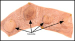
up to 84 caruncles from Merino sheep prior to mating in order to assess the impact on placentation. While fertility was not affected, the ewes in which sixty or more caruncles had been removed delivered prematurely and the lambs had reduced birth weights. The caruncle removal was compensated by an increased size of the cotyledons. Worthington et al. (1981) carried out similar reductions with electrocautery. They found a similar effect on fetal growth. Creasy et al. (1972) embolized fetal cotyledons with microspheres between days 96 and 125 and found significant reduction in fetal size. The affected cotyledons showed hyalinization and necrosis with marked reduction in size. This was followed by similar experiments of Duncan et al. (2000). These investigators examined the consequences of interference with placentation on fetal brain development. They identified significant damage to the white matter of the CNS in the growth-restricted lambs. There are many other, similar studies that probe fetal CNS development by restricting placental performance. For instance, Dawes & Mott (1964) altered oxygen supply to fetal lambs and studied their responses. Keunen et al. (1997) found no CNS damage with cord occlusion of up to ten minutes, but Ball et al. (1994) found that seizures took place when there was severe cord occlusion for 90 minutes. Ikeda et al. (1998) sought to explain the CNS lesions after cord occlusion by brain lipid peroxidation. Mallard et al. (1992) found primarily hippocampal lesions after occlusion, while Clapp et al. (1988) found mostly white matter damage after intermittent occlusion of the cord's vessels. Gardner et al. (2002) used sheep placentation to study the effect of periodic cord occlusion. They found that the number of caruncles did not decrease, but their weights decreased and appearances changed.

Gilbert et al. (1996) showed that technetium crossed the ovine placenta more rapidly than the smaller sodium ion. Ovine urine production was studied by Ross, et al. (1988) and by many other investigators who are listed by these authors. Ross et al. found in this carefully executed protocol (days after surgical canulations, standing ewes, 4-hour experiment, careful 
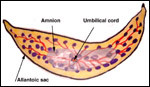
electrolyte measurements) that the amount of allantoic fluid was greater than that of amniotic fluid; that fetal urination is greater into the allantoic sac (through urachus) than the amnionic sac (through urethra); that transmembranous flow probably exists; that lung fluid participates in amnionic fluid composition and swallowing in disposing of it. These are difficult experiments whose interpretation is complicated by the presence of two large fluid-filled sacs in the ovine placenta. They are not directly transferable to human amnionic fluid metabolism for that reason. With Barron we catheterized the urachus in an instrumented sheep that was suspended in a water bath. We found that urine production decreased markedly over a few hours when all urine from the urachus was collected externally. Its osmolality also increased and much fructose was contained in the urine. Urination increased rapidly when water was injected into the allantoic cavity. We conjectured that the water was rapidly absorbed by the allantoic circulation and rehydrated the water-deprived fetus. Matsumoto et al. (2000) ligated the fetal esophagus and urachus and were unable to produce polyhydramnios, as had been their aim. This suggested to them that transmembranous transport must be large.

More recently, Daneshmand et al. (2003) investigated the regulation of amnionic fluid volume in sheep and suggested that vascular endothelial growth factor increases intramembranous absorption by microvesicular transport.

17) Other resources

Cell strains of various sheep species are available from CRES at the Zoological Society of San Diego.


18) Other remarks - What additional Information is needed?

Relatively few noninfectious placental lesions have been described in sheep. More knowledge is also needed on umbilical cords. Mossman (1987) pointed out that more information is needed on the insertion of the cords 
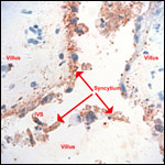
of multiple gestations. Because there is still some uncertainty as to the true origin of the syncytial giant cells, additional studies are warranted.

Acknowledgement

Most of the animal photographs in these chapters come from the Zoological Society of San Diego. I appreciate also very much the help of the pathologists at the San Diego Zoo.

References

Alexander, G.: Studies of the placenta of the sheep (Ovis aries L.). Placental size. J. Reprod. Fertil. 7:289-305, 1964.

Alexander, G.: Studies on the placenta of the sheep (L.). Effect of surgical reduction in the number of caruncles. J. Reprod. Fertil. 7:307-322, 1964.

Arvy, L. and Pilleri, G. The Cordon Ombilical (Funis umbilicalis). Verlag Hirnanatom. Instit., Ostermundigen, Switzerland, 1976.

Assheton, R.: VI. The morphology of the ungulate placenta, particularly the development of that organ in the sheep, and notes upon the placenta of the elephant and hyrax. Phil. Trans. Roy. Soc. London, Series B 198:143-220, 1906.

Ball, R.H., Parer, J.T., Caldwell, L.E. and Johnson, J.: Regional blood flow and metabolism in ovine fetuses during severe cord occlusion. Amer. J. Obstet. Gynecol. 171:1549-1555, 1994.

Barron, D.H.: Some aspects of the transfer of oxygen across the syndesmochorial placenta of the sheep. The Yale J. Biol. Med. 24:169-190, 1951.

Bautzmann, H. and Schroder, R.: Vergleichende Studien uber Bau und Funktion des Amnions. Das Amnion der Säuger am Beispiel des Schafes (Ovis aries). Z. Zellforsch. 43:48-63, 1955.

Bourne, G.L.: The Human Amnion and Chorion. Lloyd-Luke, London, 1962.

Bunch, T.D., Foote, W.C. and Spillett, J.J.: Sheep-goat hybrid karyotypes. Theriogenology 6:379-385, 1976.

Clapp, J.F., Peress, N.S., Wesley, M. and Mann, L.I.: Brain damage after intermittent partial cord occlusion in the chronically instrumented fetal lamb. Amer. J. Obstet. Gynecol. 159:504-509, 1988.

Creasy, R.K., Barrett, C.T., Swiet, M.de, Kahanpää, K.V. and Rudolph, A.M.: Experimental intrauterine growth retardation in the sheep. Amer. J. Obstet. Gynecol. 112:566-573, 1972.

Daneshmand, S.S., Cheung, C.Y. and Brace, R.A.: Regulation of amniotic fluid volume by intramembranous absortion in sheep: Role of passive permeability and vascular endothelial growth factor. Amer. J. Obstet. Gynecol. 188:786-793, 2003.

Davies, J.: Correlated anatomical and histochemical studies on the mesonephros and placenta of the sheep. Amer. J. Anat. 91:263-299, 1952.

Davies, J. and Wimsatt, W.A.: Observations on the fine structure of the sheep placenta. Acta anat. 65:182-223, 1966.

Dawes, G.S. and Mott, J.C.: Changes in O2 distribution and consumption in foetal lambs with variation in umbilical blood flow. J. Physiol. 170:524-540, 1964.

Dent, J., McGovern, P.T. and Hancock, J.L.: Immunological implications of ultrastructural studies of goat x sheep hybrid placentae. Nature 231:115-117, 1971.

Duncan, J.R., Cock, M.L., Harding, R. and Rees, S.M.: Relation between damage to the placenta and the fetal brain after late-gestation placental embolization and fetal growth restriction in sheep. Amer. J. Obstet. Gynecol. 183:1013-1022, 2000.

Gardner , D.S., Ward, J.W., Giussani, D.A. and Fowden, A.L.: The effect of a reversible period of adverse intrauterine conditions during late gestation on fetal and placental weight and placentome distribution in sheep. Placenta 23:459-466, 2002.

Gardner, D.S., Ward, J.W., Giussani, D.A. and Fowden, A.L.: The effect of a reversible period of adverse intrauterine conditions during late gestation on fetal and placental weight and placentome distribution in sheep. Placenta 23:459-466, 2002.

Gilbert, W.M., Newman, P.S., Eby-Wilkens, E. and Brace, R.A.: Technetium Tc 99m rapidly crosses the ovine placenta and intramembranous pathway. Amer. J. Obstet. Gynecol. 175:1557-1562, 1996.

Gray, A.P.: Mammalian Hybrids. Second edition. A Check-List with Bibliography. Commonwealth Agricultural Bureaux, Farnham Royal, Slough, UK, 1972.

Houston, F., Foster, J.D., Chong, A., Hunter, N. and Bostock, C.J.: Transmission of BSE by blood transfusion in sheep. Lancet 356:999-1000 (and 955-956 for Comments), 2000.

Hyde, S.R. and Benirschke, K.: Gestational psittacosis: Case report and literature review. Modern Pathol. 10:602-607, 1997

Ikeda, T., Murata, Y., Quilligan, E.J., Parer, J.T., Doi, S. and Park, S-D.: Brain lipid peroxidation and antioxidant levels in fetal lambs 72 hours after asphyxia by partial umbilical cord occlusion. Amer. J. Obstet. Gynecol. 178:474-478, 1998.

Johnson, G.A., Burghardt, R.C., Joyce, M.M., Spencer, T.E., Bazer, F.W., Pfarrer, C. and Gray , C.A. : Osteopontin expression in uterine stroma indicates a decidualization-like differentiation during ovine pregnancy. Biol. Reprod. 68:1951-1958, 2003.

Keisler, D.H.: Sheep and Goats. In, Encyclopedia of Reproduction. E. Knobil and J.D. Neill, eds. Vol. 4, pp. 479-492. Academic Press, San Diego, 1999.

Keunen, H., Blanco, C.E., Reempts, J.L.H.van and Hasaart, T.H.M.: Absence of neuronal damage after umbilical cord occlusion of 10, 15, and 20 minutes in midgestation fetal sheep. Amer. J. Obstet. Gynecol. 176:515-520, 1997.

King, G.J., Atkinson, B.A. and Robertson, H.A.: Implantation and early placentation in domestic ungulates. J. Reprod. Fertil. Suppl. 31:17-30, 1982.

Kleemann, D.O., Walker, S.K., Hartwich, K.M., Fong, L., Seamark, R.F., Robinson, J.S. and Owens, J.A.: Fetoplacental growth in sheep administered progesterone during the first three days of pregnancy. Placenta 22:14-23, 2001.

Lawn, A.M., Chiquoine, A.D. and Amoroso, E.C.: The development of the placenta in the sheep and goat: An electron microscopic study. J. Anat. 105:557-578, 1969.

Lee, C.S., Gogolin-Ewens, K. and Brandon, M.R.: Comparative studies on the distribution of binucleate cells in the placentae of deer and cow, using monoclonal antibody, SBU-3. J. Anat. 147:163-179, 1986.

Liggins, G.C.: The foetal role in the initiation of parturition in the ewe. CIBA Fndt. Symp. On Foetal Anatomy. G.E.W. Wolstenholm & M. O'Connor, eds. Pp. 218-231, 1969.

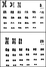
Long, S.E. and Williams, C.V.: Frequency of chromosomal abnormalities in early embryos of the domestic sheep (Ovis aries). J. Reprod. Fertil. 58:197-201, 1980.

Ludwig, K.S.: Zur Feinstruktur der materno-fetalen Verbindung im Placentom des Schafes (Ovis aries L.). Experientia 18:212-213, 1962.

Ludwig, K.S.: Zur vergleichenden Histologie des Allantochorion. Rev. Suisse Zool. 75:819-831, 1968.

Makowski, E.L.: Maternal and fetal vascular nets in placentas of sheep and goats. Amer. J. Obstet. Gynecol. 100:283-288, 1968.

Mallard, E.C., Gunn, A.J., Williams, C.E., Johnston, B.M. and Gluckman, P.D.: Transient umbilical cord occlusion causes hippocampal damage in the fetal sheep. Amer. J. Obstet. Gynecol. 167:1423-1430, 1992.

Matsumoto, L.C., Cheung, C.Y. and Brace, R.A.: Effect of esophageal ligation on amniotic fluid volume and urinary flow rate in fetal sheep. Amer. J. Obstet. Gynecol. 182:699-705, 2000.

Miyazaki, H., Imai, M., Hirayama, T., Saburi, S., Tanaka, M., Maruyama, M., Matsuo, G., Meguro, H., Nishibashi, K., Inoue, F., Djiane, J., Gertler, A., Tachi, S., Imakawa, K. and Tachi, C.: Establishment of feeder-independent cloned caprine trophoblast cell line which expresses placental lactogen and interferon tau. Placenta 23:613-630, 2002.

Mossman, H.W.: Vertebrate Fetal Membranes. MacMillan, Houndmills, 1987.

Naaktgeboren, C. and Zwillenberg, H.H.L.: Untersuchungen uber die Auswuchse am Amnion und an der Nabelschnur bei Walen und Huftieren, mit besonderer Berucksichtigung des europäischen Hausrindes. Acta morphol. Neerlando-Scand. 4:31-60, 1961.

Nowak, R.M.: Walker's Mammals of the World. 6th ed. The Johns Hopkins Press, Baltimore, 1999.

Osgerby, J.C., Gadd, T.S. and Wathes , D.C. : The effects of maternal nutrition and body condition on placental and foetal growth in the ewe. Placenta 24:236-247, 2003.

Puschmann, W.: Zootierhaltung. Vol. 2, Säugetiere. VEB Deutscher Landwirtschaftsverlag Berlin, 1989.

Reynolds, S.R.M.: The proportion of Wharton's jelly in the umbilical cord in relation to distention of the umbilical arteries and vein, with observations on the folds of Hoboken. Anat. Rec. 119:365-377, 1952.

Ross, M.G., Ervin, M.G., Rappaport, V.J., Youssef, A., Leake, R.D. and Fisher, D.A.: Ovine fetal urine contribution to amniotic and allantoic compartments. Biol. Neonat. 53:98-104, 1988.

Silverstein, A.M., Prendergast, R.A. and Kraner, K.L.: Homograft rejection in the fetal lamb: The role of circulating antibody. Science 142:1172-1173, 1963.

Silverstein, A.M., Thorbecke, G.J., Kraner, K.L. and Lukes, R.J.: Fetal response to antigenic stimulus. III. ?-globulin production in normal and stimulated lambs. J. Immunol. 91:384-395, 1963.

Smith, H.A., Jones, T.C. and Hunt, R.D.: Veterinary Pathology. Lea & Febiger, Philadelphia, 1972.

Ward, J.W., Wooding, F.B.P. and Fowden, A.L.: The effect of cortisol on the binucleate cell population in the ovine placenta during late gestation. Placenta 23:451-458, 2002.

Wilsmore, A.J., Izzard, K.A., Wilsmore, B.C. and Dagnall, G.I.R.: Breeding performance of sheep infected with Chlamydia psittaci (ovis) during their preceding pregnancy. Vet. Rec. 130:68-70, 1990.

Wimsatt, W.A.: New histological observations on the placenta of the sheep. Amer. J. Anat. 87:391-458, 1950.

Wooding, F.B.P.: Role of binucleate cells in fetomaternal cell fusion at implantation in the sheep. Amer. J. Anat. 170:233-250, 1950.

Wooding, F.B.P.: Frequency and localization of binuclear cells in the placentome of ruminants. Placenta 4:527-540, 1983.

Wooding, F.B.P., Flint, A.P.F., Heap, R.B. and Hobbs, T.: Autoradiographic evidence for migration and fusion of cells in the sheep placenta: Resolution of a problem in placenta classification. Cell Biol. Internat. Reports 5:821-827, 1981. See also: Cell Biol. Int. Rep. 5:821-827, 1981. See also: Cell Biol. Int. Rep. 5:821-827, 1981.

Wooding, F.B., Flint , A.P., Heap, R.B., Morgan, G., Buttle, H.L. and Young, I.R.: Control of binucleate cell migration in the placenta of sheep and goat. J. Reprod. Fertil. 76:499-512, 1986.

Wooding, F.B., Flint, A.P., Heap, R.B., Morgan, G., Buttle, H.L. and Young, I.R.: Control of binucleate cell migration in the placenta of sheep and goat. J. Reprod. Fertil. 76:499-512, 1986.

Worthington, D., Piercy, W.N. and Smith, B.T.: Effects of reduction of placental size in sheep. Obstet. Gynecol. 58:215-221, 1981.


back to top

© 2004. All rights reserved.
welcome | home | intro | placentation | glossary | author contact me: kbenirsc@ucsd.edu