 |
| (Clicking
on the thumbnail images will launch a new window and a larger version
of the thumbnail.) |
| Last updated: July 31, 2004. |
Saiga tatarica tatarica
Order: Artiodactyla
Family: Bovidae
1) General Zoological Data
The saiga that once extended from Poland to Siberia is now, once again, critically endangered, so states the IUCN publication of April, 2004. Having plummeted from millions to number now 50,000 the animal is still poached for its horn (alleged to be aphrodisiac) and for meat in Russia . It now extends in Southern Russia through the grassland, although attempts at its protection are made in several of the new countries of that region. The subspecies S. t. mongolica is the most threatened of the two subspecies (Mix, 1995). Saigas are strange animals whose phylogenetic origin is still under discussion. Thenius (1969) placed it between gazelles and caprines and referred to the findings of fossils as far distant from Russia as in France , England and even Alaska . Recently, the study of mitochondrial DNA and other molecular means have been employed. They have shown that the rDNA data consistently place the enigmatic genera Pelea, Pantholops and Saiga (Gatesy et al., 1997).
The name saiga is equivalent to the Russian antelope (Gotch, 1979). It is a most conspicuous animal because of its large and inflatable nose with its unusual internal structure. The reason for the existence of this extraordinary nose has often been debated; it is now presumed to be useful in filtering dust and, especially, for warming the air in extreme winters. Males have short, ringed horns (lyrate), females lack horns. The animals' weights range from 21 to 50 kg and they form large groups that range widely. Although the Tierpark Berlin (Pohle, 1974), and also the San Diego Zoo, have kept groups of saigas, they have never done really well in zoos and most eventually died out.
 |
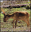
Male saiga (Photographed by Brent Huffman www.ultimateungulate.com). |
 |
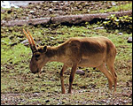
Female saiga (Photographed by Brent Huffman www.ultimateungulate.com). |
The length of gestation is 140 days; the first conception is usually a singleton, twins follow thereafter. The neonatal weight is 3,500 g according to Bannikov et al. (1967) and life expectancy in the wild is 10-12 years. Jones (1993) indicated longevity of 8 years 10 months in captivity.
3) Implantation
I cannot find any reference of descriptions on early stages of gestation.
4) General Characterization of the Placenta
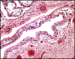
This is a polycotyledonary, chorioepithelial placenta without trophoblast invasion. I have only the placenta of a set of female twins that were born at San Diego Zoo's Wild Animal Park in May, 1984. Unfortunately I have no gross pictures of this placenta. These were two completely separate placentas, each weighing 900 g. Each sac had 40 rather thin and flat cotyledons that were arranged in rows; the cotyledons measured up to 5 cm in diameter and 0.6 cm in thickness.
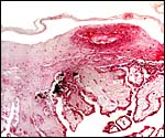 |
Surface of delivered saiga placenta with subchorial pigment deposits in trophoblast. |
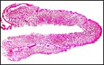 |
Low power view of one complete cotyledon. |
5) Details of fetal/maternal barrier
The villi are covered with a single layer of cuboidal trophoblast among which occasional binucleate cells are found. In addition, numerous deposits of dark yellow/brown pigment were present beneath the chorionic plate, undoubtedly similar to those of other species' hemophagous organs.
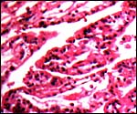 |
Villi of one twin placenta. |
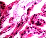 |
Binucleate trophoblast at arrow. |
6) Umbilical cord
The umbilical cords were short, not spiraled, and torn; each contained four vessels and an allantoic duct. Small squamous plaques were present on their surfaces. There were numerous small amuscular blood vessels in this cord, many of which surrounded the larger vessels. Tiny vessels could be pursued into the lumen of the larger vessels.
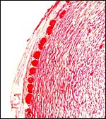 |
Surface of one umbilical artery with conspicuous small vessels; some lumens can also be seen in the muscular wall. |
7) Uteroplacental circulation
There are no publications and I have no such specimens.
8) Extraplacental membranes
The allantoic sac is richly vascularized.
 |
Edge of cotyledon with trophoblast extending on the membranes to the next cotyledon. |
9) Trophoblast external to barrier
None.
10) Endometrium
I have no such specimens and there are no publications.
11) Various features
There is no subplacenta and trophoblast presumably does not extend into the uterus.
12) Endocrinology
I am not aware of any endocrine studies to have been published.
13) Genetics
The saiga possesses 60 acrocentric chromosomes that are extremely similar to those of the domestic goat (Wurster & Benirschke, 1968). Hybrids are unknown.
 |
Edge of cotyledon with trophoblast extending on the membranes to the next cotyledon. |
14) Immunology
I know of no published studies.
15) Pathological features
There are few publications on this species' pathology. Dukes et al. (1992) identified Mycobacterium paratuberculosis in gut and nodes of saigas, fed it to young sheep and saw them develop the disease. Baumeister et al. (1983) identified rotaviruses from the feces of captive saigas and many other species. Minar & Lobachev (1981) found larvae of Pallasiomyia antilopum in saigas and Grokhovskaia (1969) found wild saigas to be parasitized by Hyalomma scupense . Griner (1983) autopsied four saigas and found malnutrition (in a youngster), pneumonia and, in one case, coccidioidomycosis (often found at San Diego Zoo's Wild Animal Park ) as causes of death. Trichuris was also found in the intestines of one animal. Of 249 specimens for which the cause of death has been reported, 108 were from trauma (M.L. Jones, personal communication, 1996).
16) Physiologic data
Because the horn is being smuggled as an alleged aphrodisiac, its precise composition (for forensic recognition) has been studied in some detail. Thus it has become possible through the studies of Zhai et al. (2000) and Hashiguchi et al. (2001) to identify it precisely. The keratin base is typically (and exceptionally) interrupted by mineral deposits of octacalcium phosphate. Hagey et al. (1997), who studied the composition of bile acids in bovids, found that the saiga had a 99.8% taurine conjugation pattern, similar to a rupicaprine specimen, but different from most other Bovidae.
17) Other resources
Cell strains from skin biopsies are available from CRES at San Diego Zoo by contacting Dr. Oliver Ryder at oryder@ucsd.edu.
18) Other remarks What additional Information is needed?
Observations on early stages of development and endocrine studies are badly needed.
Acknowledgement
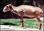
The animal photographs in this chapter come from the internet and were produced by Brent Huffman (www.ultimateungulate.com).
References
Bannikov, A., Zhirnov, L.V., Lebedeva, L.S. and Fandeev, A.A.: Biology of the Saiga. Israel Progr. Sci. Transl., Jerusalem , 1067.
Baumeister, B.M., Castro, A.E., McGuire-Rodgers, S.J. and Ramsay, E.C.: Detection and control of rotavirus infection in zoo animals. J. Amer. Vet. Med. Assoc. 183:1252-1254, 1983.
Dukes, T.W., Glover, G.J., Brooks, B.W., Duncan, J.R. and Swendrowski, M.: Paratuberculosis in saiga antelope ( Saiga tatarica ) and experimental transmission to domestic sheep. J. Wildl. Dis. 28:161-170, 1992.
Gotch, A.F.: Mammals Their Latin Names Explained. Blandford Press, Poole , Dorset , 1979.
Griner, L.A. : Pathology of Zoo Animals. Zoological Society of San Diego , San Diego , California , 1983.
Grokhovskaia, I.M.: Parasitization of the antelope ( Saiga tatarica L.) by Hyalomma scupense Sch. Ticks. Med. Parazitol (Mosk.) 38:493-494, 1969. (In Russian).
Hagey, L.R., Gavrilkina, M.A. and Hofmann, A.F.: Age-related changes in the biliary bile acid composition of bovids. Can. J. Zool. 75:1193-1201, 1997.
Hashiguchi, K., Hashimoto, K. and Akao, M.: Morphological character of crystalline components present in saiga horn. Okajimas Folia Anat. Jpn. 78:43-48, 2001.
IUCN Bulletin, Gland , Switzerland . April 15, 2004.
Jones, M.L.: Longevity of ungulates in captivity. Intern. Zoo Yearbk. 32:159-169, 1993.
Minar, J. and Lobachev, V.S.: Warble flies (Hypodematidae, Oestridae) of some wild animals from Mongolia and Tuva. Folia Parasitol. (Praha) 28:89-91, 1981.
Mix, H.: Kann die Mongolische Saiga überleben? Kritische Aussichten für die grossen Wildtiere der Mongolei. Mitteilungen der Zoologischen Gesellschaft für Arten- und Populationsschutz e.V. 11(2):1-4, 1995.
Nowak, R.M.: Walker 's Mammals of the World. 6 th ed. The Johns Hopkins Press, Baltimore, 1999.
Pohle, K.: Haltung und Zucht der Saiga-Antilope (Saiga tatarica ) im Tierpark Berlin . Zool. Garten NF 44:387-409, 1974.
Thenius, E.: Stammesgeschichte der Säugetiere (einschliesslich der Hominiden). In, Handbuch der Zoologie. J.-G. Helmcke, D. Starck, and H. Wermuth, eds. Vol. 8, # 48. 2(1) 369-722. Walter de Gruyter & Co. Berlin , 1969.
Wurster, D.H. and Benirschke, K.: Chromosome studies in the superfamily Bovoidea. Chromosoma 25:152-171, 1968.
Zhai, Y.J., Wang, R.X., Ji, J.Y. and Wang, G.: Pharmacognostical identification of antelope horn (Cornu saiga tataricae) and its adulterants. Zhongguo Zhong Yao Za Zhi. 25:334-337, 2000. (In Chinese).