 |
| (Clicking
on the thumbnail images will launch a new window and a larger version
of the thumbnail.) |
| Last updated: June 13, 2011. |
South African Meerkat
Suricata suricatta
Order: Carnivora
Family: Herpestidae (Viverridae)
General Zoological Data
The Name (also Mierkat) is Afrikaans of Dutch origin and refers to a marsh-living cat. The animals are home to South Africa and Botswana, are mostly insectivorous and they live in large subterranean but diurnal colonies. They weigh about 700 g and live up to 14 years. Their coats are yellowish and several dark stripes (8-10) run across their backs. In zoological gardens they are favorite animals. This is especially true for children who love the ‘sentry’ animal on its hind legs that guards the access to the colony of some 20 animals.
Aside from insects, meerkats may consume lizards, small snakes, scorpions, mice, small birds etc., and it is known that they are mostly immune to the scorpion venom. These animals belong to a very rich group of carnivore and there are at least 6 subspecies in Africa (Dücker & Dathe, 1972).
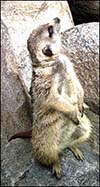
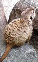
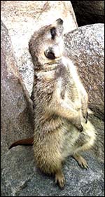 |
Meerkat at San Diego Zoo. |
 |
2. General Gestational Data
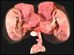
Pregnancy lasts about 11 weeks, occurs usually only once a year, and up to 4-5 newborns are conceived. They weigh from 25-30 g at birth and are naked and altricial. They come first above ground after 21 days of age but are still being well-guarded by 'baby-sitters'. This pregnancy under consideration was near term but the two newborns merely weighed 15 g; one was contained in each uterine horn. Sexual maturity is considered to occur at one year of age.
 |
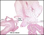
Twin 15 g meerkats in different uterine horns with amnions intact. At the very periphery the allantoic sac encloses the amnion. Yolk sac remnants are at the mesometrial side and dark yellow/orange. Cervix below. |
 |
Another view of the twins with their lobulated placentas behind. |
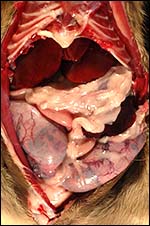 |
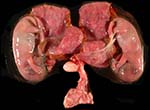
Opened dam with twins in utero at bottom. |
3. Implantation
Early implantations have not been studied and with the exception of a superficial listing of the features of term placentas by Mossman (1987) no descriptions of the placenta have been published. In general though, the placenta is very similar to that of the cat (Amoroso, 1961).
4. General Characterization of the placenta
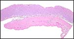
Each apparently zonary placenta measured approximately 7 x 3 cm and was 3 mm in thickness. The cord insertion must have been either marginal or velamentous as the placental surface vessels are of very fine-caliber and large fetal vessels are also found in the membranes. Marginal hematomas do not exist, nor is there the green discoloration, as was already pointed out by Strahl (1905, and also by Grosser, 1927). At one edge, however, some orange-yellow discoloration can be seen as shown in the first photograph and this is iron-staining negative. The placenta is labyrinthine with an endotheliochorial interface. Neither the uterus nor the maternal vessels are infiltrated by trophoblast. A large allantoic sac exists which encloses the amnionic cavity.
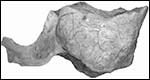 |
This is a photo of the fetal surface of the placenta after fixation in Bouin's solution. It shows the thin nature of the placenta and its surface with very thin blood vessels that appear to arise from the placental margin. |
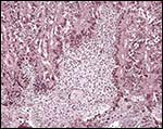
5. Details of fetal/maternal barrier
No details have apparently been published but the interface of suricatta is endothelio-chorial and thus similar to other carnivore and especially to that of the cat. The major portion of the placental tissue is labyrinthine, while near the uterine basal portion a narrow spongy layer is seen. This is also quite similar to that seen in the cat (Parkes, 1961, Chapter 15, Fig. 15.54b).
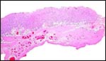 |
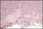
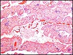
Implanted placenta with rich uterine circulation that is free of trophoblastic infiltration. |
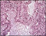
 |
Thin disk implanted on myometrium. The endometrial remnants and debris lie between the purple placenta and red myometrium. |
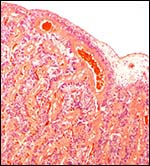 |
Fetal surface of implanted placenta with red blood in fetal blood vessels. |
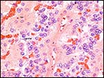 |
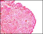
Higher magnification of trophoblastic/endothelial interface. Central in this section is a fetal vessel, bordered by blue trophoblast that is focally syncytial. |
 |
Trophoblast (blue nuclei) of fetal "villi" adjoin debris and maternal endothelium. |
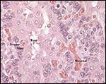 |
Interface of endothelio-chorial placenta of Suricata suricatta. Fetal vessels are carried in the allantois-derived connective tissue and the trophoblast is focally syncytial. |
 |
Interface of endothelio-chorial placenta with the trophoblast labeled. |
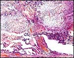
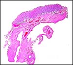 |
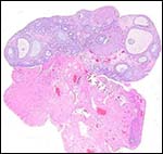
Low power of implanted placenta with cross section of Fallopian tube at bottom. |
 |
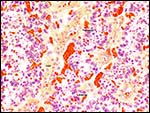
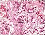
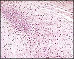
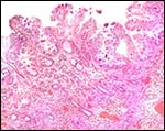
Implantation site with large pale cellular regions of maternal connective tissue origin that are bordered by blue syncytial trophoblast. Focal areas of minute calcification are present in their centers. Note the cellular debris of endometrium beneath the placenta. |
 |
Implantation site of junctional zone. Trichrome stain. VERY light blue connective tissue is present in the central cellular masses that also contain a fragment of calcification. |
 |
Implantation site. The large pale areas above are maternal connective tissue covered with syncytium, trichrome stain (see previous slide). |
 |
Base of placenta with endometrial gland remnants and debris below. |
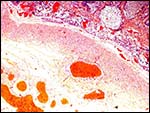 |
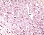
Myometrial/placental and placental labyrinthine zone without trophoblastic infiltration into uterine vessels or uterine musculature. |
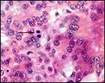 |
Trophoblastic mitosis at arrow. |
6. Umbilical cord
The cords of these two fetuses were approximately 3 cm long and had no twists. They had a marginal insertion into the placentas as is seen in the first photograph. Two arteries and veins were found and there is an allantoic sac that is clad with very thick urothelium.
 |
Insertion from the allantoic duct into allantois; in addition, yolk sac remnants and amnion are visible. |
 |
Central in the umbilical cord is the allantoic duct. |
 |
Higher magnification of allantoic duct epithelium. |
 |
|
 |
7. Uteroplacental circulation
There are no publications on the uterine and placental circulations.
 |
Unopened pregnant uterus to show the very thin blood vessels on surface. Cervix is below. |
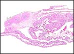
8. Extraplacental membranes
A large allantoic sac with fetal vessels is present and composed of very thins connective tissue, in addition to the amnionic sac which lies central to the allantois.
| Allantoic and amnionic membranes with allantoic blood vessel in the center at the bottom. | |
 |
Membranes with large fetal vessels above. |
 |
Yolk sac surface. |
9. Trophoblast external to barrier
There is no trophoblastic infiltration into myometrium or the maternal blood vessels.
10. Endometrium
Mossman (1937) considered that no decidua exists; it certainly does not exist in the floor of the implanted placenta, but on the membranes it appears to be focally present.
 |
Membranes with yolk sac epithelium and the brownish discolored content. The blue areas scattered through the yolk sac epithelium are capillaries filled with red blood cell precursors. |
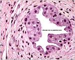
 |
Uterine epithelium with glands beneath. |
11. Various Features
No other features are described.
12. Endocrinology
The ovarian bursa was briefly described by Mossman & Duke (1973) as being 'complete with a porelike orifice similar to that of mustelids'.
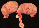
 |
One ovary with two corpora lutea. |
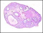 |
The other ovary with one corpus luteum. |
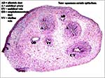
13. Genetics
Meercats have 36 chromosomes (Todd, 1966; Hsu & Benirschke, Vol. 1, Folio 30, 1967). With the exception of a minute Y-chromosome, most chromosomes are mediocentric or submetacentric elements.
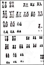 |
36 chromosomes of meerkat, from Hsu & Benirschke, 1967. |
14. Immunology
There are no published publications on immunological features except that the animals are apparently immune to scorpion toxins.
15. Pathological features
None have been described even in the comprehensive text by Griner (1983).
16. Physiologic data
None is known, except that the animals are described as having exceptionally good vision so that they can recognize a potentially threatening bird (Vulture) at high altitude and the emit a warning sound.
17. Other resources
Cell cultures are frozen in the San Diego Zoo's "Frozen Zoo" and can be obtained by requesting them from Dr. Oliver Ryder (oryder@ucsd.edu) at the Institute for Conservation Research at the San Diego Zoo.
18. Other remarks – What additional information is needed?
No good information exists on early implantation, pathology, physiology and endocrinology.
Acknowledgement
This tissue was kindly provided by Dr. Rebecca Papendick and April Gorow.
References
Amoroso, E.C.: Placentation, Chapter 15 in: Parkes, A.S.: Marshall’s Physiology of Reproduction. Little, Brown and Co. Boston1961.
Dücker, G. and Dathe, H.: Schleichkatzen und Erdwölfe; chapter 5 of Vol. XII. Kindler Verlag, Zürich, 1972.
Griner, L.A.: Pathology of Zoo Animals. Zoological Society of San Diego, 1983.
Grosser, O.: Frühentwicklung, Eihautbildung und Placentation des Menschen und der Säugetiere. J.F. Bergmann, München, 1927.
Hsu, T.C. and Benirschke, K.: An Atlas of Mammalian Chromosomes. Springer-Verlag, New York.1:Folio 30, 1967.
Mossman, H.W.: Vertebrate Fetal Membranes. MacMillan, Houndmills, 1987.
Mossman, H.W. and Duke, K.L.: Comparative Morphology of the Mammalian Ovary. University of Wisconsin Press, 1973.
Parkes, A.S.: Marshall’s Physiology of Reproduction. Little, Brown and Co. Boston1961.
Strahl, H.: Beiträge zur vergleichenden Anatomie der Placenta. (Lemuriden, Viverra civetta, and Centetes ecaudatus) Abhandl. Senckenbergsche Naturf. Gesell. Frankfurt 27:263-319, 1905.
Todd, N.B.: The karyotypes of the lesser Indian mongoose (Herpestes javanicus Geoffroy), the mierkat (Suricata suricatta Desmarest) and comments on the taxonomy and karyology of the Viverridae. Mammalian Chromosomes Newsletter No. 21:154, 1966.