(Clicking on the thumbnail images below will launch a new window and a larger version of the thumbnail.)
 
Last updated:
May 18, 2007.
Greater Indian Rhinoceros
Rhinoceros unicornis

Order: Perissodactyla.
Family: Rhinocerotidae.

1) General zoological data of species


As the name suggests, this species of the five rhinoceroses comes from India and is the largest. Adult Indian rhinoceroses weigh approximately 2,000-4,000 kg. A comprehensive review addresses and illustrates all aspects of Indian rhinoceros questions (Rüedi, 1984). Zoogeographical and evolutionary data are to be found by Groves (1972, 1975).

2) General gestational data

The gestational length of the Indian rhinoceros is 462-491 days (Laurie et al., 1983). Since writing this chapter, a new calf was born to a multiparous female with good breeding dates; her gestation was 491 days long. That placenta weighed 6,650 g.; furthermore, a primiparous female delivered a healthy calf whose placenta weighed 3,550 g and measured 25 cm in greatest diameter. The remarkable aspect of that placenta was the enormous size of the hippomanes. They are depicted below; they weighed 368 g. With the exception of size (female black rhinos weigh about 1,600 kg, the Indian rhino weighs <1,600 kg) that is reflected in the membranes, no significant difference was found in the morphology of placentas from white, black and Indian rhinoceroses (Please see the chapter on white rhinoceros). They implant in both uterine horns, the fetus being located mostly in one horn, and the placenta extending into the other. The weight at birth is 40-80 kg, and females mature at about 3 years. One young is born only.

The placental weight at term, including the membranes but without umbilical cord, varies between 4,200 and 7,600 g in our large experience. The most recently born Indian rhinoceros in San Diego had a placental weight of 6,600 g. It measured 270 cm from side to side, 100 cm in greatest width and was very thin, less that 0.5 cm.
In May, 2007, a term stillborn fetus was delivered of a primigravid female at the Wild Animal Park of the San Diego Zoo; in contrast to most other deliveries, this did not occur in a “Boma” but in the enclosure. It weighed 57 kg and had a 4,600 g placenta attaché with very short umbilical cord. The hippomanes attached weighed 180 g, were flattened and measured 18x8.5x2.5 cm.
   
  Two Greater Indian rhinoceroses at the San Diego Wild Animal Park.
     
  Stillborn term calf.
     
 
3) Implantation

Early implantational stages were depicted by the sonographic study of Radcliffe et al. (1997). The earliest stage shown occurred 15 days after ovulation, with embryonic definition visible on day 23, and heartbeat on day 26.

The exact time of implantation, however, has not yet been determined. The placenta is diffusely villous, with larger streak-like areas of their absence. This is one reason for the organ to be called "Placenta villosa diffusa incompleta". These bare areas are also referred to as "streets (Strassen)". They show up extremely well when the placenta is trans-illuminated. They generally follow the larger blood vessels.
   
  Maternal aspect of rhinoceros placenta with white "Strassen" along major vessels.
     
  This is a photograph of a portion of the placenta of an Indian rhinoceros, slightly trans-illuminated. It shows broad areas of absent villous (red) regions, so-called "streets" follow the major fetal blood vessels.
     
 
The organ implants in both uterine horns, with the fetus located mostly in one horn, and the placenta extending to both sides.
   
  Schematic representation of rhinoceros placentation
     
 
4) General characteristics of placenta

The placentas are very large and very thin. One weighing 7,600 g is depicted here. It measured 190 cm in length, 100 cm in greatest width, and 50 cm in smallest diameter. It was 2 mm in thickness. The placenta of a term Indian rhinoceros weighed 5,300 g, with an additional 950 g of amnionic and allantoic membranes. It measured 230 cm in greatest width. Another recently obtained placenta from a term, surviving Indian rhinoceros weighed 5.500 g, had dimensions of 270 x 98 cm and a 5 cm cord but with four blood vessels. In yet another additional specimen from a term, surviving infant, the umbilical cord had three vessels and the weight of the placenta was 5,000 g; yet another weighed 3,550 g, this one from a primipara. These are diffuse placentas without cotyledons, and of an epitheliochorial barrier character. Many placentas have on their maternal surfaces band-like connective tissue areas that are bare of villi. They usually follow the lesser curvature of the placenta and the larger blood vessels; they have been referred to as “Strassen” (streets) in the Suisse literature. They were depicted earlier.

In December, 2005 a stillborn female calf weighing 77 kg was delivered of an experienced female; it could not be resuscitated. The placenta weighed 6,300 g, was 280 cm long and had the usual characteristics with a 5 cm cord and three vessels. The remarkable aspect of this gestation was the course of the umbilical vein in the fetal abdomen. It was hugely dilated (up to 10 cm) shortly after entering the abdomen, filled with fresh clot and, when dissected further, there was no obstruction or another feature that could explain the aneurysmal distention.


  Fetal surface of rhinoceros placentas with short umbilical cord.


The cord was relatively short; one of our recent specimens had a 25 cm long cord with 4 cm thickness. The last-born placenta had a longer cord; its allantoic portion was 36 cm, the amnionic portion 40 cm long, and there were three vessels plus the large allantoic duct. It measured 5 cm in width. There are generally three umbilical vessels and a large allantoic duct. There is a large, heavily vascularized allantoic sac. Hippomanes of the last two Indian rhinoceros placentas were green, pasty, and only one lump was present. In another specimen, 180 g were found in an Indian rhinoceros placenta.


Exceptionally large hippomanes (368 g).

     
  Section of hippomanes with crystalloid inclusions.
     
  The hippomanes in polarized light microscopy.
     
 
In addition, there were numerous round, yellowish squamous patches projecting on the amnionic surface of one Indian rhinoceros placenta that measured 0.5-1 cm. They were absent in another specimen. Naaktgeboren & Zwillenberg (1961) have discussed these structures at great length, for numerous species, but especially the cow.
   
  Amnionic surface of Indian rhinoceros placenta with round nodules
     
 
Leaf-like and folded villi were alluded to by Ludwig et al. (1965), representing thinner and others, taller epithelial structures. Histochemical reactions showed only minor differences (Ludwig & Müller, 1965). Occasional binucleated trophoblastic cells have been described, as are also found in the horse and many ungulates. But there is no uterine invasion by trophoblast. The electronmicroscopic study (Ludwig & Villiger, 1965) identified similarities to equine placentas and numerous trophoblastic transport vesicles were seen.

The allantoic sac is anchored to the chorion by thin connective strands (Dolinar et al., 1965). While the amnionic epithelium is very thin and flat, the allantoic sac is lined by cuboidal to columnar epithelium. The allantois is diffusely vascularized.
   
  Higher magnification of white rhinoceros villi.
     
  Higher power of the villous surface of an Indian rhinoceros placenta with "Strassen".
     
  Structure of amnion/allantois (left), chorion and villi of a mature Indian rhinoceros placenta at right.
     
  Another view of the placental surface (right) with villous branches extending left.
     
 
A complete review of all publications on three species of rhinocerotidae placentas was published by Benirschke & Lowenstine, 1995. This report contained details in tabular form. It is printed at the end of the chapter for the convenience of the reader.

5) Details of barrier structure

This is a typical diffuse epithelial-chorial (appositional) placenta without invasion of the endometrium.

6) Umbilical cord

The umbilical cord is short, usually with the delivered placenta come only 5-6 cm. The portion remaining on the fetus is usually also short and has only one twist. In the stillborn fetus where both fetus and placenta were available, the cord was 5 cm long, had no twists and contained 2 arteries and one vein, in addition to many smaller vessels and a large allantoic duct. The cord of one of our Indian rhinoceros placentas was 10 cm long, another was 25 x 4 cm. But the cord has not been measured in utero.

   
  Umbilical cord of stillborn fetus.
     
  Fetal surface of Southern white rhinoceros placenta.
     
 
A remarkably long umbilical cord (57.8 cm) in an Indian rhinoceros was described by Dolinar et al. (1965). The umbilical cord has 4 large umbilical vessels and a very large number of small vessels that provide the circulation for the allantois. It also has a large allantoic duct. A vitelline duct is absent. The surface of umbilical cords has squamous plaques.

7) Uteroplacental circulation

No descriptions are available.


8) Extraplacental membranes

Only an allantoic sac is present that is diffusely fused to the amnion and, less
completely, to the chorion. Free membranes do no exist. Numerous allantoic vessels supply the allantois.

   
  Amnion at left (flat epithelium) allantois right (columnar epithelium).
     
 
9) Trophoblast external to barrier

There is no trophoblast beyond the villous structures.

10) Endometrium

Decidualization has not been described, but hemosiderin is found in post partum uteri, and many have cystic glands.

11) Various features

The endometrium has hemosiderin-laden macrophages after gestations, suggesting that there is some bleeding during parturition. No implanted placenta has been observed, all those seen by us and described in the literature had been delivered. Their preservation is thus somewhat impaired.

12) Endocrinology

The estrous cycle was defined by Radcliffe et al. (1997), in a combined sonographic and endocrine study, as being 31-35 days. Estrus lasts less than 24 hours. Numerous other studies of estrogens and progesterone have been performed and are quoted by Miller (1983).

Schwarzenberger et al. (2000) provided a detailed analysis of the ovarian cycle in this species by employing fecal steroid measurements. They suggested from these studies that pregnane metabolites are reliable indicators of the corpus luteum activity. Such determinations allow the observer to diagnose pregnancy, while 17-oxo-androstenes and estrogens were indicators of the follicular phase.

Neonatal gonads display pronounced testicular interstitial cell, or ovarian lutein, activity, not unlike those of horses. While no gonadotropins have yet been identified, it is likely that some are present during gestation because of the luteinization of fetal ovaries and stimulation of testicular interstitial cells of neonates. These may be define some similarities to the equine gestation, but hormones have yet to be defined.

Reproductive parameters in zoological gardens have been delineated for the black rhinoceros by Carlstead et al. (1999). A workshop on white rhinoceros steroid determinations and induced ovulation was held in San Diego (Patton et al., 1998) and is available from that institution. Another comprehensive conference volume on rhinoceros biology is available from a 1991 conference (Ryder, 1993).

The preservation of fecal samples for steroid study has been discussed many times and was controversial when samples had been imported for study. Now, Galama et al. (2004) have published a study that indicates reliable preservation of steroids when stored in methanol or after drying in a solar box cooker. Admittedly, the study was done in black rhinoceros, but should be equally applicable to other species.

13) Genetics

The chromosome number of the Greater Indian Rhinoceros (as well as the white and Sumatran rhinoceroses) is 2n=82 (Hansen has reported a chromosome number of 84). The X-chromosome is the only metacentric element. Many other elements have small short arms, even the Y-chromosome (Hsu & Benirschke, 1973). Three animals of the Northern white rhinoceros had only 81 elements. Mitochondrial DNA was studied in different animals and in different subspecies; these are reports by Ashley et al. (1990), Harley & O'Ryan (1993), and by George et al. (1993). Only the black rhinoceros has a chromosome number of 2n=84. Allozyme variation was studied in African and Indian rhinoceroses by Mereniender et al. (1989).

Karyotypes of male and female Indian rhinoceroses (Hsu & Benirschke, 1973).


14) Immunology

No studies are known to us.

15) Pathological features

Veterinary problems, diseases and numerous procedures, were summarized by Silberman & Fulton (1979), and a complete veterinary bibliography was compiled by Miller (1983). It includes references to reproductive laboratory data as well. Dermal problems and infections appear to be the commonest ailments, including pox (Schaller & Pilaski, 1979).

A major problem in the black rhinoceroses is hemolytic anemia (Chaplin, et al., 1986; Miller & Boever, 1982; Paglin et al., 1986).

We have reported huge vacuoles in the trophoblast of a normal placenta from an Indian rhinoceros. This area overlay a region of endometrial degeneration and the vacuoles do not represent the trophoblastic transport vesicles.

The abnormal distention of the intra-abdominal portion of the umbilical vein is shown below.

The remarkably distended portion of the umbilical vein is shown at the arrow.


  Indian rhinoceros chorionic surface with abnormally vacuolated trophoblast at right, overlying debris.
     
 
16) Physiological data

Whatever relevant physiologic data are available have been provided in review form by Silberman & Fulton (1979), and as bibliography by Miller (1983).

17) Other resources

Cell strains of four species are available through CRES at the Zoological Society of San Diego by calling on Dr. Oliver Ryder at oryder@ucsd.edu; none are available of the exceptionally rare Javan rhinoceros.

18) Other aspects of interest:

Additional literature copied for convenience here. (Because this reference is so difficult to obtain in print, it is here reprinted without the figures.)

THE PLACENTA OF THE RHINOCEROTIDAE

By Kurt Benirschke, M.D. and Linda J. Lowenstine, D.V.M., Ph.D.
Departments of Pathology, University of California, San Diego and the San Diego Zoo (Verh. Ber. Erkr. Zootiere (Dresden). 37:15-23, 1995).

Introduction

It is rarely possible to examine the placenta of rhinoceroses except following the birth of offspring in Zoological Gardens. It is therefore not surprising that the only specimens described so far have come from the Basel Zoo through the efforts of Ernst Lang. Only the Indian rhinoceros' placenta has been fully studied, yet it would be of considerable interest to know more about the structure of the placenta of all five species of these animals. For one thing, the fact that this mammalian family is composed of four genera is astounding, and one may thus wonder whether this taxonomic oddity is reflected in significant placental differences. Further, if ever one may wish to practice embryo transfer for artificial rearing of a severely endangered form, the function of the placenta is critical and must be better understood. This knowledge must of course extend beyond the analysis of anatomical features, but they are a first beginning. This paper describes the placenta of three species of rhinoceros; it cannot be anticipated presently that Javan and Sumatran rhinoceros placentas will become available soon. The former is not in captivity and the latter animals have not bred thus far in zoos that hold them.

The placentas of the other four species of rhinoceros, other than that of the Indian rhinoceros, have heretofore not been fully described. It is further important to recall that Mossman (1987) correctly stated in his extensive monograph on comparative placentation, that the rhinoceros placenta has been described only from delivered term pregnancies. None has yet been studied in utero, attached to the endometrium. It is important that the in situ placenta of rhinoceroses be observed so as to achieve answers to some critical questions posed in this paper. Mossman described the placenta of the Indian rhinoceros (Rhinoceros unicornis), the only studied organ at the time of his study, to be an "omphaloplacenta, i.e. a disc of bilaminar omphalopleure", and viewed it as being similar to that of the tapir. Previous authors quoted by Mossman had considered the organ to be intermediate between that of the horse and cattle.

Ludwig (1962) was the first to describe the microscopic features of an African rhinoceros (Diceros bicornis L.) and considered it to be a "placenta villosa diffusa". It was composed of broad-based villous tufts with leaf-shaped branches. Ludwig and Müller (1965) studied the afterbirth of an Indian rhinoceros (Rhinoceros unicornis L.) from the Basel Zoo histochemically. Because of those areas that were found to be free of villi (the a-villous "streets"), they described this organ to be a "Placenta villosa diffusa incompleta". Perhaps the previous specimen of which Ludwig had been able to obtain only small samples had similar villus-free areas but they had not been submitted for study. Two different types of villi were described for the first time: "leaf-like villi" and "folded villi". The epithelium of the former was cuboidal to flat, and the chorionic plate was covered by a more cylindrical epithelium. Conversely, the folded villi were covered by tall cylindrical trophoblast. A variety of histochemical reactions were possible on this unusually well-preserved tissue. These authors also compared their findings with those in cattle placentas and found great similarities. They also identified occasional binucleate trophoblastic cells and sought similarities to the diplokaryocytes of cattle placentas.

In their description of the fine structure of the placenta of an Indian rhinoceros, Ludwig and Villiger (1965) found that the epithelium of the leaf-like villi is identical to that of equidae. An exception were the "diplokaryoctes" which had many more mitochondria and endoplasmic reticulum. Many transport vesicles were found in the trophoblastic epithelium.

Dolinar et al. (1965) observed the freshly delivered placenta of another Indian rhinoceros and were also able to obtain very fresh tissue for histology. It came from a term pregnancy with a 68 kg male infant. The placenta weighed 4,960 g, had an amnionic cavity of approximately 78 liter content, a 57.8 cm umbilical cord and resembled the organ described for tapir and black rhinoceros. The villus-free "streets" were well depicted as were the fibrous strands that connect allantois to chorion. These thin fibers also attach to and perhaps anchor the large muscular vessels that course within the space between allantois and chorion.

Diplokaryocytes were again noted and the tall epithelium of the villus-free chorion was likened to the focal scars of horse placentas that are believed to overlie the uterine glands and may absorb "uterine milk". Amnionic pearls or hippomanes were not identified.

Details concerning the weights and some reproductive and gestational features of the Indian rhinoceros can be found in Lang's paper (1967). He described the average weight of six neonates to be 65.6 kg, and the average gestational length as being 477 days.

Macroscopic Observations

At the Zoological Parks of San Diego and San Pasqual we have been able to collect the placentas from the following species:

Indian rhinoceros (Rhinoceros unicornis L.) # 16
Black rhinoceros (Diceros bicornis L.) # 6
Southern white rhinoceros (Ceratotherium s. simum L.) # 6

The available weights of neonates and placentas of these animals and those obtained from the literature are summarized in Table 1. The placenta of rhinoceroses is very thin and large, and it conforms roughly to the bicornuate uterus of the animal. The overall length reaches 230 cm in an Indian rhinoceros, with maximal circumference of up to 120 cm. One horn of the bicornuate placenta (Figs. 1,2) is considerably larger and contains the fetus than the other horn which is largely filled with membranes and fluid. The umbilical cord inserts nearly at the junction of the two sides, also near the site of rupture during delivery, the cervical outpouching of the sac. The fetus is contained within the amnionic sac whose membrane is intimately fused with the allantoic membrane (Fig. 1). The reflected allantois, in turn, is fused with the chorionic membrane on whose outside reside the trophoblastic epithelium and villi. The entire thickness of the placenta is never greater than 3 mm, and often it is thinner. This is especially true in the larger streets ("Strassen") of avillous membranes which were described especially well by Dolinar et al. (1965). When we witnessed the delivery of an Indian rhinoceros at night, the dam lay on her side and the amnionic sac containing the fetus slid out slowly and without great maternal effort. The cord ruptured spontaneously; thereafter the dam stood up, exhibited Flehmen, and then turned to the amnion-enclosed fetus and proceeded to lick off the sac, thus freeing the neonate from its enclosure. It is for this reason that the amnionic sac often comes separately and that the placenta is delivered at a later time without amnion. It also explains the incomplete presence of umbilical cords in submitted specimens.

The umbilical cord severs spontaneously during the birth of rhinoceroses, most often near its fetal end. The length of most umbilical cords of the specimens described was not available to us, most of it remained on the neonate. It was also not measured as it was not made available and seemed of little importance. Only Dolinar et al. (1965) gave an accurate measurement for the entire length of one umbilical cord of an Indian rhinoceros at term. It was 57.8 cm long. Aside from the three or four large allantoic vessels and the numerous tiny allantoic vessels, the umbilical cord contains a huge allantoic duct that connect dome of bladder with the allantoic cavity. In an attempt to assess aspects of placental efficacy, Kloosterman and Huidekoper (1954) have calculated a "placental coefficient", the ratio of placental to fetal weight. They found for the human development that this ratio declined from 2 at 8 weeks gestation to 0.13-0.19 at term (40 weeks). The importance of this ratio is difficult to validate exactly and that is true even more so in these rhinoceros specimens. It lies at 0.089 in the Indian rhinoceros, at 0.115 in the Black rhinoceros, and in the White rhinoceros it is at 0.075. Because of some frequently contaminating dirt and because of the excessively large quantity of amnion present in these animals, this placental/fetal ratio is probably meaningless for our specimens. In other species, a fetal to cotyledonary weight ratio has been used to infer some functional capacity of the placenta, but even this has given rise to much inconclusive discussion (Dixon and Robertson, 1969). Such figures that have been arrived at are probably of little us. Nevertheless, in order to provide as much information as possible, the weights available to us for fetus and placentas are listed in Table 1. They may be useful in future comparative studies.

In several species the amnion and/or allantois possesses "pustules", projections on the surface of the membranes or the umbilical cord. They may be very pronounced and were seen in only few of the rhinoceros placentas. One such case is illustrated in Figure 3, the allantoic surface of an Indian rhinoceros. There were about 20 such 0.5-2 cm, slightly pedunculated nodules located towards the end of the smaller horn. Their brown surfaces were smooth and the content was soft. Microscopically, they are epithelium-lined that has keratin production. Perhaps when these nodules are degenerating they produce part of the hippomanes. We were not impressed by a marked difference in the villous structures described (leaf-like or folded), either in their macroscopic or microscopic details.

Microscopic Observations

The placenta of rhinoceroses is an epithelial-chorial organ without invasion of the maternal tissues by the trophoblast. Nevertheless, in endometria available to us from post partum uteri it is evident that some endometrial hemorrhage must occur after delivery, since hemosiderin-laden macrophages are abundant in the endometrial stroma. We found no significant histological differences among the microscopic features of the three species of rhinoceros examined here. Villus-free areas of chorion (Figs.1,4) were found in all our specimens. These are membranous areas that Dolinar et al. (1965) speculated lie over endometrial glands and that may serve to absorb uterine milk. They likened this feature to the anatomy of the horse placenta, as depicted by Amoroso (1952). In situ observations have not yet been made and will be of interest in the future. This is especially so as these areas in the horse placenta are much smaller and less abundant than in the rhinoceroses studied.

The simply branched villi (Fig. 4) contain few elements other than the fetal vessels, but there was occasional yellow-green pigment within the villous connective tissue whose origin is uncertain. Macrophages, similar to the Hofbauer cells of primate placentas, were not identified. The trophoblastic cover of the villi is usually tall columnar, with basal nuclei and prominent brush border (microvilli) toward the endometrium. Occasional binucleate cells ("diplokaryocytes") are found but they were not especially prominent in any species. Therefore, we hesitate to reiterate the analogy to bovine placentas made in previous studies, especially so because only so few of these cells were found. Moreover, the villi of rhinoceros placentas are fairly uniformly distributed, excepting the villus-free streets, and they have no similarity to the cotyledonary structures of bovidae. We found no trophoblastic mitoses. In many areas the cylindrical epithelium becomes more cuboidal without there being any obvious special localization of these changes. There were a few remarkably enlarged vacuoles in some areas of the placenta of an Indian rhinoceros (Fig. 5). They extended into the superficial villous stroma and originated in the trophoblast of the placenta. Many vacuoles had an eosinophilic proteinaceous content and projected slightly on the epithelial surface and were found primarily in the non-villous chorionic "streets". The nature of these large vacuoles is uncertain, even though transport vesicles have been described electronmicroscopically (Ludwig and Villiger, 1965). The vacuoles that we observed in this case were much larger than the usual transport vacuoles of trophoblast, and they were mostly confined to the superficial villous connective tissue but apparently originated from the trophoblastic cytoplasm. In no case were the villous capillaries protruding into the trophoblastic cover, such as is described for human syncytiocapillary membranes. The vacuoles do not represent such a vascular adaptations as they never contained blood.

Amnion and allantois are densely adherent in the thin sac that encloses the fetus (Fig. 6). The amnionic epithelium is flat and that of the allantois mostly cuboidal but it is also usually not very well preserved. An abundance of capillaries and larger vessels course in the allantoic connective tissue; none are present in the amnion, or they are stray from the allantois. Allantois and chorion are more loosely connected and, especially when pulled apart, they leave characteristic thin connective tissue strands (Fig. 7). The area in between these two sheets is also the position of the very muscular fetal placental surface vessels that carry blood from cord to the villous branches and that run in the chorionic membrane.

Contrary to earlier observations, keratinized squamous metaplasia was found on the cord of an Indian rhinoceros, and typical squamous keratin "pearls" were observed in amnion, on the cord surface, and in allantoic areas of the placenta of the placenta of a black rhinoceros (Fig. 8). They are the apparent equivalents of amnionic "pustules" described for so many animals (Ramsey, 1975; Benirschke and Calle, 1994). In contrast to the findings of earlier students, we found several animals to possess large hippomanes, mostly in the narrow horn, and weighing up to 180 and 168 g, and measuring 14 x 6 cm (Table 1). They were olive-green or brownish and had an abundance of polarizing crystals within the mostly acellular debris. The crystals are presumably derived from urinary products. The centers of these protrusions were soft and they either attached to the allantoic surface or lay free.

Conclusions

The large rhinoceros placenta consists of a thin membrane of allantochorion with rather diffusely scattered villi. Occasional villous-free regions are prominent findings of all species and are referred to as "streets". No significant differences were found in the structure of the placenta from these three species of rhinoceros examined, irrespective of their generic differences in classification. Before one can conclude that the placentas are sufficiently similar that inter-specific (-generic) transfer of embryos may be possible, more detailed biological studies are needed. For instance, no information on placental hormone production is yet available. While the rhinoceros fetal gonads resemble those of horse fetuses, it would be premature to deduce that rhinoceros placentas have similar endocrine activities to stimulate fetal tissues.


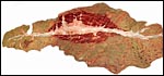
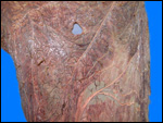
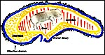
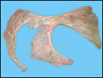
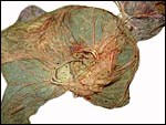
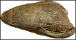
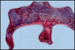
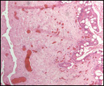
Since then we have seen two more placentas of Indian rhinoceroses. One weighed 7,000 g, had a healthy newborn, had hippomanes and measured 304 cm in greatest length, 132 and 54 cm in largest and smallest diameters and had a 10 cm cord attached. The other also was at term, had a healthy neonate and weighed 6,600 g. It measured 270 x 100 cm and had a 10 cm cord attached.


Combined References

Cell strains of four species of rhinoceros are available from the “Frozen zoo” at the Zoological Society of San Diego: www.FrozenZoo@sandiegozoo.org

Amoroso, E.C.: Placentation. In, Marshall's Physiology of Reproduction. A.S. Parkes, ed., 3rd ed., Vol. II. London. Longmans, Green, 1952.

Ashley, M.V., Melnick, D.J. and Western, D.: Conservation genetics of the black rhinoceros (Diceros bicornis): I. Evidence from the mitochondrial DNA of 3 populations. Conserv. Biol. 1:71-77, 1990.

Baumgartner, K. and Schaftenaar,W.: Fecal progesterone, estrogen, and androgen metabolites for nonivasive monitoring of reproductive function in the female Indian rhinoceros, Rhinoceros unicornis. Gen. Compar. Endocrinol. 119:300-307, 2000.

Benirschke, K. and Calle, P.P.: The placenta of the Beluga whale (Delphinapterus leuca). Verh. Ber. Erkg. Zootiere 36:309-314, 1994.

Benirschke, K. and Lowenstine, L.J.: The placenta of the rhinocerotidae. Verh. Ber. Erkr. Zootiere (Dresden). 37:15-23, 1995.(Attached at end)

Bennett, C. and Kleiman, D.G.: Black rhinoceros (Diceros bicornis) in U.S. Zoos: II. Behavior, breeding success, and mortality in relation to housing facilities. Zoo Biol. 18:35-52, 1999.

Carlstead, K., Fraser, J., Bennett, C. and Kleiman, D.G.: Black rhinoceros (Diceros bicornis) in U.S. Zoos: II. Behavior, breeding success, and mortality in relation to housing facilities. Zoo Biol. 18:35-52, 1999.

Chapin, H., Malecek, A.C., Miller, R.E., Bell, C.E., Gray, L.S. and Hunter, V.L.: Acute intravascular hemolytic anemia in the black rhinoceros: Hematologic and immunohematologic observations. Amer. J. Vet. Med. 47:1313-1320, 1986.

Dixon, H.G. and Robertson, W.B.: The growth of the conceptus and its blood supply. In, Foetus and Placenta. Klopper, A. and E. Diczfalusy, eds. Oxford and Edinburgh: Blackwell, pp. 1-32, 1969.

Dolinar, Z.J., Ludwig, K.S. und Müller, E.: Ein weiterer Beitrag zur Kenntnis der Placenten der Ordnung Perissodactyla: Zwei Geburtsplacenten des Indischen Panzernashorns. (Rhinoceros unicornis L.). Acta Anat. 61:331-354, 1965.

Galama, W.T., Graham, L.H. and Savage, A.: Comparison of fecal storage methods for steroid analysis in black rhinoceros (Diceros bicornis). Zoo Biol. 23:291-300, 2004.

George, M. Jr., Chemnick, L.G., Cisova, D., Gabrisova, E., Stratil, A. and Ryder, O.A.: Genetic differentiation of white rhinoceros subspecies: diagnostic differences in mitochondrial DNA and serum proteins. In, Proc. Intern. Conference on Rhinoceros Biology and Conservation, San Diego, CA 1991, pp. 105-113.

Groves, C.P.: Ceratotherium simum. In, Mammalian Species. 8:1-6, 1972. Amer. Soc. Mammalogy.

Groves, C.P.: Taxonomic notes on the white rhinoceros. Ceratotherium simum (Burchell, 1817). Säugetierk. Mitteil. 23:200-212, 1975.

Groves, C.P.: Phylogeny of the living species of Rhinoceros. Z. zool. System. Evol. 21:293-313, 1983.

Hansen, K.M.: Q-bands of some chromosomes of white rhinoceros (Diceros simus). Hereditas 82:205-208, 1976.

Harley, E.H. and O'Ryan, C.: Molecular genetic studies of southern African rhinoceros. In, Proc. Intern. Conference on Rhinoceros Biology and Conservation, San Diego, CA 1991, pp. 101-104.

Heinichen, I.G.: Karyological studies on southern African perissodactyla. Kodoe 13:51-108, 1970.

Houck, M.L., Ryder, O.A., Váhala, J., Kock, R.A. and Oosterhuis, J.E.: Diploid chromosome number and chromosomal variation in the white rhinoceros (Ceratotherium simum). J. Hered. 85:30-34, 1994.

Hsu, T.C. and Benirschke, K.: An Atlas of Mammalian Chromosomes. Vol. 7: Folio 340, 1973. Springer-Verlag, New York.

Hungerford, D.A., Chandra, H.S. and Snyder, R.L.: Somatic chromosomes of a black rhinoceros (Diceros bicornis Gray 1821). Amer. Naturalist 101:357-358, 1967.

Kloosterman, G.J. and Huidekoper, B.L.: The significance of the placenta in obstetrical mortality. A study of 2.000 births. Gynaecologia 138:529-550, 1954.

Lang, E.M.: Geburt eines Panzernashorns, Rhinoceros unicornis, im Zoologischen Garten Basel. Säugetierk. Mitt. 5:69-70, 1957.

Lang, E.M.: Einige biologische Daten vom Panzernashorn (Rhinoceros unicornis). Rev. Suisse Zool. Genève 74:603-607, 1967.

Laurie, W.A., Lang, E.M. and Groves, C.P.: Rhinoceros unicornis. In, Mammalian Species # 211, pp.1-6, 1983. Amer. Soc. Mammalogy.

Ludwig, K.S.: Zur Kenntnis der Geburtsplacenten der Ordnung Perissodactyla. Acta Anat. 49:154-167, 1962.

Ludwig, K.S. und Villiger, W.: Zur Ultrastruktur der Blattzottenepithelien in der Placenta des Indischen Panzernashorns (Rhinoceros unicornis L.). Acta Anat. 62:593-605, 1965.

Ludwig, K.S. und Müller, E.: Zur Histochemie der Placenta des Panzernashorns (Rhinoceros unicornis L.). Acta Anat. Suppl. 115:155-159, 1965.

Mereniender, A.M., Woodruff, D.S., Ryder, O.A., Kock, R. and Váhala, J.: Allozyme variation and differentiation in African and Indian rhinoceroses. J. Hered. 80:377-382, 1989.

Miller, R.E.: Veterinary Bibliography for Rhinoceros. A.A. Balkema Publ., Amsterdam, 1983.

Miller, R.E. and Boever, W.J.: Fatal hemolytic anemia in the black rhinoceros: Case report and a survey. J.A.V.M.A. 181:1228-1231, 1982

Morales,J.C., Andau, P.M., Supriatna, J., Zainuddi, Z.Z. and Melnick, D.J.: Mitochondrial DNA variability and conservation genetics of the Sumatran rhinoceros. Conserv. Biol. 11:539-543, 1997.

Mossman, H.W.: Vertebrate Fetal Membranes. MacMillan Company. Houndmills, 1987.

Naaktgeboren, C. and Zwillenberg, H.H.L.: Untersuchungen über die Auswüchse am Amnion und an der Nabelschnur bei Walen und Huftieren, mit besonderer Berücksichtigung des europäischen Hausrindes. Acta Morphol. Neerl.-Scand. 4:31-60, 1961.

Paglin, D.E., Valentine, W.N., Miller, R.E., Nakatani, M. and Brockway, R.A.: Acute intravascular hemolysis in the black rhinoceros: Erythrocyte enzymes and metabolic intermediates. Amer. J. Vet. Res. 47:1321-1325, 1986.

Patton, L., Czekala, N. and Lance, V.: Workshop on problems associated with the low rate of reproduction among captive-born female Southern White Rhinoceros (Ceratotherium simum simum). Zoological Society of San Diego, 1998.

Radcliffe, R.W., Czekala, N.M. and Osofsky, S.A.: Technical Report. Combined serial ultrasonography and fecal progestin analysis for reproductive evaluation of the female white rhinoceros (Ceratotherium simum simum): Preliminary results. Zoo Biol. 16:445-456, 1997.

Ramsey, E.: The Placenta of Laboratory Animals and Man. Holt, Rinehart and Winston, NY 1975.

Rüedi, D. The great Indian rhinoceros. Chapter 18, pp. 171-190. In, One Medicine, Ryder, O.A. and M.L. Byrd, eds. Springer-Verlag, New York, 1984.

Ryder, O.A., ed.: Rhinoceros Biology and Conservation. Zoological Society of San Diego, 1993 (Proc. of Conference, 1991).

Schaller, K. and Pilaski, J.: Pocken bei Breitmaulnashörnern (Ceratotherium s. simum) im Zoologischen Garten Münster. Zool. Garten 49:169-184, 1979.

Schwarzenberger, F., Rietschel,W., Vahala, J., Holeckova, D., Thomas, P., Maltzan, J., Baumgartner, K. and Schaftenaar, W.: Fecal progesterone, estrogen, and androgen metabolites for nonivasive monitoring of reproductive function in the female Indian rhinoceros, Rhinoceros unicornis. Gen. Compar. Endocrinol. 119:300-307, 2000.

Silberman, M.S. and Fulton, R.B.: Medical problems of captive and wild rhinoceros - a review of the literature and personal experiences. J. Zoo Anim. Med. 10:6-16, 1979.

Wurster-Hill, D.H. and Benirschke, K.: The chromosomes of the Great Indian Rhinoceros (Rhinoceros unicornis L.). Experientia 24:511, 1968.


back to top

© 2007. All rights reserved.
welcome | home | intro | placentation | glossary | author | contact