 |
| (Clicking
on the thumbnail images will launch a new window and a larger version
of the thumbnail.) |
| Last updated: June 23, 2009 |
Red-fronted Gazelle
Eudorcas (Gazella) rufifrons
Order: Artiodactyla
Family: Bovidae (Antilopinae)
1) General Zoological Data
This species, also named the “Korin Gazelle”, was discussed by Groves (1985). He considered that the Thomson’s gazelle was a subspecies of Eudorcas rufifrons, as did Rebholz & Harley (1999). Others have also disagreed with older convention and considered both to be ‘good species’ (Nowak, 1999). The animals exist in the sub-Saharan desert from Senegal to Ethiopia and they are considered to be ‘vulnerable’ by IUCN because of excessive hunting. These animals are not commonly seen in zoological gardens and no larger colonies exist.
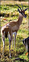
This pregnant animal from the San Diego Zoo died while being captured for translocation. This species has an expected longevity of 8-11 years. Jones (1993), who differentiated between 3 subspecies of red-fronted gazelles, reported the longevity for the Sudanese form as being 13 ½ years.
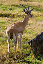 |
Gazella rufifrons at San Diego Zoo. |
2) General Gestational Data
Dittrich (1968) suggested that the length of gestation is between 187 and 189 days, similar to that of the Thomson gazelle. G. rufifrons normally produces 1 offspring (Hayssen et al., 1993). The neonatal weight is around 1,200 g; adults are between 20 and 35 kg.
3) Implantation
The placenta and fetus were located in the right uterine horn but the placenta (with numerous tiny cotyledons) extended into the left horn. The male fetus weighed 700 g and the uterus with placenta weighed 550 g. This is an epitheliochorial placenta, although the development of a ‘symplasma’ (Krölling, 1931) suggested to that author a ‘syndesmochorial’ placentation. Binucleated cells in the trophoblastic layer are abundant. These cells were investigated in great detail by Wimsatt (1951). He declared them to being ‘postmitotic’ and to have similar functions as the syncytiotrophoblast of ‘deciduate placentas’. Numerous studies have since been done on this unique binucleate cell and the placental lactogen production has been affirmed (Wooding, 1982). In fact, Atkinson et al. (1993) studied in detail the glycoprotein nature secreted by these binucleated cells from early gestation on. There is no infiltration of the uterine mucosa, the villous tissue merely interdigitates with the caruncular epithelium.
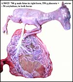 |
Uterus, placenta and fetus of this specimen. |
 |
Uterus unopened. |
4) General Characterization of the Placenta
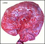
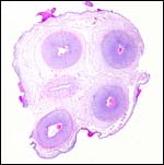
Approximately 50 cotyledons were counted, measuring from 0.4 cm (left horn) to 4 cm. Krölling (1931) found 60 cotyledons that remained ‘flat’ throughout gestation.
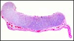 |
One large “flat” cotyledon of the right horn with attached uterine wall. |
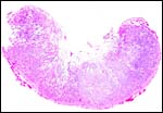 |
One of the smaller cotyledons in the left uterine horn. |
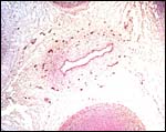
5) Details of fetal/maternal barrier
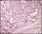
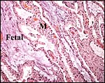 |
The somewhat edematous-appearing cotyledon interdigitates with the maternal tissue (M). |
 |
Section from the margin of the large cotyledon with endometrium. |
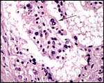 |
Binucleate cell at arrow. |
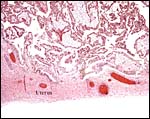
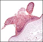
 |
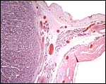
Uterus below from which extends the endometrium between villi. |
 |
Another view of the uterine wall beneath the large cotyledon. |
 |
Same uterine wall. |
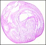
6) Umbilical cord
The umbilical cord measured 9 cm in length, had four vessels and a large allantoic duct. Tiny vessels accompanied the allantoic duct but were absence from the remainder of the cord. Its surface had numerous keratinized squamous pearls and the cord had a minimal left twist.
 |
Cross section of the umbilical cord. |
 |
The allantoic duct with vessels of the umbilical cord. |
 |
Areas of squamous metaplasia of the cord. |
7) Uteroplacental circulation
No further details are available.
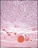
8) Extraplacental membranes
 |
One of the small numbers of ‘hippomanes’. |
There is no decidua and the amnion is delicate and avascular. The allantois is covered with very thin epithelium. A few pearls of ‘hippomanes’ were found.
9) Trophoblast external to barrier
None are present.
10) Endometrium
There is no decidual formation.
11) Various features
Subplacenta and metrial glands are absent.
12) Endocrinology
The finding of interstitial cell stimulation in even this tiny embryo suggests that LH or some other hormone must circulate in the fetus. Its origin may be placental, but there has been no evidence produced to affirm this and this seems to be unlikely at this point. No endocrine studies have been reported in this species. It is likely, however, that relaxin is produced by the binucleate cells, as in other ruminants.
13) Genetics
This gazelle possesses 58 chromosomes that are very similar to those of Thomson’s gazelle (Effron et al., 1976; O’Brien et al., 2006). More details of the relationship between various gazelles can be found in the detailed review of banded chromosomes by Vassart et al. (1995). They described the X/autosome translocation that characterizes these gazelles and considered the karyotype of red-fronted gazelles and Thomson’s gazelle to be similar if not identical. Rebholz & Harley (1999) grouped G. thomsoni as a subspecies of G. rufifrons on the basis of mtDNA sequence studies.
Gray (1972) listed hybridization with three other gazelles (Bennett’s, Thomson’s, and Heuglin’s gazelles. The former 2 did not survive; the last animal was reported in the IZY of 1969.
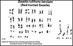 |
Karyotype from a red-fronted gazelle at San Diego Zoo, CRES and published by O’Brien et al. (2006). |
14) Immunology
I know of no immunological studies.
15) Pathological features
Griner (1983) does not list Eudorcas in his exhaustive review of the pathology of zoo species but among Thomson’s gazelles, trauma was the principal cause of death.
16) Physiologic data
No data are known to me.
17) Other resources
Cell strains of this gazelle species are available from CRES at San Diego zoo by contacting Dr. Oliver Ryder at: oryder@ucsd.edu.
18) Other remarks – What additional Information is needed?
A term placenta has not yet been available for study.
Acknowledgement
The animal photographs in this chapter and the karyotype come from the Zoological Society of San Diego.
References
Atkinson, Y.H., Gogolin-Ewens, K.J., Hounsell, E.F., Davies, M.J., Brandon, M.R. and Seamark, R.F.: Characterization of placentation-specific binucleate cell glycoprotein possessing a novel carbohydrate. J. Biol. Chem. 268:26679-26685, 1993.
Dittrich, L.: Keeping and breeding gazelles at Hannover Zoo. Intern. Zoo Ybk. 8:139-143, 1968.
Effron, M., Bogart, M.H., Kumamoto, A.T. and Benirschke, K.: Chromosome studies in the mammalian subfamily Antilopinae. Genetica 46: 419-444, 1976.
Gray, A.P.: Mammalian Hybrids. A Check-list with Bibliography. 2nd edition. Commonwealth Agricultural Bureaux Farnham Royal, Slough, England, 1972.
Griner, L.A.: Pathology of Zoo Animals. Zoological Society of San Diego, San Diego, California, 1983.
Groves, C.P.: An introduction to the gazelles. Chinkara 1:4-10, 1985.
Hayssen, V., van Tienhoven, A. and van Tienhoven, A.: Asdell’s Patterns of Mammalian Reproduction: a Compendium of Species-specific Data. Comstock/Cornell University Press, Ithaca, 1993.
Jones, M.L.: Longevity of ungulates in captivity. Intern. Zoo Yearbk. 32:159-169, 1993.
Krölling, O.: Über den Bau der Antilopenplazentome. Ztschr. Mikrosk.-Anatom. Forschung 27:211-232, 1931.
Nowak, R.M.: Walker’s Mammals of the World. 6th ed. The Johns Hopkins Press, Baltimore, 1999.
O’Brien, S.J., Menninger, J.C. and Nash, W.G., eds.: Atlas of Mammalian Chromosomes. Wiley-Liss; A. John Wiley & Sons, Inc. Hoboken, N.J., 2006.
Rebholz, W. and Harley, E.: Phylogenetic relationships in the bovid subfamily Antilopinae based on mitochondrial DNA sequences. Mol. Phylogenet. Evol. 12:87-94, 1999.
Vassart, M., Séguéla, A. and Hayes, H.: Chromosomal evolution in gazelles. J. Hered. 86:216-227, 1995.
Wimsatt, W.A.: Observations on the morphogenesis, cytochemistry, and significance of the binucleate giant cells of the placenta of ruminants. Amer. J. Anat. 89:233-282, 1951.
Wooding, F.B.: The role of the binucleate cell in ruminant placental structure. J. Reprod. Fertil. Suppl. 31:31-39, 1982.