 |
|
(Clicking
on the thumbnail images below will launch a new window and a larger
version of the thumbnail.)
|
| Last updated: August 20, 2007 |
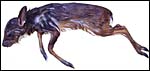
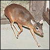
Neotragus pygmaeus
Order:
Artiodactyla
Family: Bovidae
1)
General Zoological Data
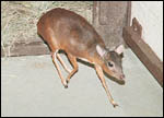
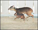
This animal, one of the three or perhaps more species
of "dwarf antelopes", has the distinction of being the smallest
antelope extant. The name Royal Antelope is said to derive from the local
Liberian designation as being the "King of the Hares". Royal
antelopes are West African animals and are rarely seen in zoos. They are
very shy animals and thus difficult to photograph and perhaps do not serve
as good exhibition specimens. Some of the related species of dwarf antelopes
are more commonly seen in zoological gardens, especially the suni, Neotragus
moschatus. Neotragus is often used synonymously with Nesotragus. The longevity
of Royal antelopes is 6 years and 8 months, according to Jones (1993).
The details of the evolution of bovidae has been controversial. It is
reviewed in some detail by Matthee & Robinson (1999). They suggested
that the strictly African neotragini arose 12 MYA but then stated that
"the assessment of evolutionary relationships in the dwarf antelope
(Neotragini) has been troubled by many symplesiomorphic morphological
characters, all possibly linked to their small size (Gentry, 1992)".
They studied these controversies by analyzing the mitochondrial cytochrome
b gene. It should be pointed out, however, that they did not have material
from the Royal antelope available; it was restricted to suni, oribi, grysbok,
steenbok, dik-diks and klipspringer. Of this group, only klipspringer
and suni failed to follow a monophylogenetic grouping. These anomalies
are discussed at some length and will require further work for resolution.
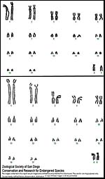
Chromosomally, these animals also present some challenges with their cryptic
chromosomal variation and with chromosome numbers ranging from 2n=30 in
the steenbok to 2n=60 in oribi and klipspringer. Much more study is required
before clarity of species designation is had.