 |
|
(Clicking
on the thumbnail images below will launch a new window and a larger
version of the thumbnail.)
|
| September 12, 2004. |
Pudu pudu(a)
Order: Artiodactyla
Family: Cervidae
1) General Zoological Data
The pudu, a South American deer, is the smallest deer and, because of its similarity to brocket deer, these two species have sometimes been grouped together. There are some significant bony differences, however, that led to their separation (Groves & Grubb, 1987). The longevity of pudus is maximally 15 years 9 months according to Jones (1993); more often they die at a younger age. The name pudu is said to derive from Mapuche, people in southern Chile (Gotch, 1979). The designations Pudu pudu and Pudu puda are interchangeable and there is debate as to which is more correct. There are two species, Pudu mephistophiles and P. pudu according to the detailed taxonomic considerations of Hershkovitz (1982). The former species is darker, smaller and may be more primitive. It is from the Northwest of South America; the latter species is from Chile and both are severely endangered. Both have rudimentary antlers in males only and only the Southern pudu fawn is spotted. The maximum weight recorded is 13.4 kg; most are smaller (6-11 kg).
A number of zoos have bred the Southern pudu successfully and those institutions are listed by Hershkovitz (1982). The Northern species, however, is said to be more difficult to maintain. A successful colony exists at the Wild Animal Park of the Zoological Society of San Diego. The recent publication by Schurer & Sliwa (2002) takes issue with the species name and explains in considerable detail how the name was derived. Accordingly, we now prefer to name this Southern pudu Pudu pudu.
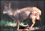 |
Adult Chilean pudu at San Diego Wild Animal Park. |
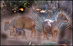 |
Pair of Chilean pudus with young at San Diego Wild Animal Park. |
Hershkovitz (1982), in his extensive review of all known characters of pudu, reported that pudus have a gestational length of 7 months (around 202-223 days) and that the single newborns weigh 35-37 oz. He also listed the weight of 370 g for an animal born in the Cologne Zoo, but Hick (1967/8; 1969) described the successful raising of a 1,000 g neonate at Cologne. A second birth resulted in a much smaller neonate. The male neonate that we observed weighed 700 g but died on its second day of life of "maternal neglect". Macnamara & Eldridge (1987) provided an overview of the reproduction in pudus and brocket deer. The birth of occasional twins, in both species, has been reported by some authors.
The recent publication by Schurer & Sliwa (2002) provides much more information and more accurate data on this species than has been available in the past. The average length of gestation observed was 210 days; the average weight (of 55 newborn animals) was 890 g, the female usually being as heavy as the males. Offspring below 600 g did not survive, nor did those weighing more than 1000 g. In addition, these authors provide information on seasonality, reproductive age and longevity.
3)
Implantation
No implanted placenta has been described and early stages are not known.
4)
General Characterization of the Placenta
The pudu placenta, like that of other deer is oligocotyledonary, villous,
and epithelio-chorial. Its placenta had only one intact cotyledon and
a second atrophying cotyledon. The larger cotyledon measured 8 x 1 cm.
The placenta weighed 110 g.
Hamilton et al. (1960) described the placentation of several other deer
species and all were found to be oligocotyledonary. In the tufted deer
(see chapter on tufted deer) I found only two cotyledons. In the general
chapter on deer, several other species are comprised with a table that
gives access to their gross morphology. Pudu has had so far the smallest
number of cotyledons I have observed. The one intact cotyledon, however,
was quite large. The smaller lobe appeared to have degenerated at least
partially before delivery. Moreover, there was some inflammation on the
placental surface.
A second specimen became available in August 2004 from a successful birth at the San Diego Zoo's Wild Animal Park . It also had only one large cotyledon (5x5.5 cm) and where the cord vessels divided, a second cotyledon may have been detached and remained in utero ; alternatively, it was also degenerated. The cord of this specimen was 6 cm long and had 4 vessels as well as an allantoic duct.
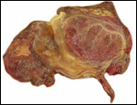 |
Pudu placenta with intact cotyledon at right and the atrophic part at left. Note that a pair of vessels branches to both sides. |
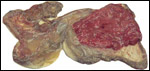 |
Maternal aspect of the same placenta with atrophied villous remnants at left. |
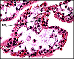
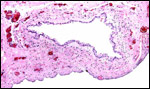
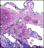
Unfortunately, the first placenta was significantly degenerated due to autolysis. Nevertheless, some trophoblast was sufficiently intact to identify the large number of binucleate cells. The trophoblast is single-layered, cuboidal and cylindrical in other regions. The stroma was edematous, as is often the case in the placentas that I receive. No maternal tissue was present. The second placenta was slightly better preserved and had pronounced binucleate cells but no yellow hemophagous trophoblastic areas under the chorion. Some villi are somewhat edematous again from autolysis.
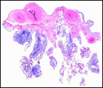 |
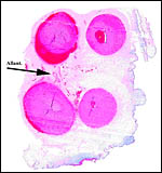
Surface of first pudu placenta with autolyzed (edematous, blue) villi. |
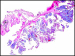 |
Surface of first pudu placenta with membrane attachment at top left. |
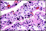 |
The villous trophoblast is cylindrical to cuboidal and, at the peripheral ends, there are numerous binucleate trophoblastic cells (arrows). |
 |
Surface of second placenta. |
 |
Binucleate cell at area, second placenta. |
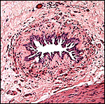
The umbilical cord was 8 cm long and 1 cm in width. It contained 4 vessels and an allantoic duct. The relatively small duct is shown next at the arrow. The cord was not spiraled. The umbilical cord of the second specimen was similar but only 6 cm long. The second placenta's umbilical cord had numerous patches of keratinizing squamous metaplasia on the surface.
 |
Umbilical cord of second specimen with allantoic duct at arrow. |
 |
Allantoic duct of second placenta's umbilical cord. |
 |
The umbilical cord of the first placenta has a relatively small allantoic duct and a few small allantoic vessels. There are two large (post partum) hemorrhages. |
 |
Allantoic duct in the umbilical cord with adjacent allantoic blood vessels. |
 |
Surface of the second placenta's umbilical cord with squamous metaplasia. |
This has not been studied.
8)
Extraplacental membranes
There is an allantoic sac but no vitelline structures were recognized.
The epithelium of the allantoic sac was much degenerated, while the amnion
was well-preserved. No decidua capsularis exists.
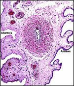 |
Membranes with degenerated allantoic epithelium at left and amnion at right. Note the vasculature in the allantoic membrane. |
Although no implanted placenta has been observed, judging from what is known of other cervidae, it is unlikely that trophoblast invasion of the endometrium occurs.
10)
Endometrium
I am not aware of any studies. In many other deer studied, however, the
endometrium between caruncles "remains intact" (Hamilton et
al., 1960).
11)
Various features
There is no subplacenta.
12)
Endocrinology
The seasonality of LH and testosterone concentrations was studied by Reyes
et al. (1993). Bartos et al. (1998) studied the hormonal levels of two
males housed together with females in order to assess dominance characteristics.
They found that IGF-1 levels (IGF = insulin growth factor) were higher
during the spring during antler-growth (in Chile: September to November),
while testosterone levels were higher during rut, and FSH levels were
lower. Prolactin was higher in the summer. Later, they showed that antler
growth is very sensitive to androgen levels (Bubenik et al., 2002). The
presence of so many binucleate cells, characteristically found in all
cervidae, suggests that they may have the same function as in other deer.
This topic (placental lactogen production) is discussed at length in the
general chapter on "deer".
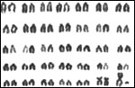
13) Genetics
The pudu has 70 chromosomes (Koulisher et al., 1972). The X-chromosome
and one pair of autosomes are metacentric, all others are acrocentric
elements. Other authors have confirmed this result (Schreiber & Dmoch,
1994). Chromosomal banding was described by Petit & deMeurichy (1989).
The pericentric heterochromatin of pudu chromosomes has a peripheral distribution
in sectioned nuclei (Berrios et al., 1999). Hybrids have not been described.
The karyotype shown next was copied from Koulisher et al., 1972),
the next one is from the San Diego Zoo.
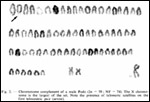 |
Karyotype from Koulisher et al., 1972. |
 |
Karyotype of male Pudu pudu from the San Diego Zoo. |
The only data known to me are the results of the pox infection reported by Junge et al. (2000). They reported the development of neutralizing antibodies following infection.
15)
Pathological features
Kohls (1969) reported infection by Ixodes taglei in Chile. Poxvirus infection
of pudu can be fatal, can lead to large skin ulcerations and to secondary
fungal infection. Junge et al. (2000) observed such an outbreak at the
St. Louis Zoo and found neutralizing antibodies to develop. They were
successful with therapy with the antiviral agent cidofovir. Pudus are
also reported to be susceptible to hoof and mouth disease.
 |
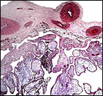
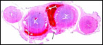
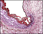
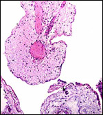
Surface chorion and amnion with degenerating villi below of the degenerating cotyledon. There are a substantial number of polymorphonuclear leukocytes in chorion and stem villi. |
 |
Section from the degenerating cotyledon showing autolyzed fetal blood and degenerating trophoblast. |
No detailed studies are known to me.
17)
Other resources
Cell strains of pudu are available from CRES
at the San Diego Zoo by contacting Dr. Oliver Ruder at oryder@ucsd.edu.
18) Other remarks - What additional Information is needed?
No implanted placenta has been observed and nothing is known about trophoblastic
invasion of the endometrium. Also, it would be interesting to known the
number of caruncles in the pudu uterus. Endocrine studies are needed.
Acknowledgement
The animal photographs in this chapter come from the Zoological Society
of San Diego. I appreciate also very much the help of the pathologists
at the San Diego Zoo.
References
Bartos, L., Reyes, E., Schams, D., Bubenik, G. and Lobos, A.: Rank dependent
seasonal levels of IGF-1, cortisol and reproductive hormones in male pudu
(Pudu puda). Comp. Biochem. Physiol. A Mol. Integr. Physiol. 120:373-378,
1998.
Berrios, S. Ayarza, E., Moreno, M. Paulos, A. and Fernandez-Donoso, R.: Non-random distribution of the pericentromeric heterochromatin in meiotic prophase nuclei of mammalian spermatocytes. Genetica 106:187-195, 1999.
Bubenik, G.A., Reyes, E., Schams, D., Lobos, A., Bartos, L. and Koerner, F.: Effect of antiandrogen cyproterone acetate on the development of the antler cycle in Southern pudu (Pudu puda). J. Exp. Zool. 292:393-401, 2002.
Gotch, A.F.: Mammals - Their Latin Names Explained. Blandford Press, Poole, Dorset, 1979.
Groves, C.P. and Grubb, P.: Relationships of living deer. Pp. 21-59, in Biology and Management of the Cervidae. C.M. Wemmer, ed. Smithsonian Institution Press, Washington, DC 1987.
Hamilton, W.J., Harrison, R.J. and Young, B.A.: Aspects of placentation in certain cervidae. J. Anat. 94:1-23, 1960.
Hershkovitz,
P.: Neotropical Deer (Cervidae). Part I. Pudus, Genus Pudu Gray.
Fieldiana. Zoology. New Series # 11, pp. 86, 1982.
Hick, U.: Gegluckte Aufzucht eines Pudus (Pudu pudu Mol.) im Kolner
Zoo. Freunde des Kolner Zoo. 10:111-117. 1967/8.
Hick, U.: Successful raising of a pudu Pudu pudu at Cologne Zoo. Int. Zoo Yearbk. 9:110-112, 1969.
Jones, M.L.: Longevity of ungulates in captivity. Int. Zoo Yearbk. 32:159-169, 1993.
Junge, R.E., Duncan, M.C., Miller, R.E., Gregg, D. and Kombert, M.: Clinical presentation and antiviral therapy for poxvirus infection in pudu (Pudu puda). J. Zoo Wild. Med. 31:412-418, 2000.
Kohls, G.M.: Ixodes taglei n. Sp. (Acarina: Ixodidae) a parasite of the deer, Pudu pudu (Wol.), in Chile. J. Med. Entomol. 6:280-283, 1969.
Koulisher, L., Tyskens, J. and Mortelmans, J.: Mammalian Cytogenetics. VII. The chromosomes of Cervus canadensis, Elaphurus davidianus, Cervus nippon (Temminck) and Pudu pudu. Acta Zool. Pathol. Antverp. 56:25-30, 1972.
Macnamara, M. and Eldridge, W.: Behavior and reproduction in captive pudu (Pudu puda) and red brocket (Mazama americana), a descriptive and comparative analysis. In, Biology and Management of the Cervidae, C.M. Wemmer, ed. Smithsonian Institution Press, Washington, DC, 1987.
Petit, P. and de Meurichy, W.: On the banding patterns of the chromosomes of the Pudu pudu (Pudu). Ann. Genet. 32:141, 143, 1989.
Reyes, E., Munoz, P., Recabarren, S., Torres, P. and Bubenik, G.A.: Seasonal variation of LH and testosterone in the smallest deer, the pudu (Pudu puda molina) and its relationship to the antler cycle. Comp. Biochem. Physiol. Comp. Physiol. 106:683-685, 1993.
Schreiber,
A. and Dmoch, R.: Chromosomes of two rare species of neotropical mammals:
Southern pudu (Pudu puda) and Bush dog (Speothos venaticus).
Z. Säugetierk. 59:317-320, 1994.
Schurer, U. and Sliwa, A.: Sudpudus (Pudu pudu) in zoologischen
Garten. Bongo 32:3-11, 2002.