| |
8)
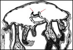
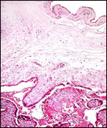
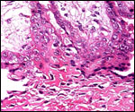
Extraplacental membranes
The amnion is pressed against the chorion in which the fetal blood vessels
are carried. The amnion is composed of a thin epithelium and a layer of
connective tissue without vessels. More peripheral to the chorionic membrane
is a thin layer of extravillous trophoblast, and then follows a small amount
of decidua capsularis. There is no allantoic sac. Of interest is that the
chorion laeve did not contain atrophic villi, as they are abundant in human
placentas. This absence is similar to the condition found in macacs and
other monkeys with bidiscoid placentas. You will find more discussion in
the macac chapter on this absence of atrophied villi in the membranes.
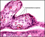
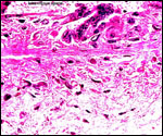
9) Trophoblast external to barrier
There is an extensive shell of invading extravillous trophoblast ("X-cells").
It infiltrates the superficial layer of the decidua basalis but does not
reach the myometrium.
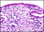
10) Endometrium
The endometrium undergoes decidual changes very much like those that occur
in human gestation. There is no basal layer of typical fibrinoid as seen
in humans (the Rohr and Nitabuch layers); instead, a broad band of decidual
necrosis is found here, as is true for other cercopithecids.
11) Various features
None.
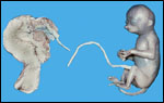
12) Endocrinology
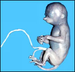
I know of no studies specifically directed to this species. Typically
though, the fetus depicted above had the usual large fetal adrenal glands
of primates that are due to the presence of a wide fetal zone (Soma &
Benirschke, 1977). The adrenal glands were even larger than the kidneys.
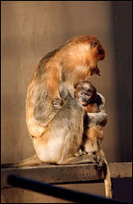
It is of interest that Wislocki (1939), who saw monochorionic twins in
this species, does not mention any freemartin effect on the female fetus.
Perhaps there were no vascular anastomoses between the twins, or perhaps
the situation is similar to that of marmosets.
13)
Genetics
The proboscis monkey has 48 chromosomes, and Giemsa banding has been done
(Chiarelli, 1966; Hsu & Benirschke, 1975; Soma & Benirschke, 1975).
Hybrids are unknown. One study on the cytochrome b mitochondrial gene
has been published by Collura et al. (1996). In studies that employ chromosome
painting, Bigoni et al. (2003) found complex rearrangements to explain
the unique karyotype of this unusual animal. It may not, as had been suspected,
be most ancestral of colobids. Further studies of related taxa are imperative.
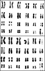 |
Male
and female karyotypes of proboscis monkey from Hsu & Benirschke,
1975. |
14)
Immunology
The deaths of proboscis monkeys at San Diego were frequently due to infection
with cryptococci, but it is not known whether this is due to an immune
deficiency.
15) Pathological features
Cryptococcosis has been a significant cause of mortality in San Diego.
Pulmonary acariasis due to Pneumonyssus simicola was also common
in the animals of our zoo (Griner, 1983). In the Frankfurt Zoo, where
many animals have been kept in the past, intestinal parasites were an
initial problem.
16)
Physiologic data
Few studies other than the brief description of the partitioned stomach
that is so characteristic of these leaf-eaters are published (Soma &
Benirschke, 1977).
17)
Other resources
Some cell strains are available from CRES
at the San Diego Zoo by contacting Dr. Oliver Ryder at: oryder@ucsd.edu.
18) Other remarks - What additional Information is needed?
Term placentas have not been described. Thus, the weight of the mature
placenta and the length of term umbilical cords are unknown. No endocrine
studies have been conducted and they are needed.
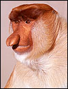
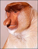
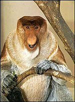
Acknowledgement
The animal photographs in this chapter come from the Zoological Society
of San Diego. I appreciate also very much the help of the pathologists
at the San Diego Zoo.
References
Bigoni, F., Stanyon, R., Wimmer, R. and Schempp, W.: Chromosome painting
shows that the proboscis monkey (Nasalis larvatus) has a derived
karyotype and is phylogenetically nested with asian colobines. Amer. J.
Primatol. 60:85-93, 2003.
Chiarelli, B.: The chromosome complement of Nasalis larvatus (Wurm
1781). Experientia 22:797, 1966.
Collura,
R.V., Auerbach, M.R. and Stewart, C.B.: A quick, direct method that can
differentiate expressed mitochondrial genes from their nuclear pseudogenes.
Curr. Biol. 6:1337-1339, 1996.
Griner,
L.A.: Pathology of Zoo Animals. Zoological Society of San Diego, San Diego,
California, 1983.
Hill,
J.P.: The developmental history of the primates. Philosoph. Trans. B.
221:45-178, 1932.
Hsu,
T.C. and Benirschke, K.: An Atlas of Mammalian Chromosomes. Springer-Verlag,
New York. Vol. 9, Folio 449, 1975.
Luckett,
W.P.: Comparative development and evolution of the placenta in primates.
Contrib. Primatol. 3:142-234, 1974.
Macdonald,
D.W.: Notes on the size and composition of groups of proboscis monkey,
Nasalis larvatus. Folia Primatol. 37:95-98, 1982.
MacKinnon,
K.: The conservation status of nonhuman primates in Indonesia. Chapter
8 in, Primates - The Road to self-sustaining Populations, K. Benirschke,
ed. Pp.99-126, Springer-Verlag, NY, 1986.
Nowak,
R.M.: Walker's Mammals of the World. 6th ed. The Johns Hopkins Press,
Baltimore, 1999.
Selenka,
E.: Entwickklung des Gibbons (Hylobates und Siamanga). Fortsetz.
Stud. Entw. Gesch. Tiere 8:173-208, 1900.
Soma,
H., Benirschke, K. and Robinson, P.: The chromosomes of the Proboscis
monkey (Nasalis larvatus). Chromosome Information Service 17:24-26.
1974
Soma,
H. and Benirschke, K.: Observations on the fetus and placenta of a proboscis
monkey (Nasalis larvatus). Primates 18:277-284, 1977.
Wislocki,
G.B.: Observations on twinning in marmosets. Amer. J. Anat. 64:445-483,
1939.
Yeager,
C.P., Silver, S.C. and Dierenfeld, E.S.: Mineral and phytochemical influences
on foliage selection by the proboscis monkey (Nasalis larvatus).
Amer. J. Primatol. 41:117-128, 1997.
|