(Clicking on the thumbnail images below will launch a new window and a larger version of the thumbnail.)
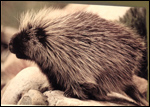
Canadian or North American Porcupine
Erethizon dorsatum

Order: Rodentia
Family: Erethizontidae

1) General zoological data of species


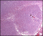
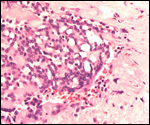
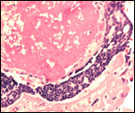
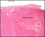
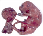
This single species (with 6 possible subspecies) is indigenous to North America and represents its only porcupine species. Its habitat is largely woods, with a strong arboreal tendency. It is strictly herbivorous with an appetite for bark and salty woods. The animal is mostly nocturnal and is not closely related to the African porcupines, both having arisen by converging evolution from other Rodentia (Wood, 1950). On the other hand, when Luckett & Mossman (1981) studied the placentas of Hystrix and another hystricognath rodent, Bathyergus, they found them to have a great similarity in their placentation with the South American hystricomorph species. This, among other aspects, has given rise to a great controversy over the possible monophyly or development by converging evolution of porcupines.

I believe, however, that the originally South American origin of this hystricomorph rodent is strongly supported by its having an X-chromosome that is, as is true for other South American rodents, of twice the size of all other rodents. While captive breeding is often successful and newborns become easily tractable, we are not aware of any colonies other than the animals that are kept in zoological gardens. Because of its preference for trees, Europeans refer to these animals as "Urson" (in German "Baumstachler"). The modified hair consists of quills with fine hooks at the end. Further references to its anatomy are listed by Hayssen et al. (1993). The longevity of American porcupines is at least 10 years.
   
  North American porcupine at San Diego Zoo.
     
 
2) General gestational data

North American porcupines are solitary animals, which produce a single precocious newborn in spring. Without delayed implantation they breed in autumn and have a long gestation of seven months (205-217 days). The ovarian activity is unusual, as is its placenta. Possible positioning into a separate suborder has been entertained (Perrotta, 1959). Depending on the stage of pregnancy, adult females weigh between 4,000 and 7,000 g. Males are much larger; they weigh up to 18 kg (Nowak & Paradiso, 1983). The newborn weight is given as between 340 and 640 g. Shadle & Ploss (1943) observed a normal newborn weighing 480. Later, Shadle (1948) provided new data on the length of gestation.

The uterus is described as being "transitional between duplex and bicornuate" by Perrotta (1959) and as having spiraled lumens (Mossman, 1987). The cervix is Y-shaped. The endometrium consists of tubular glands, some of which are branched, in the endocervix. Accessory corpora lutea develop from atretic follicles but persist only on the side of pregnancy. Mossman (1987) made other pertinent remarks on the preservation of corpora lutea. The placental weight at term is around 50 g, including the membranes and the relatively short umbilical cord. An extensive investigation of placenta and reproduction of the porcupine comes from Perrotta's detailed study (1959).

3) Implantation

Implantation occurs antimesometrially, the disk being initially mesometrially oriented at implantation, however. There is little endometrial degeneration at the site of implantation. Perrotta, who was unable to obtain later stages of the early implantation, stated that some degree of interstitial implantation is likely to occur. The blastocyst is relatively large at the time of implantation and has a protruding amnionic cavity. The exact time of implantation remains unknown. The most striking feature of hystricomorph rodents is the early inversion of their germ layers and degeneration of their parietal trophoblast. Eventually, endoderm (visceral yolk sac) epithelium forms the outside of the membranes and is there apposed to the uterine epithelium. Roberts and Perry (1974) gave a good summary of these stages. Please also see the chapter on the mouse.

Perrotta (1959) identified three types of trophoblast at the time of implantation:

1) Polar trophoblast (at implantation filling the gap in the endometrium),
2) Parietal and,
3) Covering trophoblast.

During further development, four types are distinguishable:

1) Primary,
2) Trophospongium,
3) Trophoblastic canals (through which the maternal blood flows),
4) Giant cells (they are syncytial and occur at the periphery and especially at the floor of the placental disk).

Because placental development of the hystricognath rodents is complex and difficult to understand, several diagrams are now illustrated.

   
  General situs of placental development of the porcupine.
     
  General situs of placental development of the porcupine.
     
 
4) General characteristics of placenta

Perrotta's study (1959) relied upon 200 macroscopic and 40 histologic specimens. Despite that commendable effort, very few early stages were identified.

This animal has a discoid, labyrinthine placenta with a complex lobular pattern and a hemochorial fetal-maternal relation. The placenta resembles that of the guinea pig. The placenta of the guinea pig, another South American rodent, has been well studied by Kaufmann (1981), and by Enders (1965). The latter preferred the designation of this animal as having a "hemomonochorial" maternal/fetal relationship (Enders, 1965).

My specimen measured 6 x 5-x 2 cm. It had abundant membranes. There was an area of marked necrosis and calcification in the center of the maternal surface, which corresponds to the "subplacenta" and that marks the attachment site to the decidua below. Lateral to this central calcified area there was a greenish hue due to fibrin-like material that contained hemosiderin granules in macrophages and in degenerating basal trophoblastic giant cells. Degenerating decidua and trophoblast were present in this hyalinized mass, as well as some specks of dystrophic calcification.

The basal giant cell trophoblastic layer that abuts this hyalinized floor is often markedly vacuolated and has occasional pyknotic nuclei. This was previously described by Perrotta (1959). Some cells contain additional hemosiderin, demonstrable by Prussian blue stain.

At term, the yolk sac stalk is mesometrially oriented. The inverted endodermal slpanchnopleure lies over the disk and forms a ring that is seen in the macroscopic picture of this placenta. There is a circular ring of vitelline vessels, similar to that in the guinea pig.

Invasive trophoblast such as occurs into the myometrium of humans has not been described. Giant cells penetrate the decidua and cause degenerative changes there, which eventuate in calcification. In this area, the peripheral trophoblast forms huge giant cells; many become vacuolated, accumulate iron and subsequently degenerate in the floor. It should be noted, however, that these trophoblastic cells invade in the guinea pig (Nanaev et al., 1995) and we have found it in the pacarana (Dinomys branickii). Moreover, Miglino et al. (2002) studied three other hystricomorph rodent placentas with great similarities of many features seen in porcupines.

The most complicated aspect of porcupine placentation is the inversion of the yolk sac. It comes to envelop the outside of the conceptual sac, with yolk sac endoderm located on a connective tissue sheet (the "Splanchnopleure"). We assume that it has absorptive function, but that has not been studied in the porcupine. It is so, however, in other rodents (Please see the chapter on mouse). As this vascularized splanchnopleure comes toward the edge of the placental disk, a fibrovascular "ring" develops that is illustrated here. The original yolk sac atrophies completely, as does the transitory allantois.
   
  Delivered placenta at term.
     
  Cross-section of porcupine placenta with the inverted yolk sac membranes above. Calcified subplacenta is yellow in the center and on the maternal surface. Next are the cross-sections of the placental lobules. The luxurious, undulating splanchnopleure membranes are folded on top.
     
  Sections through the edge and center of the placenta with the "subplacenta" located at the bottom.
     
  Sections through the edge and center of the placenta with the "subplacenta" located at the bottom.
     
  Edge of porcupine placenta with villous tissue below, splanchnopleure curving to left top and vascular ring (VR).
     
  One of many complex lobules of the porcupine placenta with maternal (M) and fetal (F) vessels in the center.
     
  Masses of giant cells at the floor of the porcupine placenta.
     
  Masses of giant cells at the floor of the porcupine placenta.
     
  Higher magnification of one lobule of porcupine placenta with maternal (MA) and fetal (FV) blood vessels.
     
  Attachment site of porcupine placenta - so-called "subplacenta".
     
 
5) Details of barrier structure

The fetal-maternal relationship is hemochorial, in a labyrinthine network of villi with complex maternal and fetal vascularization. Trophoblastic channels contain the maternal blood. Fetal capillaries are present in the ample connective tissue. Thus, there is a typical countercurrent blood flow. The next pictures show the intricacies of the main placental tissue.
   
  Highest magnifications of the trophoblastic and vascular arrangement of the porcupine placenta.
     
  Highest magnifications of the trophoblastic and vascular arrangement of the porcupine placenta.
     
  Another view of the complex fetal/maternal relationship. G=Giant cells; F=Fetal blood space.
     
 
6) Umbilical cord

The umbilical cord of our specimen inserted near the center of the placenta and was 13 cm in length, and 1 cm in width. It had no twists. There were two chorio-allantoic arteries and two umbilical veins, plus a large number of smaller vessels (usually seen in placentas endowed with allantoic sacs). Some of these vessels had thick muscular walls. Others had many capillary-like small vessels without any specific grouping around the remaining parts of the allantoic duct. There was a remnant of allantoic duct and, in a portion of one of the umbilical cords available to me, smooth muscle bundles accompanied the duct remnant. Another umbilical cord that I studied had a patent allantoic duct.

The surface of the cord is remarkably more loosely structured than its center. The cord surface has a thin amnion. Except for locations directly beneath the surface, little Wharton's jelly exists. There are no amnionic "callosities".
   
  Cross-sections through two umbilical cords of porcupine.
     
  Cross-sections through two umbilical cords of porcupine.
     
 
7) Uteroplacental circulation

No details have been published and no uterus was available to us.

8) Extraplacental membranes

The initial decidua capsularis is described to disappear halfway through gestation; the decidua basalis is sparse. There is no allantoic cavity and amniogenesis is by cavitation. Perrotta (1959) stated that there is fine vascularization of the amnion near the placental disk, clearly an unusual feature in most mammals. Perrotta assumed that these vessels were vitelline blood vessels. More likely, they merely border the amnion and do not provide amnionic vascularization. In any case, they are confined to a small ring.

The placenta has a bilaminar omphalopleure; the endoderm extends over a portion of the parietal trophoblast of the blastocyst that then disappears and persists only on the surface of the placental disk. There is no so-called "chorio-vitelline" placenta but the endoderm extends over the periphery with numerous closely approximated villi.

The "vascular splanchnopleure" has the complete inversion of the yolk sac. This persists to term, with numerous blood vessels (of vitelline nature) of yolk sac origin. The epithelium, endodermal-yolk-sac-derived, makes up its surface. It lies against the opposite side of the endometrial epithelium where it assumes an epitheliochorial relation between mother and fetus (Kaufmann, 1981). One must assume that much transport can occur here in this rodent placenta as well; certainly this is so in early gestation (King & Enders, 1970a). Protein-rich material locates here in rodents and presumably it is absorbed by the complex epithelial trophoblastic surface (Please see chapter on mouse). For the guinea pig, this has been well illustrated by King & Enders (1970b). Towards the embryo, this inverted yolk sac membrane abuts the connective tissue of the amnion and then the amnionic epithelium.
   
  Placental structure at the site of surface chorionic plate with maternal vessel (M), large fetal blood vessel (F.V.) and peripheral trophospongium and giant cells.
     
 
9) Trophoblast external to barrier

None.

10) Endometrium

The endometrium beneath the major placental disk of rodents becomes the decidua, as is also true in Erethizon. It must be cautioned, however, that this tissue differs from the "decidualization" induced by progesterone in human uteri, as Wynn (1965) has made very clear. It is a complex structure and may subserve some nutritional role but is not well studied in the porcupine. Perrotta (1959) showed its hyalinization and degenerative changes towards term, and our placenta had not only hyalinization and degeneration, but much dystrophic calcification within it as well.
   
  This is a section through the inverted endodermal (yolk) membrane that apposes the uterine wall with many villi and a cuboidal epithelium.
     
 
11) Various features

An accessory placenta ("subplacenta") exists in early placentation but disappears gradually later through a process of hyalinization and degeneration. It is confined to the central portion of the disk and is remarkably calcified in my specimen. The location of the subplacenta is essentially similar to that described for the guinea pig (Kaufmann, 1981; see also Kaufmann & Davidoff, 1977).

12) Endocrinology

Estrus lasts a few hours (Woods, 1973) and estrous cycling occurs between 27 and 30 days when copulation is unsuccessful. Ovulation occurs largely from the right ovary, possibly being induced (Dodge, 1967). Mossman & Judas (1949) examined the ovaries of thirty pregnant porcupines in some detail and described their findings in a highly readable paper. One follicle is ovulated but, during pregnancy numerous corpora albicantia are luteinized additionally, and only on the side bearing the normal corpus. Likewise, thecal cells external to the corpus luteum undergo the same stimulation. Thus, the ovary has ultimately a most unusual morphology with these structures that have been named "accessory corpora lutea". There is additional extensive consideration of such unusual ovarian structures and the luteinization of the "interstitial cells" of the porcupine ovary. It appears that the hystricomorph rodents have numerous other unusual anatomical presentations. They will be discussed in more detail in my consideration of the chinchilla and pacarana placentas.

I have been unable to find information on gonadotropins or estrogens in porcupine pregnancies.

13) Genetics

The North American porcupine has 42 chromosomes, 34 metacentrics, 6 acrocentrics, a large submetacentric X (12%), and medium-sized submetacentric Y (6 %). This species is related to the South American hystricomorph rodents of the family Erezithontidae and is its largest representative (Benirschke, 1968). Like other South American rodents, but in contrast to all other rodents, it possesses an X-chromosome that is approximately twice the size of the "usual" X-chromosome, comprising about 10% of the haploid set of DNA. Hybrids are unknown.

14) Immunology

I know no significant information.

15) Pathological features

The only information available is the report of an albino porcupine depicted by Sokolowsky (1926/27), and a few parasitic infections listed and available through logging on to PubMed.

I have had the opportunity also to study the placenta (and fetus) of an aborted prehensile-tailed porcupine (Coendu prehensilis bolivianus). The fetus was aborted because of maternal septicemia and abscesses were found in the placenta and there was chorioamnionitis.

16) Physiological data

I know of no studies.

17) Other resources

Cell strains are available from CRES at the Zoological Society of San Diego.

18) Other relevant data.

In 1980 the Zoological Society of San Diego lost a prehensile-tailed porcupine from Bolivia (Coendu prehensilis bolivianus) due to sepsis. The female was pregnant and a stillborn immature fetus was delivered, weighing 150 g, with a placental weight of 35 g (4.5x2 cm). There were abscesses in the villous tissue and chorioamnionitis was also present. The 14 cm umbilical cord had five (!) blood vessels, a remnant of allantoic and omphalomesenteric duct. Many small additional blood vessels were present in the umbilical cord and there were squamous "pearls" (metaplasia) on the cord surface. The implantation site and general histology were identical to those of Erethizon. The subplacenta had maternal endometrial glands attached.

   
  Prehensile-tailed porcupine abortus with placenta attached. Note the thick membranes with yolk sac epithelium. At right is the maternal side of this placenta with the light area being the subplacenta.
     
  Prehensile-tailed porcupine abortus with placenta attached. Note the thick membranes with yolk sac epithelium. At right is the maternal side of this placenta with the light area being the subplacenta.
     
  Maternal surface of the aborted prehensile-tailed porcupine. It shows extensive basal degeneration and the villous tissue is undergoing early autolysis.
     
 
References

CRES at: http://www.sandiegozoo.org/conservation/cres_home.html. Please direct your inquiries to Dr. Oliver Ryder (oryder@ucsd.edu).

Benirschke, K.: The chromosome complement and meiosis of the North American porcupine. J. Hered. 59:71-76, 1968.

Dodge, W.E.: The biology and life history of the porcupine (Erethizon dorsatum) in western Massachusetts. Ph.D. Thesis, University of Massachusetts, Amherst, 1967.

Enders, A.C.: A comparative study of the fine structure of the trophoblast in several hemochorial placentas. Amer. J. Anat. 116:29-68. 1965.

Hayssen, V., v. Tienhoven, A. and v. Tienhoven, A.: Asdell's Patterns of Mammalian Reproduction. Comstock Publishing Assoc., Ithaca and London, 1993.

Kaufmann, P: Functional anatomy of the non-primate placenta. Placenta (supplement 1) pp:13-28, 1981.

Kaufmann, P. and Davidoff, M.: The guinea pig placenta. Adv. Anat. Embryol. Cell Biol. 53:1-91, 1977.

King, B.F. and Enders, A.C.: Protein absorption and transport by the guinea pig visceral yolk sac placenta. Amer. J. Anat. 129:261-288, 1970a.

King, B.F. and Enders, A.C.: The fine structure of the guinea pig visceral yolk sac placenta. Amer. J. Anat. 127:397-414, 1970b.

Luckett, W.P. and Mossman, H.W.: Development and phylogenetic significance of the fetal membranes and placenta of the African hystricognathous rodents Bathyergus and Hystrix. Amer. J. Anat. 162:265-285, 1981.

Miglino, M.A., Carter, A.M., Santos Ferraz, R.H. dos and Fernandes Machado, M.R.: Placentation in the capybara (Hydrochaerus hydrochaeris), agouti (Dasyprocta aguti) and paca (Agouti paca). Placenta 23:416-428, 2002.

Mossman, H.W.: Vertebrate Fetal Membranes. MacMillan, Houndsmills, 1987.

Mossman, H.W. and Judas, I.: Accessory corpora lutea, lutein cell origin, and the ovarian cycle in the Canadian porcupine. Amer. J. Anat. 85:1-39, 1949.

Nanaev, A., Chwalisz, K., Frank, H.G., Kohnen, G., Hegele-Hartung, C and Kaufmann, P.: Physiological dilation of uteroplacental arteries in the guinea pig depends on nitric oxide synthase activity of extravillous trophoblast. Cell Tissue Res. 282:407-442, 1995.

Perrotta, C.A.: Fetal membranes of the Canadian porcupine, Erethizon dorsatum. Amer. J. Anat. 104:35-59, 1959.

Roberts, C.M. and Perry, J.S.: Placental development , pp. 344-350, in, The Biology of Hystricomorph Rodents. I.W. Rowlands and B.J. Weir, eds. Academic Press, N.Y. 1974.

Shadle, A.R.: Gestation period in the porcupine, Erethizon dorsatum. J. Mammal. 29:162-164, 1948.

Shadle, A.R. and Ploss, W.R.: An unusual porcupine parturition and development of the young. J Mammal. 24:492-496, 143.

Sokolowsky, A.: Der Tierpark als wissenschaftliche Forschungsst@tte. Carl Hagenbeck's Illustrierte Tier- und Menschenwelt. 1(12):259, 1926/27.

Wood, A.E.: Porcupines, Paleography, and Parallelism. Evolution 4:87-98, 1950.

Woods, C.A.: Erethizon dorsatum. Mammalian Species, # 29, 1973.

Wynn, R.M.: Electron microscopy of the developing decidua. Fertil. Steril. 16:16-26, 1965.


back to top

© 2002. All rights reserved.
welcome | home | intro | placentation | glossary | author contact me: kbenirsc@ucsd.edu