(Clicking on the thumbnail images below will launch a new window and a larger version of the thumbnail.)
Patas monkey
Erythrocebus patas

Order: Primates
Family: Cercopithecidae

1) General zoological data of species

Patas monkeys are savannah dwelling long-legged, large cercopithecids and often also referred to as "red monkeys, or red guenons" (in German: "Husarenaffe"). They have been a desirable animal species for a variety of research studies. Males weigh 7-13 kg, females 4-7 kg. They travel in groups of up to 30 animals, led by a male. This African cercopithecid has been extensively used as an experimental animal: for viral studies, for issues relating to pregnancy, in phylogenetic research, and in other experiments. Thus, after a free-ranging population was established in southwestern Puerto Rico, it has been studied extensively (Gonzalez-Martinez (1998). But animals escaped from there and they are now free-ranging. Other colonies exist in research centers as well but the species is not frequently shown in zoological parks. Protocols for captive breeding have been published by Kaplan et al. (1981).

This "red monkey" is named "patas" from "pata", a Wolof word of the people in the western Sudan where the animal lives (as well as in western parts of Uganda and Kenya - Gotch, 1979). Its range, however, is really much larger but mainly restricted to open grassland (Nowak & Paradiso, 1983). Nevertheless, the animals can climb trees well. A western and eastern form is differentiated.

The phylogeny of 26 African monkeys was studied with mitochondrial sequences by van der Kuyl et al. (1995). They found that patas monkeys, despite their structural differences, "…..are clearly related to members of the genus Cercopithecus….". Page et al. (1999) examined the cercopithecid relationships from gamma globulin DNA sequences.

2) General gestational data

Patas monkeys begin breeding at the age of two+ years and are mildly seasonal. They thrive in captivity. The length of their gestation is 167 days (other reports state 163 days), with 62% of pregnant females having live births. 28% had fetal demise, 9% had stillbirths in an experimental group (Sly et al., 1983). In their study of 501 conceptions, twins occurred in 4 pregnancies (see below). This relatively poor reproductive outcome (36% losses) is exceptional in primates and has been the topic of many investigations. The longevity of patas monkeys is around 21 years. Newborns weigh between 400 and 500 g.

   
  Typical male patas monkey (Erythrocebus patas).
     
 
3) Implantation

The placenta implants antimesometrially, is superficially invasive and consists generally of two lobes. One may be anterior, the other posterior in the simplex uterus. But other locations, and fused lobes occur. An exact timing of implantation has not apparently been described. Although occupying a separate genus, the implantation of this species' blastocyst may be similar to that of other cercopithecids, especially so since it has at least once hybridized with Cercopithecus cephus, the moustached guenon (Please see also the chapter on Kolb's monkey).

The location of the placental discs has been verified by cineradiography (Panigel et al. (1967). As Panigel showed, it is similar to other cercopithecids, which are reflected well in the studies on rhesus monkeys by Ramsey et al. (1963).

4) General characteristics of placenta

I have had the opportunity of studying three placentas of patas monkeys. All three were markedly abnormal. The placenta is typically composed of two lobes, much like that of rhesus monkeys and other cercopithecidae, with cord insertion on one lobe ("primary lobe") and blood vessels coursing in the membranes to the other lobe. The placenta is cotyledonary, with folded villi, and the feto-maternal barrier is hemomonochorial. Panigel & Brun (1968) showed by cineradiography that each lobule is pierced from the decidual floor by one spiral arteriole that infuses blood into the intervillous space.

A term placenta weighed 150 g (one lobe 110, the other 40). They respectively measured 11.5 x 1 and 7 x 0.5 cm.
   
  Term placenta of patas monkey, nearly fused. Fetal side left, maternal side below. Note the cotyledonary divisions and small white infarcts. The umbilical cord actually inserts on the smaller lobe.
     
  Term placenta of patas monkey, nearly fused. Fetal side above, maternal side left. Note the cotyledonary divisions and small white infarcts. The umbilical cord actually inserts on the smaller lobe.
     
 
5) Details of barrier structure

This is a characteristic hemochorial, villous placenta. The trophoblast of the villous surfaces consists of typical syncytiotrophoblast. There is, however, a vast difference of the larger villi when compared to those of the ape or human placenta. Specifically, beneath the syncytium of a large number of medium-sized villi is a huge accumulation of cytotrophoblast. They have the general appearance of the cells that make up the X-cell columns of anchoring villi or the extravillous trophoblast in ape placentas. When the intervillous circulation is deprived, these villi shrink, their vasculature disappears, and merely remnants of villi with this trophoblast remain. It then is often focally calcified.

The villous barrier has been depicted in excellent electronmicrographs by Panigel & Brun (1968, their fig. 6 ). It is similar to human and other primate placentas, having peripheral syncytium with brush border, cytotrophoblast, basement membranes and fetal capillary endothelium.
   
  Low power view of term villi and large villus with cytotrophoblastic excess at left. Higher magnification of terminal villi and cytotrophoblast column below.
     
  Low power view of term villi and large villus with cytotrophoblastic excess above. Higher magnification of terminal villi and cytotrophoblast column at left.
     
  Maternal floor of term placenta with anchoring villi connected to basal decidua by columns of extravillous trophoblast. Focal calcification in the center of a column.
     
 
6) Umbilical cord

One normal term placenta had a 26 cm umbilical cord that inserted on the larger lobe. It contained two arteries and one vein. Panigel & Brun (1968) demonstrated conclusively that the two arteries have an anastomosis ("Hyrtl's anastomosis") close to the cord's insertion on the primary disk. There were no secondary structures such as ducts or vessels in their or our specimens. There was a slight right twist of the cord in one of our placentas, but the others were straight. The connecting blood vessels between the two lobes are relatively thin-walled. The surface of the umbilical cord consists of a thin, squamous amnionic epithelium. Callosities do not exist.

7) Uteroplacental circulation

Maternal blood is injected from the decidua basalis vessels into the intervillous space and returned to the endometrial veins, presumably in a manner studied extensively by cineradiography by Ramsey et al. (1963). Panigel et al. (1967), who also studied patas monkeys in addition to macacs, found that this arrangement of perfusion is identical to rhesus. "Borell jets" inject the blood in an irregular manner to the centers of individual cotyledons, whence the blood dissipates among the villous trees. The maternal spiral arterioles are invaded by extravillous trophoblast similar to that found in human placentas, but this substitution of the muscular wall is perhaps less complete. Also, less fibrinoid is there deposited.
   
  Maternal spiral arteriole in the decidual floor of a mature Patas monkey placenta. Note that the muscular wall is partially replaced by extravillous trophoblast.
     
 
8) Extraplacental membranes

The inner surface is lined by cuboidal to cylindrical amnionic epithelium, depending on the distension of the sac or the physical stretching of the membranes. The amnionic epithelium is supported by a thin layer of avascular connective tissue that is passively pressed against the chorionic membrane. This thin amnion is often disrupted. The chorionic membrane carries the fetal blood vessels. Peripheral to it is a thin layer of cellular trophoblast and this abuts a moderate amount of decidua capsularis. Atrophied villi are not found in these membranes. The decidua capsularis has numerous very small maternal blood vessels. Although one might expect to find vascular lesions of "toxemia" in these vessels of affected pregnancies (similar to the "atherosis" described in human preeclampsia), this has rarely been the case in our studies.

In term placentas there is neither an allantoic sac nor a yolk sac. They have atrophied early in development.
   
  "Membranes" (chorion laeve) of mature Patas monkey placenta.
     
 
9) Trophoblast external to barrier.

Large nests of extravillous trophoblast are present between villi. Some are cystic and may also be calcified.

The placental floor shows large numbers of extravillous trophoblast. They invade the decidua basalis and also the spiral arterioles. This is well shown in figure 11 of Panigel (1969) and also shown above. Some giant cells are present in the decidua basalis that may derive from fusion of these "X-cells" or represent syncytiotrophoblast. I am uncertain about their nature. It would have to be studied with appropriate antibodies.

10) Endometrium

There is moderate and typical decidualization of the endometrium during pregnancy.

11) Various features

There are neither a subplacenta nor metrial glands.

12) Endocrinology

Pregnancy and birth were described by Goswell & Gartlan (1965), and endocrine parameters of pregnancy may be found in the paper by Winterer et al. (1985). These investigators were interested in the 6% pre-eclampsia rate that occurs in pregnancies of these animals. They found that the parameters of plasma levels of estrone, estradiol, estriol, aldosterone and progesterone arereasonably similar to those of human gestation. But, the rise in estriol to much higher levels than in rhesus was unexpected. The estrogens (and aldosterone) rose steadily through pregnancy; high levels of progesterone were present early in gestation. They fell to a nadir at 8 weeks, and then rose again. This was similar to findings in other cercopithecids and falls into the period of "rescue" of the corpus luteum. In general, the authors concluded that, because of the similarity of steroid metabolism and aldosterone secretion, the animal would be a good model for toxemia studies. None of their eight animals developed toxemia.

The menstrual cycle is 30.6 days in length and conceptions were assumed to take place when mating occurred 10-12 days after menstruation (Sly et al., 1983). No chorionic gonadotropin was detected in pregnancy, nor was it found in the animals with experimentally induced "choriocarcinoma" when novel assays to detect CG were used (Rice et al. 1981). In reading this article, however, I am not convinced that the tumors produced were true choriocarcinomas; at least their appearance is much different from human choriocarcinomas. They formed no syncytial cells and had an appearance that was more similar to cytotrophoblast, perhaps most similar to extravillous trophoblast, the "X-cells". The authors had the same impression; nevertheless, the tumors were truly malignant.

It is likely that the hormonal patterns follow similar cycles to those in the rhesus and other Old World monkeys (Atkinson et al., 1975). Here, the detectable RhCG (ascertained with more specific antibodies ---- usually, hCG and RhCG antibodies interact poorly) lasts only for the first 35 days of gestation although in the placenta it rises continuously. The RhCG is secreted to "rescue" the corpus luteum, so as to have it provide sufficient progesterone for the maintenance of the decidua. Later the placenta produces the progesterone. Nothing is known in patas monkeys about the possible secretion of "major basic protein" (MBP) by the abundant X-cells, as was the case for rhesus monkeys (Wasmoen et al., 1987). Thus, the choriocarcinoma metastases of Rice et al. (1981) might have been more profitably studied with antibodies to MBP.

13) Genetics

The patas monkey has 54 chromosomes (Baylet & Grattepanche, 1964; Hsu & Benirschke, 1974).

Lucotte & Dandieu (1983) did electrophoresis of albumin and some enzymes in the subspecies E. p. pyrrhonotus but found only minimal heterozygosity. A number of DNA sequences and mtDNA studies have been undertaken. Some are mentioned above and show patas monkeys to rightfully occupy a separate genus rank. Chabra et al. (1999) suggested from their examination of cytochrome gene polymorphisms that patas monkeys are a useful model for toxicity studies.

A male patas monkey has once hybridized at the St. Paul Zoo with a female Cercopithecus cephus (Moustached guenon), according to Gray (1972), but no further information is available on the fate of the hybrid. The latter species has a chromosome number of 2n=66; thus, fertility of the hybrid would have been unlikely.

14) Immunology

Numerous immunologic studies have been undertaken after patas monkeys were infected with specific pathogens. The results are beyond the scope of this book, however.

15) Pathological features

It is possible that superfetation may have occurred once, according to Leakey (1969). When 59 patas monkeys were subjected to weekly injections of ethylnitrosourea during timed pregnancy, seven animals died of "choriocarcinoma". These were histologically aneuploid-appearing cytotrophoblastic lesions in the peripheral tissues, but the primary tumors were not identified. Chorionic gonadotropins were absent, but may not have been studied with appropriate techniques.

There have been many spontaneous infectious diseases in patas monkeys, such as: fatal herpesvirus simiae infection (when housed next to macacs - Loomis et al., 1981) and also reported by Wilson et al. (1990); cysticercosis (Sulaiman et al. (1986); and cryptococcosis was the cause of death in a newly imported animal that was also clinically blind (Sly et al., 1977). This last animal had no antibodies against the antigen, despite widespread lesions.

Creutzfeldt-Jakob disease was experimentally induced (Espana et al., 1975), and Bibollet-Ruche et al. (1996) compared the infection with Simian immunodeficiency virus between patas and African green monkeys. Only once was a lentivirus recovered from a patas monkey, which indicated to the authors a cross-species infection.

Other pathologic features are mostly of vascular nature. Thus, Scott (1992) referred to a case of an aneurysm of the ascending aorta in a 14 year-old animal, and Griner (1983) described atherosclerosis in a patas monkey. Similarly, Mahley et al. (1980) described numerous atherosclerotic lesions of aorta and coronary arteries.

Perhaps the most interesting condition of this species, however, is the apparent frequency of placental changes that simulate the common (6-10%) human condition known as pregnancy-induced hypertension (PIH), or pre-eclampsia, also known as "toxemia of pregnancy". In human gestations, this condition is usually confined to first pregnancies and it is more prevalent in the presence of larger placentas (multiple pregnancy) and with hydatidiform moles. The delivery of the placenta abolishes all symptomatology. If untreated, PIH may lead to eclampsia and, occasionally, to maternal death. The infants are usually growth-restricted because of the placental disease that is manifest by infarcts and maternal spiral arteriolar disease (thrombosis, endothelial degeneration, atherosis, etc.) For a complete description of the lesions in human placentas the reader is referred to the large literature cited in Benirschke & Kaufmann (2000).

Palmer et al. (1979) reviewed this human disease also and then recounted the many animal models with which investigators have endeavored to recreate the symptoms of preeclampsia so that a better understanding of this still enigmatic condition can be found. They then described that the condition occurs in 6% (like human gestations) of their 98 pregnant patas monkeys, again with primiparous animals most commonly, but not exclusively, affected.

They observed striking edema (as in women), proteinuria and hypertension, the hallmarks of the human disease. While their placentas had no infarcts, the placentas were said to be "aged", as is described for placentas in human toxemia. Gille et al. (1977) had reported a severely infarcted placenta of a preeclamptic patas monkey. There was moderate arteriolar disease of the spiral arterioles as well. Since then, I have seen two additional cases with infarcts and one with abruptio placentae, a condition that is also seen complicating human preeclampsia. Thus, this animal appears to have a heightened sensitivity for this condition and may therefore be a good species for further exploration of the etiology of human PIH.

We have not seen any spontaneous abortions in this species, but a complete placenta previa was described in a primigravid animal by Wolf (1971). The female had vaginal bleeding at the end of gestation and a Cesarean section was performed; she recovered. The placenta was unusual in that it was composed of only one lobe (18.5 x 7 cm) that lay over the entire endocervical os. It had a posterior implantation. Wolf (1971) speculated that the unusual shape (single lobe) might have been responsible for this rare condition.

A 36% fetal death rate, however, was reported in the extensive experience of Sly et al. (1983). This has also been observed in another colony and may relate to the placental problems described above. The exact reason for this wastage remains unclear.

   
  This 30 g placenta comes from a patas monkey gestation that was complicated by eclampsia and renal failure. The animal recovered from this illness. The specimen was about 20% infarcted and had vascular thrombi in the decidua (Courtesy Dr. W. Karesh, then at Seattle).
     
  This 30 g placenta comes from a patas monkey gestation that was complicated by eclampsia and renal failure. The animal recovered from this illness. The specimen was about 20% infarcted and had vascular thrombi in the decidua (Courtesy Dr. W. Karesh, then at Seattle).
     
  Histologic appearance of the previous placenta's maternal spiral arteriole with mural thrombosis and "atherosis" (cholesterol-laden macrophages in the artery).
     
  Another spiral arteriole with mural thrombosis and partial trophoblastic substitution of its wall.
     
  Serial sections of formalin-fixed placenta from patas monkey pregnancy whose baby survived but was growth restricted. The mother was bleeding. Note the numerous white (yellow) infarcts and intervillous thromboses. There was also evidence of abruptio placentae.
     
 
16) Physiological data

General laboratory data (hematology, etc.) became available from the studies by Sly et al. (1978). Kessler et al. (1983) studied hematologic values in the free-ranging Puerto Rico animals. Paterson (1975) showed that a wide variation of biochemical parameters exists in patas monkeys. Thermoregulation was studied by Gisolfi et al. (1983), which included some heart rate measurements as well.

17) Other resources

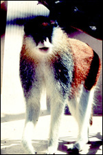
There is a large number (+/- 150) of free-ranging patas monkeys on Puerto Rico. They have escaped from the former breeding colony and are considered to be a "pest" now. Other colonies are held in a few laboratories referred to by Sly et al. (1983).

Cell lines are available from CRES at the Zoological Society at San Diego.

18) Other relevant features

Mattison & King (1983) have proposed this animal and other cercopithecids for the experimental study of fetoscopy and fetal blood sampling. When pregnant patas monkeys were given zidovudine (AZT - used for treatment of AIDS) in doses equivalent to human therapy, the fetuses (heart and muscle) and their placentas showed a dose-dependent damage of their mtDNA (Gerschenson & Poirier, 2000).

Twinning is not very common and it is rarely successful. Sly et al. (1983) reported that 4 sets of twins were observed among 501 gestations. Of these, one set aborted at midgestation, one had a mummified fetus with a liveborn infant, one liveborn had a stillborn twin, and two twins were stillborn. Sex and zygosity were not mentioned.
Conaway (1969) described adrenal rests at the ovarian hilus, a frequent finding in many other species and also occurring near testes. The interesting aspect of these rests is that they never have any medullary tissue.

In view of the large proportion of "X-cells" in Patas monkey placentas it would be useful to search for major basic protein MBP in them, ascertain whether the human antibodies label them and perhaps reexamine the "choriocarcinomas" described earlier.

References

Atkinson, L.E., Hotchkiss, J., Fritz, G.R., Surve, A.H., Neill, J.D. and Knobil, E.: Circulating levels of steroids and chorionic gonadotropin during pregnancy in the rhesus monkey, with special attention to the rescue of the corpus luteum in early pregnancy. Biol. Reprod. 12:335-345, 1975.

Baylet, R. and Grattepanche, H.: Sur les chromosomes des cercopithecidae Papio papio, Macaca mulatta, Cercopithecus aethiops, Erythrocebus patas. C.R. Soc. Biol. 158:1382, 1964.

Benirschke, K. and Kaufmann, P.: The Pathology of the Human Placenta. 4th edition. Springer-Verlag, NY, 2000.

Benirschke, K. and Miller, C.J.: Anatomical and functional differences in the placenta of primates. Biol. Reprod. 26:29-53, 1982.

Bibollet-Ruche, F., Galat-Luong, A., Cuny, G., Sarni-Manchado, P., Galat, G., Durand, J.P., Pourrut, X. and Veas, F.: Simian immunodeficiency virus infection in a patas monkey (Erythrocebus patas): Evidence for cross-species transmission from African green monkeys (Cercopithecus aethiops sabaeus) in the wild. J. Gen. Virol. 77:773-781,
1996.

Cell strains: CRES at http://www.sandiegozoo.org/conservation/cres_home.html. Please direct your inquiries to Dr. Oliver Ryder (oryder@ucsd.edu).

Chabra, S.K., Reed, C.D., Anderson, L.M. and Shiao, Y.H.: Comparison of the polymorphic regions of the cytochrome P45 CYP2E1 gene of humans and patas and cynomolgus monkeys. Carcinogenesis 20:1031-1034, 1999.

Conaway, C.H.: Adrenal cortical rests of the ovarian hilus of the patas monkey. Folia Primatol. 11:175-180, 1969.

Espana, C., Gajdusek, D.C., Gibbs, C.J. Jr. and Lock, K.: Transmission of Creutzfeldt-Jakob disease to the patas monkey (Erythrocebus patas) with cytopathological changes in in vitro cultivated brain cells. Intervirology 6:150-155, 1975-1976.

Gerschenson, M. and Poirier, M.C.: Fetal patas monkeys sustain mitochondrial toxicity as a result of in utero zidovudine exposure. Ann. N.Y. Acad. Sci. 918:269-281, 200o.

Gille, J.H., Moore, D.J. and Sedgwick, C.J.: Placental infarction: A sign of preeclampsia in a patas monkey (Erythrocebus patas). Lab. Anim. Sci. 27:120-121, 1977.

Gisolfi, C.V., Wall, P.T. and Mitchell, W.R.: Thermoregulatory responses to central injections of excess calcium in monkeys. Amer. J. Physiol. 245:R76-82, 1983.

Gonzalez-Martinez, J.: The ecology of the introduced monkey (Erythrocebus patas) population of southwestern Puerto Rico. Amer. J. Primatol. 45:351-365, 1998.

Goswell, M.J. and Gartlan, J.S.: Pregnancy, birth and early infant behaviour in the captive patas monkey Erythrocebus patas. Folia Primatol. 3:189-200, 1965.

Gotch, A.F.: Mammals - Their Latin Names Explained. Blandford Press, Poole, Dorset, 1979.

Gray, A.P.: Mammalian Hybrids. Second edition. A Check-List with Bibliography. Commonwealth Agricultural Bureaux, Farnham Royal, Slough, UK, 1972.

Griner, L.A.: Pathology of Zoo Animals. Zoological Society of San Diego, 1983.

Hsu, T.C. and Benirschke, K.: An Atlas of Mammalian Chromosomes, Vol. 8, Folio 399, 1974. Springer-Verlag, NY.

Kaplan, J.R., Anthony, M. and Wood, J.: Domestic breeding of patas monkeys (Erythrocebus patas). Lab. Anim. Sci. 31:409-412, 1981.

Kessler, M.J., Phoebus, E.C., Rawlins, R.G., Turnquist, J.E. and London, W.T.: Blood values of free-ranging patas monkeys (Erythrocebus patas). J. Med. Primatol. 12:209-217, 1983.

Kuyl, A.C.v.d., Kuiken, C.L., Dekker, J.T. and Goudsmit, J.: Phylogeny of African monkeys based upon mitochondrial 12S rRNA sequences. J. Molec. Evol. 40:173-180, 1995.

Leakey, L.S.: Presumed super-foetation in an Erythrocebus patas monkey. Nature 223:754, 1969.

Nowak, R.M. and Paradiso, J.L.: Walker's Mammals of the World, Vol. II. 4th edition. The Johns Hopkins University Press, Baltimore, 1983.

Loomis, M.R., O'Neill, T., Bush, M. and Montali, R.J.: Fatal herpesvirus infection in patas monkeys and a black and white colobus monkey. J. Amer. Vet. Med. Assoc. 179:1236-1239, 1981.

Lucotte, G. and Dandieu, S.: Electrophoretic polymorphism in Erythrocebus patas. (In French). Folia Primatol. 40:197-204, 1983.

Mahley, R.W., Johnson, D.K., Pucak, G.J. and Fry, D.L.: Atherosclerosis in the Erythrocebus patas, and Old World monkey. Amer. J. Pathol. 98:401-424, 1980.

Mattison, D.R. and King, J.C.: Development of a nonhuman primate model for fetoscopy. J. Med. Primatol. 12:319-330, 1983.

Page, S.L., Chiu, C. and Goodman, M.: Molecular phylogeny of Old World monkeys (Cercopithecidae) as inferred from gamma-globulin DNA sequences. Molec. Phylogenet. Evol. 13:348-359, 1999.

Palmer, A.E., London, W.T., Sly, D.L. and Rice, J.M.: Spontaneous preeclamptic toxemia of pregnancy in the patas monkey (Erythrocebus patas). Lab. Anim. Sci. 29:102-106, 1979.

Palmer, A.E., London, W.T., Sly, D.L. and Rice, J.M.: Toxemia of pregnancy (preeclampsia, eclampsia, hypertensive disorder of pregnancy). Chapter 88, pp. 213-215, in, Spontaneous Animal Models of Human Disease. Vol. 1. E.J. Andrews, B.C. Ward and N.H. Altman, eds. Academic Press, NY 1979.

Paterson, M.: Biochemical parameters in the patas monkey. Lab. Anim. 9:21-31, 1975.

Panigel, M., Brun, J.-L. and Pascaud, M.: Étude angiographique de la circulation utero-placentaire chez les singes Cynomolgus (Macaca) irus et Erythrocebus patas. Bull. l'Assoc. Anat., 52nd Cong. Paris-Orsay, pp. 965-957, 1967.

Panigel, M. and Brun, J.-L.: Anatomie vasculaire, histologie et ultrastructure du placenta a la fin de la gestation chez certains primates: Macaca (Cynomolgus) irus et Cercopithecus (Erythrocebus) patas. Bull. l'Assoc. Anatom. 53rd Cong., Tours, # 142, pp.1270-1286, 1968.

Panigel, M.: Structure et ultrastructure compares de la membrane placentaire chez certains primates non humains (Galago demidovii, Erythrocebus patas, Macaca irus (fascicularis), Macaca mulatta et Papio cynocephalus). Bull. l'Assoc. Anatom. 54thCong., Sofia # 145, pp.320-337, 1969.

Ramsey, E.M., Corner, G.W. and Donner, M.W.: Serial and cineradiographic visualization of maternal circulation in the primate (hemochorial) placenta. Amer. J. Obstetr. Gynecol. 86:213-225, 1963.

Rice, J.M., Williams, G.M., Palmer, A.E., London, W.T. and Sly, D.L.: Pathology of gestational choriocarcinoma induced in patas monkeys by ethylnitrosourea given during pregnancy. Placenta Suppl. 3:223-230, 1981.

Scott, G.B.D.: Comparative Primate Pathology. Oxford University Press, Oxford, 1992.

Sly, D.L., London, W.T., Palmer, A.E. and Rice, J.M.: Disseminated cryptococcosis in a patas monkey (Erythrocebus patas). Lab. Anim. Sci. 27:694-699, 1977.

Sly, D.L., London, W.T., Palmer, A.E. and Rice, J.M.: Growth and Hematologic development of the patas monkey (Erythrocebus patas) to one year of age. J. Med. Primatol. 7:156-164, 1978.

Sly, D.L., Harbaugh, S.W., London, W.T. and Rice, J.M.: Reproductive performance of a laboratory breeding colony of patas monkeys (). Amer. J. Primatol. 4:23-32, 1983.

Sulaiman, S., Williams, J.F. and Wu, D.: Natural infections of vervet monkeys (Cercopithecus aethiops) and African red monkeys (Erythrocebus patas) in Sudan with taeniid cysticerci. J. Wildl. Dis. 22:586-587, 1986.

Wasmoen, T. L., Benirschke, K. and Gleich, G.J.: Demonstration of immunoreactive eosinophil granule major basic protein in the plasma and placentae of non-human primates. Placenta 8:283-292,1987.

Wilson, R.B., Holscher, M.A., Chang, T. and Hodges, J.R.: Fatal herpesviruses simiae (N virus) infection in a patas monkey (Erythrocebus patas). J. Vet. Diagn. Invest. 2:242-244, 1990.

Winterer, J., Palmer, A.E., Cicmanec, J., Davies, E., Harbaugh, S. and Loriaux, D.L.: Endocrine profile of pregnancy in the patas monkey (Erythrocebus patas). Endocrinology 116:1090-1093, 1985.

Wolf, R.H.: Placenta previa in a patas monkey (Erythrocebus patas). Folia Primatol. 14:80-83, 1973.


back to top

© 2002. All rights reserved.
welcome | home | intro | placentation | glossary | author contact me: kbenirsc@ucsd.edu