| |
11)
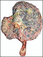
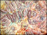
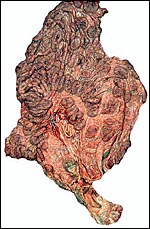
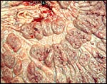
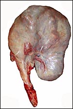
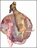
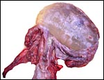
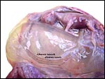
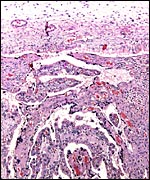
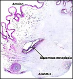
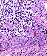
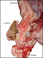
Various features
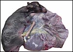
No subplacenta exists but the endometrium of the caruncles show an extensive
vascular development.
12)
Endocrinology
Patton et al. (2001) studied the control of aggression in a bachelor herd
of fringe-eared oryx by administering melengestrol. Fecal androgen excretion
decreased significantly, as did aggression. The corpora lutea were all in the right ovary which was suitably enlarged; in the third pregnancy the ovary measured 4 cm in greatest diameter, while the left was 2 cm.
13)
Genetics
The fringe-eared oryx has 58 chromosomes (Kumamoto et al., 1999). They
are very similar in karyotype to those of the other forms of the genus
Oryx. A hybrid of fringe-eared oryx and a beisa oryx (Oryx gazella
beisa) was reported by Gray (1972).
 |
Giemsa-banded
chromosomes of Fringe-eared oryx (2n=58). |
14)
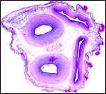
Immunology
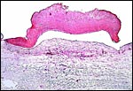
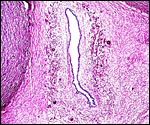
No studies are known to me. It is important, however, to point out that
most animals, including this oryx, produce enormous quantities of a very
thick, tenacious mucus in the endocervical canal during pregnancy. This
is essential as it protects against ascending infection from the vagina.
The mucus contains many cytokines, antibodies and is also a physical barrier
(see Hein et al., 2001 for a study of human cervical mucus).
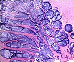 |
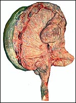
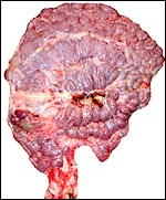
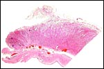
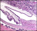
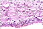
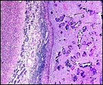
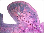
This
is a section of the endocervix from the second specimen showing intense
mucus production. |
15)
Pathological features
Griner (1983) had extensive experience with oryx species and reported
most deaths as being due to trauma, some cases of "bloat" and
hemangiomas.
16)
Physiologic data
There have been no studies.
17)
Other resources
Numerous cell strains of this and related oryx species are available from
CRES
through contacting Dr. O. Ryder at oryder@csd.edu.
18)
Other remarks - What additional Information is needed?
Virtually no endocrinology has been done in this species and the weight
and appearance of term placentas are unknown. Early implantational stages
should be studied.
Acknowledgement
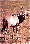
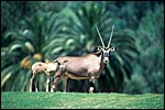
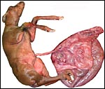
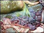
The animal photographs in this chapter come from the Zoological Society
of San Diego. I appreciate also very much the help of the pathologists
at the San Diego Zoo.
References
Gray, A.P.: Mammalian Hybrids. A Check-list with Bibliography. 2nd edition.
Commonwealth Agricultural Bureaux Farnham Royal, Slough, England, 1972.
Griner,
L.A.: Pathology of Zoo Animals. Zoological Society of San Diego, San Diego,
California, 1983.
Grubb,
P.: Order Artiodactyla, pp. 377-414, in Mammal Species of the World, 2nd
ed., D.E. Wilson and D.A.M. Reeder, eds. Smithsonian Institution Press,
Washington, 1993.
Hein, M., Helmig, R.B., Schønheyder, H.C., Ganz, T. and Uldbjerg,
N.: An in vitro study of antibacterial properties of the cervical mucus
plug in pregnancy. Amer. J. Obstet. Gynecol. 185:586-592, 2001.
Kumamoto,
A.T., Charter, S.J., Kingswood, S.C., Ryder, O.A. and Gallagher, D.S.:
Centric fusion differences among Oryx dammah, O. gazella,
and O. leucoryx (Artiodactyla, Bovidae). Cytogenet. Cell Genet.
86:74-80, 1999.
Patton, M.L., White, A.M., Swaisgood, R.R., Sprout, R.L., Fetter, G.A.,
Kennedy, J., Edwards, M.S., Rieches, R.G. and Lance, V.A.: Aggression
control in a bachelor herd of fringe-eared oryx (Oryx gazella callotis)
with melengestrol acetate: Behavioral and endocrine observations. Zoo
Biol. 20:375-388, 2001.
|