(Clicking on the thumbnail images below will launch a new window and a larger version of the thumbnail.)
 
Last updated:
March 21, 2007.
Okapi
Okapia johnstoni

Order: Artiodactyla
Family: Giraffidae

1) General Zoological Data


The okapi is a rare, beautiful animal from the dense forests of the Congo. Its position in nature has been disputed ever since it was discovered in1900. While it was initially considered to be a species of zebra, this was soon declared erroneous and an artiodactyl status was recognized. For numerous reasons, the okapi was then placed into the family of Giraffidae. Not only are okapis grouped here because of their long necks ("short-necked giraffe"), but also because of some structural similarities of organs and skeleton. There are, however, significant differences between these two Giraffidae as well. This was pointed out by Colbert's detailed osteological examination (1939). Also, their chromosome number differs markedly. Their reproductive organs and, especially that of fetuses, are markedly different, as will be explained.
The dubious status of the okapi as a ‘giraffid' species has led us to publish those data that speak against this classification; rather, we see this to be a relative of the nilgai antelope (Benirschke & Hagey, 2006).

Thenius & Hofer (1960) discussed the origin of giraffes and okapis in some detail. They concluded that the separation of these forms occurred perhaps as early as in the Miocene. Hamilton (1978) stated that okapis have no good fossil record, and Colbert (1938) regarded the animal as a "living fossil". He believed, however, that it represented a primitive giraffe ancestor. This relationship has also been discussed in great detail in the comprehensive review of Gijzen (1959) on the okapi. She emphasized that some of its features are more comparable to bovid species, especially to the nilgai (Boselaphus) antelope. Spinage (1968) also suggested that many of the okapi's anatomic features were more similar to other bovid species than to the giraffes. Moreover, their secretive behavior, as forest animals, differs markedly from the steppe-living giraffes. The okapi is now confined to the dense forest of the Republic of Congo.

Males have skin-covered "horns" similar to those of giraffes; females have only small tuberosities at that location. There is an extensive discussion of these features by Colbert (1938). It is generally considered that okapis evolved in Eurasia and subsequently became sequestered to Africa. Here, it now only lives in the dense forest of the Congo. The food preferences of okapis were discussed by Lang (1956) and also in the book by Spinage (1968).

In addition to the uncertainty of taxonomy, two subspecies of okapi have been described. Some of these forms were even proposed for species ranking (Spinage, 1968). There are now numerous okapi colonies in European zoological parks and in the USA. The animals have bred well in captivity after their initial reproductive and disease problems were understood. A studbook is kept (Gijzen, 1977; v. Puijenbroeck, 1987). Several bibliographies have been accorded this animal, presumably because of the interest the species has had for science (Anonymus, 1963; de Bois & van Elsacker, 1988). Much insight may be gleaned from the publications at an International Symposium on the Okapi (Symposium, 1978). Individual animals can be identified by their characteristic striping (Gijzen & Smet, 1977).

Adult okapis weigh between 200 and 250 kg. (Nowak, 1999). Neonates weigh around 16 -20 kg. Longevity is at least 33 years.
   
  Okapi female at San Diego Zoo.
     
  The markings of the stripes are highly characteristic for each individual animal. It can thus be identified.
     
  Female okapi at San Diego Zoo.
     
  2) General Gestational Data

The length of pregnancy was determined by Schwarzenberger et al. (1993) with the use of fecal pregnanediol measurements. In three animals, it was 423, 424, and 431 days, respectively. Okapis have singleton pregnancies - twins must be exceedingly uncommon. Newborns have a weight around 16 kg. Rabb (1978) reported the births of four okapis weighing between 32 and 43 lbs. (13-19 kg.). Raphael (1988) reported the weight of 18.5 kg in a male neonate. Gijzen (1958) gave weights in Africa of between 20-24 kg. Loskutoff et al. (1988) described the internal access to the bicornuate uterus of okapi and giraffe by instrumentation. In that investigation they determined the length of the uterus, number of caruncles and considered the feasibility of interspecific embryo transfer.
Since then, I have had the opportunity of studying one uterus of an adult okapi that died from pneumonia, nonpregnant. The horns had 48 caruncles of varying sizes.
   
 
Uterus of nonpregnant okapi with 45 caruncles in one opened horn (circled area).


3) Implantation

This has not been recorded in the literature and I have no personal information. Only the publication by Naaktgeboren (1966) shows an immature implantation, but timing of implantation remains unknown.

4) General Characterization of the Placenta

I have been able to examine several placentas of okapis but have only recently obtained a complete placenta. It is shown next. It weighed 2,650 g and had 63 cotyledons. Other placentas had between 40 and 60 cotyledons, the largest of which was 10 cm in diameter. The cotyledons were arranged in four rows. Naaktgeboren (1966) identified 47 cotyledons in an implanted gestation. 31 cotyledons were present in the right horn, 16 in the left. The placental weights of placenta examined by me were from 1,000 to 1,430 g. The folded villi are sparsely-branched and resemble those of deer species (Hradecky et al., 1987).

Term placenta of successful okapi gestation.


The only reported implanted placentation is the note on a pregnant uterus by Naaktgeboren (1966). The length of pregnancy of that female was about 6 months and he remarked that the uterus resembled the gestational uterus of a cow. The fetus lay in the right uterine horn, which was also the side of the corpus luteum (28 x 35 mm). The gravid horn was 50 cm in width and 112 cm in length; the left horn was 23 cm wide and 56 cm long. The right horn contained 31 cotyledons, the left had 16. He was unable to identify areolae, even though he had observed them in giraffes. Naaktgeboren was emphatic about the pigmentation of the chorion in this placenta, which was not melanin. Small verrucae with keratinization were present on the amnionic surface and on the umbilical cord. There were no hippomanes.

5) Details of fetal/maternal barrier

The okapi has a polycotyledonary, epitheliochorial placenta. In general, the villous structure is similar to that of other bovid species. In contrast to the giraffe, there are many more binucleate cells in the villous epithelium. They are especially concentrated on the tips of the villi. Fetal villous capillaries often bulge into the trophoblastic cover. In between the cotyledons, the trophoblastic epithelium is multilayered.

Several of the placentas I examined had brown pigment in the trophoblast below the chorionic plate, as was stated earlier. Naaktgeboren (1966) was unable to identify areolae.
   
  Tips of several villi where they dissociated from caruncle. Numerous binucleate cells are in the trophoblast.
     
  Tip of villus with binucleate cell at arrow and interdigitating fetal capillaries.
     
  Surface of okapi villus with binucleate cells at arrows. Note the fetal vessels, some capillaries indent the trophoblast.
     
  6) Umbilical cord

One umbilical cord that I measured was 30 cm long. Nouvel (1958) reported that the cord was 20 cm long in his specimen and had ruptured near the umbilicus. There are four vessels and an allantoic duct in the cord; the cord is not spiraled (Naaktgeboren, 1966). At 6 months gestation, the cord was 13.5 cm long. Gijzen & Mortelmans (1962) determined a cord length of 25 cm with a neonate of 18 kg, and 21 cm with a 25 kg animal. Foci of squamous metaplasia (verrucae) are on the surface.
   
  Inflammation in umbilical cord of neonate who died in two days.
     
  7) Uteroplacental circulation

This has not been studied.

8) Extraplacental membranes

   
  Membrane between the cotyledons and overlying the areolae.
     
  There is no decidua capsularis. The thin amnion has a flat layer of squamous epithelium with many small foci of squamous metaplasia. Naaktgeboren (1966) and I have not observed any hippomanes. There is a large allantoic sac. Vitelline remnants do not occur in the term placenta.

9) Trophoblast external to barrier

There is no invasion of the uterus by trophoblast.

10) Endometrium

The endometrium has four rows of caruncles, approximately 50 per horn (Loskutoff et al., 1988).

11) Various features

Since okapi dams immediately eat their placentas and no term implanted placenta has been studied, no evidence of a subplacenta or metrial glands are known to exist.

12) Endocrinology

In contrast to the ovarian morphology of neonatal giraffes, the neonatal okapi ovary is composed of primordial follicles only. There is no evidence of follicle formation or luteinization, as was pointed out previously (Benirschke, 1978). The adrenal gland is also much smaller than that of neonatal giraffes. The fetal testes show no interstitial cell stimulation. (See the chapter on Giraffe).

Loskutoff et al. (1982), and Schwarzacher et al. (1993) have studied the progesterone production in urine and feces, respectively. Their results indicate that ovulation and pregnancy can be accurately diagnosed by these pregnanediol steroid levels. Infertile cycles lasted about 14.5 days. Pregnancy can be ascertained when, three weeks after copulation, the urinary pregnanediol levels remain significantly elevated. Using the fecal sampling technique, Schwarzenberger et al. (1993) were able to determine the length of pregnancy in three animals as being 423, 424, and 431 days, respectively. Estrogen metabolites were recorded by Loskutoff et al. (1987). The amounts secreted into the urine, however, were too low and thus not useful for the determination of estrus or pregnancy. I am not aware of any accurate gonadotropin measurements.

   
  Neonatal ovary of two-day-old okapi. In contrast to the ovaries of giraffe neonates, there is no follicular stimulation at all. The gonad is packed with primordial ova.
     
  13) Genetics

The okapi chromosome number is 44, 45, or 46 in different animals (Ulbrich & Schmitt, 1969; Hösli & Lang, 1970; Koulisher, 1978). The fact that so many animals with 2n=45 have been identified, suggested that this karyotype may also exist in the wild (Benirschke et al., 1983). This has been established with certainty from the study of a wild-caught male (Petit & de Meurichy, 1986). Fusion of the acrocentric elements #8 and #21 from such a progenitor stock with 2n=46 is likely to have taken place in Zaire. It has now been verified in a specimen from Zaire by special banding techniques (Petit & de Meurichy, 1986). Moreover, Vermeesch et al. (1996) identified a specimen with a further reduction of chromosome number to 2n=44. They compared the fusion events with giraffes and also with the nilgai antelope. Although numerically, the nilgai antelope is similar, some specific karyotypic differences exist from the okapi karyotype (Benirschke et al., 1983; Vermeesch et al., 1996). It is desirable that more detailed comparisons are made in the future between these two species' chromosomes. A study of nilgai chromosomes was compared with that of cattle (Gallagher et al., 1998), but not with giraffidae. The giraffes, on the other hand, have only 30 chromosomes.
Hybrids of okapis with other species are not known. De Bois et al. (1990) analyzed mortality with respect to parental relationship. They found a somewhat higher neonatal mortality in newborns when inbreeding coefficients were high and suggested some degree of "inbreeding depression" to be a possible cause.


Karyotype of female okapi. 46 chromosomes.
   
Karyotype of male okapi with 45 chromosome due to incomplete fusion of the last shown autosomes.

14) Immunology

No studies are known to me.

15) Pathological features

The monograph of Gijzen (1959) provides much information of the causes of deaths of early imports, and of the animals in Epulu, Leopoldsville, and other stations in Africa. Numerous parasites played a major role in the mortality. We have seen disseminated fungal septicemia with extensive CNS involvement in an adult okapi.

One neonate that succumbed soon after birth had an acute inflammation of the umbilical cord (see photo above). Although the pigment deposits in the neonatal endometrium (see below) appear as though they should be pathological, they were seen in two otherwise normal infants. They did not stain with the Prussian blue method and are thus not iron-containing. Thus, the nature of this endometrial pigment remains unknown. It may be a physiologic feature, the significance of which is still uncertain.

In providing a protocol of electroejaculation, Rüedi et al. (1984) diagnosed in a 19 year-old male with cryptorchidism and testicular atrophy with significantly altered semen morphology.

Zwart et al. (1971) identified cowpox infection in five okapis with typical skin lesions. It ahd occurred at other zoos as well. One animal died, the others healed spontaneously with small scars. The origin of the virus was not elucidated.

Lang (1956) observed intestinal infection with Oesophagostomum, Trichostrongylus and Haemonchus spp. Many other reports of parasitic infections exist.

Benirschke summarized the published information on okapi pathology in 1978a. Until then, many cases of rectal prolapse and/or rectal "stenosis" had been observed. This malady was identified as being secondary to the excessive licking of dams, with injury to the anal region. It resulted in proctitis and peritonitis. Modification of enclosures and avoidance of boredom has eliminated this pathology. This disease was further described by Griner (1983), who also reported death from nephritis in an okapi. Raphael (1988) found the neonatal dermatitis and hyperthermia of a male neonate to respond dramatically to a transfusion of blood from another okapi.
   
  Neonatal uterus shows very minimal evidence of glands and caruncles, contrary to the giraffe. It also has much pigment (not hemosiderin) deposition in the endometrium.
     
  Higher magnification of neonatal endometrium with pigment at arrows.
     
  In two adult okapi females I have identified marked chronic inflammation of the endometrium, most striking in the caruncles that may have been responsible for some relative infertility. Numerous plasma cells infiltrated the caruncles but no organisms were seen.
   
  Endometrium of okapi with chronic endometritis, mostly confined to caruncle.
     
  Surface of caruncle 11 days following abortion. The surface is covered with exudate, fibrin and there is necrosis of the surface of the caruncle.
     
  16) Physiologic data

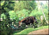
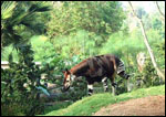
Since this animal is a browser, living in dense forest, the types of vegetation consumed have been of interest. Gijzen's monograph (1959) provides a good list of plants consumed. Rüedi et al. (1984) provided information on the collection of semen from okapis. Rabb (1978) provided hematologic and chemical data.

Lang (1956) described the maintenance of okapis at Epulu, the rapid "domestication" of the animals, their ability to jump a 1.1 m high fences, and the adoption of young by non-lactating females.

17) Other resources

Many cell lines are available from CRES at San Diego Zoo from its "Frozen Zoo" and may be accessed by contacting Dr. O. Ryder (oryder@ucsd.edu).

18) Other remarks - What additional Information is needed?

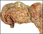
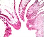
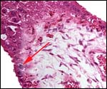
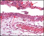
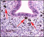
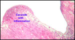
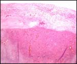
The reason for the pigmentary deposits in the neonatal endometrium is obscure and needs an explanation. Are the amounts of placental lactogen similar to the giraffe and are there placental/fetal gonadotropins? The 
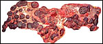
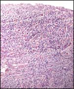
marked difference in the structure of fetal ovaries when compared with the giraffe neonate needs an explanation. The collection of normative data on okapis was emphasized earlier (Benirschke, 1978b).

Acknowledgement

I appreciate also very much the help of the pathologists at the San Diego Zoo.


References

Anonymus: Okapi bibliography. Okapi 1-20, 1963.

Benirschke, K.: General survey of okapi pathology. Acta Zool. Pathol. Antv. 71:63-78, 1978a.

Benirschke, K.: Concluding remarks on the desirability of collecting normative data on okapi. Acta Zool. Pathol. Antv. 71:131-134, 1978b.

Benirschke, K. and Hagey, L.R.: Should the okapi be listed as a giraffid species? Zool. Garten 76:197-198, 2006.

Benirschke, K., Kumamoto, A.T., Cousin, E.F.H.M. and de Boer, L.E.M.: Further observations on the chromosome of the okapi. (Okapia johnstoni) 25th Intern. Sympos. Erkrank. Zootiere, Vienna, 1983. Akademie-Verlag, Berlin. Pp. 363-372, 1983.

Bois de, H. and van Elsacker, L.: Bibliography of the okapi Okapia johnstoni - "Another 20 years later". Acta Zool. Pathol. Antv. 80:65-84, 1988.

Bois de, H., Dhont, A.A. and Puijenbroeck van, B.: Effects of inbreeding on juvenile survival of the okapi Okapia johnstoni in captivity. Biol. Conserv. 54:147-155, 1990.

Colbert, E.H.: The relationships of the okapi. J. Mammal. 19:47-64, 1938.

Gallagher, D.S., Davis, S.K., de Donato, M. Burzlaff, J.D., Womack, J.E., Taylor, J.F. and Kumamoto, A.T.: A karyotypic analysis of the nilgai, Boselaphus tragocamelus (Artiodactyla: Bovidae). Chromosome Res. 6:505-513, 1998.

Geurden, L.M.G.: Les facteurs physio-pathologiques influençant l'acclimatation de l'okapi. Bull. S.R. de Zoologie d'Anvers. # 3:1-21, 1953.

Gijzen, A.: Notice sur la reproduction de l'okapi Okapia johnstoni (Sclater) au Jardin Zoologique d'Anvers. Bull. S.R. de Zoologie d'Anvers. # 8:1-62, 1958.
Gijzen, A.: Das Okapi. (Neue Brehm-Bucherei). A. Ziemsen Verlag. Wittenberg Lutherstadt, 1959.

Gijzen, A.: Studbook of the okapi. Acta Zool. Pathol. Antv. 68:1-47, 1977. (continued by C. Kruyfhooft).

Gijzen, A. and Mortelmans, J.: Notice complémentaire sur l'okapi Okapia johnstoni (Sclater), sur sa reproduction et ses maladies au Jardin Zoologique d'Anvers. Bull. S. R. Zool. D'Anvers # 301-66, 1962.

Gijzen, A. and Smet, S.: L'okapi en captivité depuis sa découverte en 1900 (description par Sclater en 1901) jusqu'au 31 décembre 1970. Un pedigree pour l'okapi (Okapia johnstoni) (Sclater, 1901). Chronique des okapis anversois. "Zoo". S.R. Zool. D'Anvers. 37:pp13, 1972.

Griner, L.A.: Pathology of Zoo Animals. Zoological Society of San Diego, San Diego, California, 1983.

Hamilton, W.R.: Fossil giraffes from the Miocene of Africa and a revision of the phylogeny of the Giraffoidea. Phil Trans. Roy. Soc. London B. 283(996):165-229, 1978.

Hösli, P. and Lang, E.M.: A preliminary note on the chromosomes of the Giraffidae: Giraffa camelopardalis and Okapi johnstoni. Mamm. Chromos. Newsl. 11:109-110, 1070.

Hradecky, P.: Placental morphology in African antelopes and giraffes. Theriogenology 20:725-734, 1983.

Hradecky, P., Benirschke, K. and Stott, G.G.: Implications of the placental structure compatibility for interspecies embryo transfer. Theriogenology 28:737-746, 1987.

Koulisher, L.: Mammalian chromosomes. IX. The chromosomes of a female specimen of Okapia johnstoni. Acta Zool. Pathol. Antv. 71:87-92, 1978.

Lang, E.M.: Haltung und Brunst von Okapia in Epulu. Säugetierk. Mitt. IV (2):49-52, 1956.

Lang, E.M.: Einige Beobachtungen an Okapia johnstoni. Acta Tropica 13:254-258, 1956.

Loskutoff, N.M., Ott, J.E. and Lasley, B.L.: Urinary steroid evaluations to monitor ovarian function in exotic ungulates: I. Pregnanediol-3-glucoronide immunoreactivity in the okapi (Okapia johnstoni). Zoo Biol. 1:45-53, 1982.

Loskutoff, N.M., Kasman, L.H., Raphael, B.L., Ott-Joslin, J.E. and Lasley, B.L.: Urinary steroid evaluations to monitor ovarian function in exotic ungulates: IV. Estrogen metabolism in the okapi (Okapia johnstoni). Zoo Biol. 6:213-218, 1987.

Loskutoff, N.M., Raphael, B.L., Dorn, C.G., Nemec, L.A., Calle, P.P., Petric, A.M. and Kraemer, D.C.: Comparative reproductive traits of the okapi and giraffe: implications for intraspecific and intergeneric embryo transfer. Acta Zool. Pathol. Antv. 80:29-42, 1988.

Naaktgeboren, C.: Notiz über einen trächtigen Uterus von Okapia johnstoni (Sclater, 1901). Z. Säugetierk. 31:171-176, 1966.

Nouvel, J.: Remarques sur la fonction genital et la naissance d'un okapi. Mammalia 22:107-111, 1958.

Nowak, R.M.: Walker's Mammals of the World. 6th ed. The Johns Hopkins Press, Baltimore, 1999.

Petit, P. and de Meurichy, W.: On the chromosomes of the okapi Okapia johnstoni. Ann. Génét. 29:232-234, 1986.

Puijenbroeck v., B.: Studbook of the okapi Okapia johnstoni (Sclater). Royal Society of Antwerp, 1987.

Rabb, G.B.: Birth, early behavior and clinical date on the okapi. Acta Zool. Pathol. Antv. 71:93-105, 1978.

Raphael, B.L.: Neonatal illness characterized by dermatitis, hyperthermina and anemia in an okapi. Acta Zool. Pathol. Antv. 80:43-52, 1988.

Rüedi, D., Küpfer, U., Matern, B., Klöppel, G. and Heldstab, A.: Semen collection in the okapi. J. Zoo Med. 15:3-4, 1984.

Schwarzenberger, F., Patzl, M., Francke, R., Ochs, A., Biter, R., Schaftenaar, W. and de Meurichy, W.: Fecal progestagen evaluations to monitor the estrous cycle and pregnancy in the okapi (Okapia johnstoni). Zoo Biol. 12:549-559, 1993.

Spinage, C.A.: The Book of the Giraffe. Houghton Mifflin Company, Boston, 1968.

Symposium on the okapi. Acta Zool. Pathol. 71:1-134, 1978.

Thenius, E. and Hofer, H.: Stammesgeschichte der Säugetiere. Springer-Verlag, Berlin, 1960.

Ulbrich, F. and Schmitt, I.: Die chromosome von Okapia johnstoni. Acta Zool. Pathol. Antv. 49:123-124, 1969.

Vandermander, A.: Apercu succinct de nos connaissances actuelles au sujet de l'okapi (Okapia johnstoni Scl.). Bull. S.R. de Zoologie d'Anvers #3: 22-46, 1953.

Vermeesch, J.R., de Meurichy, W., van den Berghe, H., Marynen P. and Petit, P.: Differences in the distribution and nature of the interstitial telomeric (TTAGGG)n sequences in the chromosomes of the giraffidae, okapi (Okapia johnstoni), and giraffe (Giraffa camelopardalis): evidence for ancestral telomeres at the okapi polymorphic rob(4;26) fusion site. Cytogenet. Cell Genet. 72:310-315, 1996.

Zwart, P., Gispen, R. and Peters, J.C.: Cowpox in okapis Okapia johnstoni at Rotterdam zoo. Br. Vet. J. 127:20-24, 1971.


back to top

© 2007. All rights reserved.
welcome | home | intro | placentation | glossary | author contact me: kbenirsc@ucsd.edu