 |
| (Clicking
on the thumbnail images will launch a new window and a larger version
of the thumbnail.) |
| Last
updated: Feb.6, 2004. |
Tragelaphus angasi(i)
Order: Artiodactyla
Family: Bovidae
1) General Zoological Data
The nyala is one of the complex group of Tragelaphinae whose properties have been considered in other chapters of this site. Aside from the shy nyala (and "mountain nyala") they include the greater and lesser kudus, eland, sitatunga and bushbuck (Nowak, 1999). Their cytogenetics are complex but their monophyly among bovidae has been affirmed by studies of Matthee & Robinson (19999) and Matthee & Davis (2001). The designation Tragelaphus derives from Gr. tragos=he-goat, and elaphos=deer; angasi comes from George French Angas [1822-1886] and English explorer. "Nyala" is Swahili for this animal (Gotch, 1979). Tragelaphinae may have derived from presumed common ancestors with the nilgai and subsequently immigrated to Africa from Asia. Their ultimate origin is still disputed, however. Speculations and recent findings are summarized by Thenius (1969). A detailed description of the behavior, distribution, group size and other details on nyala has been provided by Tello & van Gelder (1975). This animal lives in wet forests south of the Sahara desert, is somewhat secretive and is more readily seen at night. The species is widely distributed in South Africa and its Game Parks, and is also well represented in many zoos. Males weigh up to 126 kg; females are lighter colored and weigh up to 68 kg. Juvenile males look like females and are not so dark as when they are adult. Longevity is 15+ years according to Crandall (1965) and Jones (1993).
 |
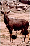
Male nyala at San Diego Wild Animal Park. |
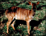 |
Female nyala at San Diego Wild Animal Park. |
Nowak (1999) indicates a gestational length of 7 months with births usually occurring in August, but Brand (1963) indicates a gestation of only 6 months. Mentis (1972) indicates a gestational length of 252 days. Most births are singletons, occasional twins have been observed. Neonates weigh around 5 kg.
3) Implantation
Early implantation stages have not been described. This specimen is probably the youngest specimen available and must be in the first trimester of gestation. The dam died from trauma in January, 2004. The bicornuate uterus has four rows of caruncles on which the 50 cotyledons attach. The placenta is very similar to that of other Tragelaphinae which may, in part, explain the apparent ease of hybridization in this subfamily (see chapter on bongo). It is noteworthy, however, that far fewer cotyledons were found in this species.
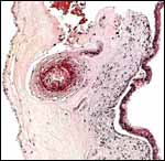
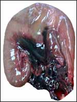 |
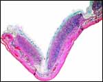
Pregnant uterus of the specimen described here also showing the abdominal hemorrhage. |
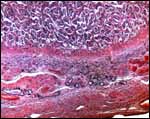
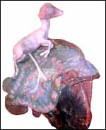 |
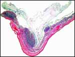
The opened uterus with immature fetus and four rows of cotyledons exposed. |
A single specimen was available to me. It came from a mid-pregnant female that died from massive abdominal hemorrhages that were presumed to be due to trauma. The female fetus was 14 cm in crown-rump length and weighed 106 g. The 50 cotyledons were arranged in four rows and measured between 2 and 0.8 cm in diameters and 2 mm in thickness. This is typical polycotyledonary, epitheliochorial placenta without maternal tissue invasion.
 |
Two adjacent implanted cotyledons. |
 |
Another group of implanted cotyledons and overlying membranes. |
The barrier is epithelio-chorial with single layer of cuboidal trophoblast that is often interrupted by numerous binucleate cells. Preservation was insufficient to decide whether fusion with maternal endometrial epithelium was present. Villi and endometrium are intimately intertwined.
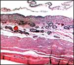
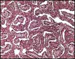 |
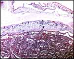
Low-power aspect of implanted placenta. The villi are the rounded structures amidst the contiguous maternal caruncular structures. |
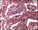 |
Higher magnification of the villi with numerous large binucleate trophoblastic cells. |
 |
Fetal surface of the cotyledon. |
The umbilical cord of this specimen measured 6 cm in length and had four blood vessels. It was not spiraled and was covered with numerous small yellowish granules of squamous metaplasia. In addition there are numerous small blood vessels, mostly around the central allantoic duct. The duct is lined with a single layer of flat epithelium.
 |
Two cross sections of this animal's umbilical cord with central allantoic duct and granules of squamous metaplasia mostly located only on the upper surface. |
 |
Higher magnification of the central portion of the umbilical cord with allantoic duct and its small vessels, as well as foci of squamous metaplasia (top). |
This has not been studied in detail.
8) Extraplacental membranes
The avascular amnion was diffusely covered with yellowish granules of squamous metaplasia. The allantois filled approximately ½ of the chorionic cavity and contained clear allantoic fluid (urine). No remnant of yolk sac was identified and there were no hippomanes.
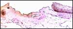 |
Membranes with thin amnion (and squamous metaplasia) at top and allantois membrane below with its small vessel (left). |
 |
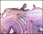
Intercotyledonary membrane with chorion at left and trophoblastic epithelium at right. |
There is no trophoblastic infiltration into maternal tissues.
 |
Maternal aspect of the implanted cotyledon with remaining endometrial glands at center, bottom showing sharp division between fibrous endometrium and villous structures. |
The uterine morphology of nyalas has been referred to by Hradeck? (1982) with the cervical canal as being similar to that of cattle with a bicornuate uterus. No true decidua develops in this species. The endometrial glands remain to term.
 |
Intercotyledonary membrane apposition to the endometrium. |
There is no subplacenta, and other unusual features are not identified.
12) Endocrinology
I have been unable to find any publications.
13) Genetics
Tragelaphinae have complex, compound chromosomes. Thus, female nyalas have 56 chromosomes, all of which are acrocentrics with the exception of the large, first compound metacentric element, while males have 55 chromosomes due to an Y-autosome translocation (Wurster & Benirschke, 1968). At the time of its first publication, this unpaired submetacentric element was found to have a large pericentric segment that was a late-labeling stretch and was assumed to be the compound Y-chromosome, with the late label representing the Y portion. Other Tragelaphinae with unusual sex chromosomes were then studied and since this publication only the paper by Petit et al. (1994) gives significant further insight. It comprises studies of several species with different banding procedures (see the chapters on the other tragelaphines - bongo, kudu, and eland). This odd, unpaired chromosome was then shown to be a translocation element of Y/13 that is also present in other tragelaphines. The sitatunga has an additional X/13 translocation. I show here the banded karyotype of a male and the unbanded karyotype of a typical female nyala. Other studies are those by Buckland & Evans (1978), Gallagher & Womack (1992), Benirschke et al. (1982) and those that have been quoted in the chapters on bongo, lesser kudu, greater kudu, sitatunga and eland. I have long considered that the ancestral bovid stock had 60 chromosomes (the monophyly was shown by DNA studies of Matthee & Davis, 2001), and that subsequent Robertsonian fusions and, thereafter, inversions and other translocations reduced the chromosome number. Indeed, that the fusions were instrumental in speciation has been supported by the considerations of others that should be consulted (Baker & Bickham, 1986; Gallagher & Womack, 1992). Buckland & Evans showed that the compound Y chromosome of tragelaphines is constructed of the Y and the # 13 autosome with banding pattern of # 13 of cattle. Thus the # 14 hypothesized by me in the past, as shown in the first diagram below, should be replaced by # 13, at least adhering to conventional numbering schemes. The second diagram that has also been shown in other chapters is thus more likely to be accurate. But much more specific study, perhaps best with FISH or chromosome painting needs to be done before the realities are disclosed.
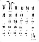 |
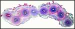
Karyotype of male nyala with 2n=55 to show the large compound # 2 and unusual sex chromosomes. |
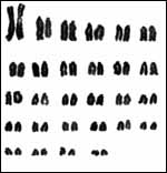 |
Karyotype of female nyala with 2n=56. The compound #2 is shown as the first element here. |
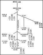 |
Early putative relationship of Tragelaphinae. |
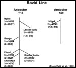 |
Similar putative relationships as constructed by Petit et al. (1995). |
I have not found any publications.
15) Pathological features
Numerous parasites have been identified in nyalas. (Boomker et al., 1991,
1996; and Boomker, 1986). Horak et al. (1983) identified the burden with
ixodid ticks of nyalas and other species in Kruger National Park. Liu
et al. (1982) described many cases of skeletal and myocardial degeneration,
associated with calcification in nyala but detected normal selenium levels.
"White muscle disease" is a well-known feature of tragelaphine
(Heldstab & Ruedi, 1980). Keep & Basson (1973) described a case
of a testicular teratoma. A variety of viruses have also been identified
in nyalas: rotavirus (Baumeister et al., 1983); bovine diarrhea antibodies
(Anderson & Rowe, 1998). A case of spongiform encephalopathy was described
in a nyala by Jeffrey & Wells (1988). These authors later transmitted
the agent (protein) to mice (Jeffrey et al., 1992). Kirkwood & Cunningham
(1994) summarized all cases of BSE in captive wild animals of the British
Isles.
16) Physiologic data
The basic hematological values of antelopes, including the nyala, have
been summarized in a contribution by Pospisil et al. (1984).
17)
Other resources
Cell strains of nyala fibroblast and other Tragulidae are available from
the "Frozen Zoo" at CRES,
San Diego Zoo by contacting Dr. Oliver Ryder at oryder@ucsd.edu.
18) Other remarks - What additional
Information is needed?
Early stages of implantation, immunologic and endocrine data are lacking.
Acknowledgement
The animal photographs in this chapter come from the Zoological Society
of San Diego. I appreciate also very much the help of the pathologists
at the San Diego Zoo.
References
Anderson, E.C. and Rowe, L.W.: The prevalence of antibody to the viruses
of bovine virus diarrhea, bovine herpes virus 1, rift valley fever, ephemeral
fever and bluetongue and to Leptospira sp. in free-ranging wildlife in
Zimbabwe. Epidemiol. Infect. 121:441-449, 1998.
Baker, R.J. and Bickham, J.W.: Speciation by monobrachial centric fusions. Proc. Natl. Acad. Sci. USA 83:8245-8248, 1986.
Baumeister, B.M., Castro, A.E., McGuire-Rodgers, S.J. and Ramsay, E.C.: Detection and control of rotavirus infections in zoo animals. J. Amer. Vet. Med. Assoc. 183:1252-1254, 1983.
Benirschke, K., Kumamoto, A.T., Esra, G.N. and Crocker, K.B.: The chromosomes of the bongo, Taurotragus (Boocerus) eurycerus. Cytogenet. Cell Genet. 34:10-18, 1982.
Boomker, J.: Paracooperia horaki n.sp. (Nematoda: Trichostrongylidae) from the nyala Tragelaphus angasi Gray, 1849. Onderstepoort J. Vet. Res. 53:161-165, 1986.
Boomker, J., Horak, I.G. and Flamand, J.R.: Parasites of South African wildlife. XII. Helminths of Nyala, Tragelaphus angasii, in Natal. Onderstepoort J. Vet. Res. 58:275-280, 1991.
Boomker, J., Booyse, D.G., Watermeyer, R. De Villiers, I.L., Horak, I.G. and Flamand, J.R.: Parasites of South African wildlife. XIV. Helminths of nyalas (Tragelaphus angasii) in the Mkuzi Game Reserve, KwaZulu-Natal. Onderstepoort J. Vet. Res. 63:265-271, 1996.
Brand, D.J.: Records of mammals bred in the National Zoological Gardens of South Africa during the period 1908-1960. Proc. Zool. Soc. London 140:617-659, 1963.
Buckland, R.A. and Evans, H.J.: Cytogenetic aspects of phylogeny in the Bovidae, G-banding. Cytogenet. Cell Genet. 32:64-71, 1978.
Crandall, L.S.: Record of African antelopes in the New York Zoological Park. Int. Zoo Yearb. 5:52-55, 1965
Gallagher, D.S. and Womack, J.E.: Chromosome conservation in the bovidae. J. Hered. 83:287-298, 1992.
Gotch, A.F.: Mammals - Their Latin Names Explained. Blandford Press, Poole, Dorset, 1979.
Heldstab, A. and Ruedi, D.: The occurrence of myodystrophy in zoo animals at the Basle Zoological Garden. Pp. 27-34, In, The Comparative Pathology of Zoo Animals, R.J. Montali and G. Migaki, eds., Smithsonian Institution Press, Washington, D.C., 1980.
Horak, I.G., Potgieter, F.T., Walker, J.B., De Vos, V. and Boomker, J.: The ixodid tick burdens of various large ruminant species in South African nature reserves. Onderstepoort J. Vet. Res. 50:221-228, 1983.
Hradeck?, P.: Uterine morphology in some African antelopes. J. Zoo An. Med. 13:132-136, 1982.
Jeffrey, M. and Wells, G.A.: Spongiform encephalopathy in a nyala (Tragelaphus angasi). Vet Pathol. 25:398-399, 1988.
Jeffrey, M., Scott, J.R., Williams, A. and Fraser, H.: Ultrastructural features of spongiform encephalopathy transmitted to mice from three species of bovidae. Acta Neuropathol. 84:559-569, 1992.
Jones, M.L.: Longevity of ungulates in captivity. Intern. Zoo Yearbk. 32:159-169, 1993.
Keep, M.E. and Basson, P.A.: A testicular teratoma in a nyala (Tragelaphus angasi Gray, 1848). J. South Afr. Vet. Assoc. 44:288, 1973.
Kirkwood, J.K. and Cunningham, A.A.: Epidemiological observations on spongiform encephalopathies in captive wild animals in the British Isles. Vet. Rec. 135:296-303, 1994 (and 135:440, 1994).
Liu, S., Dolensek, E.P., Herron, A.J., Stover, J. and Doherty, J.G.: Myopathy in the nyala. J. Amer. Vet. Med. Assoc. 181:1232-1236, 1982.
Matthee, C.A. and Davis, S.K.: Molecular insights into the evolution of the family bovidae: a nuclear DNA perspective. Mol. Biol. Evol. 18:1220-1230, 2001.
Matthee, C.A. and Robinson, T.J.: Cytochrome b phylogeny of the family bovidae: resolution within the alcelaphine, antilopini, neotragini, and Tragelaphini. Mol. Phylogenet. Evol. 12:31-46, 1999.
Mentis, M.T.: A review of some life history features of the large herbivores of Africa. The Lammergeyer 16, 1-89, 1972.
Nowak, R.M.: Walker's Mammals of the World. 6th ed. The Johns Hopkins Press, Baltimore, 1999.
Petit, P., Vermeesch, J.R., Marynen, P. and DeMeurichy, W.: Comparative cytogenetic study in the subfamily Tragelaphinae. Proc. 11th Europ. Coll. Cytogenet. Domest. Anim. Pp.109-113, 1994.
Pospisil, J., Kase, F., Vahal, J. and Mouchova, I.: Basic haematological values in antelopes - II. The Hippotraginae and the Tragelaphinae. Comp. Biochem. Physiol. A 78:799-807, 1984.
Tello, J.L.P.L and van Gelder, R.G.: The natural history of nyala Tragelaphus angasi (Mammalia, Bovidae) in Mozambique. Bull. Amer. Museum Natural History 155(#4):321-386, 1975.
Thenius, E.: Stammesgeschichte der Säugetiere (einschliesslich der Hominiden). In Handbuch der Zoologie, J.G. Helmcke, D. Starck and H. Wermuth, eds. Vol. 8/2:369-722, 1969, Walter de Gruyter & Co., Berlin.
Wurster, D.H. and Benirschke, K.: Chromosome studies in the superfamily Bovoidea. Chromosoma 25:152-171, 1968.