| (Clicking
on the thumbnail images will launch a new window and a larger version
of the thumbnail.) |
| Last updated: Feb 17, 2006. |
Moschus m. moschiferous
Order: Artiodactyla
Family: Moschidae
1) General Zoological Data
It is now generally agreed that Moschus should not be comprised among the Cervidae but that it should have its own family rank. Nevertheless, the taxonomy of this deer has been and remains somewhat controversial, as was well detailed by Nowak (1999). While Grubb (1982) formerly considered the various forms to be comprised of only one species, Groves et al. (1995) and also Nowak quoting Groves and others, suggested that five or six species exist and they reviewed the literature extensively and provided their own data (without chromosome findings, however). In addition, of Moschus moschiferous they suggested that three subspecies exist. The animal under consideration here is from a Siberian import to Zoo Leipzig. At Leipzig and here, at least, successful breeding has taken place
Musk-deer are from the mountainous regions of China , Russia and the Himalayans. Their most notable feature is the saber-like upper canine teeth and the abdominal musk gland in males that produces a desirable odoriferous secretion (used for perfumes) which has been collected at great expense and detriment to the species. The animals possess neither antlers nor a gall bladder. The name derives from the Greek moskhos = musk (Gotch, 1979).
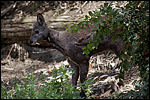 |
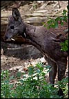
Siberian Musk deer at the San Diego Zoo. |
2) General Gestational Data
The gestation is said to last 185-198 days (Green, 1987; Nowak, 1999) with one or two offspring produced that weigh ~500 g. The longevity is 13 years and 11 months (Weigl, 2005). Adults weigh between 7 and 17 kg (Hayssen et al., 1993). Mating takes place in the winter, with births occurring in June-September.
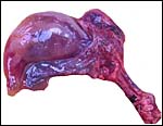 |
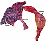
Implanted gestational sac in the right uterine horn. |
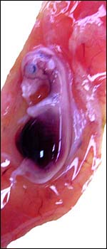 |
Embryo in its allantochorionic sac. |
3) Implantation
As is true of cervids, this musk deer had implanted in the right horn and had a mesometrial location. Timing of implantation is so far unknown.
4) General Characterization of the Placenta
Mossman (1987) mentions Weldon (1884) as having described the placenta of Moschus as being diffuse in character, but that is not so. All deer species have cotyledonary numbers between 5 and 8 and are thus all ‘oligocotyledonary'. This musk deer placenta had 7 oval cotyledons.
I have had only this one specimen available, that of a very young implanted singleton pregnancy of a deer whose leg had to be amputated and that died soon thereafter. The female fetus of this gestation was 4.5 cm long and was contained in its membranes. Unfortunately, the fetus and placenta were somewhat autolyzed. Because of the different structure of the placental cotyledons and uterine caruncles, however, the specimen is here depicted.
This is an oligocotyledonary placenta with elongated cotyledons and with their seven corresponding caruncles. The right horn was occupied by this small fetus and was filled with a large quantity of yellow allantoic fluid. The fetus was attached to the placenta by a non-spiraled 2.5 cm cord.
This uterus had seven elongated caruncles and, correspondingly, there are very thin cotyledons distributed over the allantochorionic sac. These measured 7 x 05. cm in diameters and were exceedingly thin at this stage of development.
5) Details of fetal/maternal barrier
The placenta is superficially implanted on the caruncles, is epitheliochorial and not invasive. There is a very large number of binucleate trophoblastic cells, even at this young age. The villi branched only twice at this young stage. The trophoblast is cuboidal.
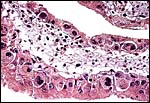 |
Villus at higher magnification with numerous binucleate trophoblast. |
6) Umbilical cord
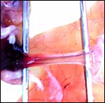
The cord of this small embryo was only 2.5 cm long without spirals and possessed four vessels and a central allantoic duct. Even at this young stage of development there is a profusion of tiny additional blood vessels, mostly allantoic in nature.
7) Uteroplacental circulation
This has not been studied.
8) Extraplacental membranes
There is a large cylindrical allantoic sac that surrounds the amnionic cavity that is more spherical at this stage.
9) Trophoblast external to barrier
No invasion occurs.
10) Endometrium
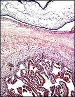
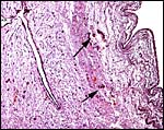
The endometrium of this bicornuate uterus has a single row of seven caruncles. There is no decidua.
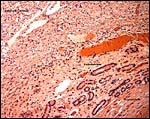 |
Uterine wall with endometrial glands and autolyzed caruncle (top left). |
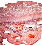 |
Full thickness of uterus with autolyzed caruncle at top, endometrial glands in the center and myometrium below. |
11) Various features
There is no subplacenta.
12) Endocrinology
A single 1 cm large corpus luteum was found in the right ovary. No endocrine data are known to me.
 |
The right ovary had a single corpus luteum as well as a parovarian cyst. |
13) Genetics
This species has 58 chromosomes that are all acrocentrics. Chi et al. (2005) have recently done comparisons of banding homologies among Chinese muntjacs, musk deer and gayal. Hybridization with other species has not been reported, except that occurring among the putative subspecies from Siberia , all of which have the same chromosome number.
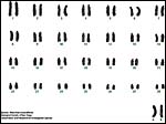 |
Chromosomes of a female musk deer from CRES. |
14) Immunology
No studies have been done.
15) Pathological features
Griner (1983) does not mention this species as having come to autopsy during his tenure at San Diego Zoo.
16) Physiologic data
I am not aware of any data.
17) Other resources
Cell lines are available from CRES at the San Diego Zoo and also from Kunming 's cell bank.
18) Other remarks – What additional Information is needed?
It is apparent that no term placenta has been studied, thus its weight is unknown and this is desirable to be ascertained.
Acknowledgement
The animal photograph in this chapter comes from the Zoological Society of San Diego.
References
Chi, J., Fu, B., Nie, W., Wang, J., Graphodatsky, A.S. and Yang, F.: New insights into the karyotypic relationships of Chinese muntjac (Muntiacus reevesi), forest musk deer (Moschus berezovskii) and gayal (Bos frontalis). Cytogenet. Genome Res. 108:310-316, 2005.
Hayssen, V., van Tienhoven, A. and van Tienhoven, A.: Asdell's Patterns of Mammalian Reproduction: a Compendium of Species-specific Data. Comstock/Cornell University Press, Ithaca, 1993.
Gotch, A.F.: Mammals – Their Latin Names Explained. Blandford Press, Poole, Dorset , 1979.
Green, M.J.B.: Scent-marking in the Himalayan musk deer (Moschus chrysogaster). J. Zool. Series B 1:721-737, 1987.
Griner, L.A. : Pathology of Zoo Animals. Zoological Society of San Diego, San Diego, California, 1983.
Groves, C.P., Wang, Y. and Grubb, P.: Taxonomy of musk-deer, genus Moschus (Moschidae, Mammalia. Acta Theriologica Sinica 15:181-197, 1995.
Grubb, P.: The systematics of Sino-Himalayan musk-deer (Moschus), with particular reference to the species described by Hodgson B H. Säugetierkundl. Mitteil. 30:127-135, 1982.
Mossman, H.W.: Vertebrate Fetal Membranes. MacMillan, Houndmills, 1987.
Weigl, R.: Longevity of Mammals in Captivity from the Living Collections of the World. E. Schweizertbart'sche Verlagsbuchhandlung (Nägele und Obermiller), Stuttgart , 2005.
Weldon, W.F.R.: Note on placentation of Tetraceros quadricornis. Proc. Zool. Soc. London pp.2-6, 1884.
