(Clicking on the thumbnail images below will launch a new window and a larger version of the thumbnail.)
House (Domestic, Laboratory) Mouse
Mus musculus

Order: Rodentia
Family: Muridae

1) General zoological data of species


This chapter is written primarily for those pathologists and investigators who find the complex placenta of rodents very difficult to understand. Because of this, essential parts of the mouse gestational sac have occasionally been discarded inadvertently. An example is the membranous dome (the inverted yolk sac placenta) through which there is much transport of various substances. Such tissues should always be included when murine experimental studies are conducted. This applies especially to studies that investigate embryonic growth, exposure to pathogens and other substances such as immunoglobulins. The chapter, therefore, differs in its composition and is more richly illustrated.

Mice are distributed worldwide. There are many different species, which often, but not always, have different chromosome numbers (2n = 22-40). Numerous breeding colonies exist in most laboratories. This is especially so for the Bar Harbor Laboratories in Maine, with the largest experience in genetic diseases and various anomalies.

2) General gestational data

The length of gestation in the mouse generally is 19 days, with the developmental stages often referred to as being: "xx dpc" (= xx days post coitum). Litter size in mice varies from 4 to 12 embryos. Occasionally, because of a fetal demise, an empty space is found in the typically bicornuate uterus. The white mice used for these pages were of the following weights: at 14 days pregnant - 38 g, after removal of uterus - 30 g; at 18 days pregnant - 57 g, after removal of the uterus - 34 g. The 18-day mouse fetus weighed 1.8 g with placenta and measured 2 cm in length with the placental disk measuring 2 x 0.6 cm. It had a 1.5 cm long umbilical cord.

   
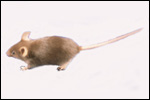
  C57/Bl/6 mouse (Courtesy Dr. E. Masliah).
     
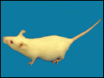
  Swiss Webster albino mouse - 13 dpc.
     
 
3) Implantation

The implantation of most rodents, including that of the mouse, occurs initially antimesometrially, with the embryonic disk occupying a mesometrial location. It is a superficially invasive placenta (it is "interstitial"), with a "folding" type of amniogenesis. The major portion of the placenta becomes discoid and has both - a larger labyrinthine (hemochorial) structure - and a smaller region, the trophospongium. Both of these areas are especially confusing. In addition to these two areas, there is the portion of the gestational sac represented by the inverted yolk sac. It forms the dome of the gestational sac.
Most mouse placentas are separated one from another, even when they were "stuffed" into uterine horns with the intent of causing chimerism. Heinecke & Grimm (1971), however, reported that fusion did occur in a very small number of implantations, perhaps as a result of damage to blastocyst or endometrium.

All these structures are substantially different. In early stages, the yolk sac is well developed. Later, however, it becomes inverted, so that its endodermal epithelial layer comes to lie opposite the endometrium on the uterine wall. The conceptus' membranous sac, lying opposite to the disk, thus becomes the "visceral yolk sac placenta".

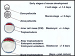
In the early stages of the developing mouse blastocyst, the trophectoderm represents the major portion (70-80% of cells) of the future conceptus. A smaller number of inner cells (the currently much debated "stem cells") that represent the future fetus, lies within the blastocyst. The initial attachment of the blastocyst, which is derived from the trophectoderm - the "mural" trophectoderm, occurs when trophoblast invades the endometrium.The implanting blastocyst is then still positioned deep in an endometrial crypt. This proliferating trophoblast forms a "cone" herein that is often referred to as the "Träger" (= carrier). When the opening of the endometrial crypt closes this area becomes the egg chamber ("Eikammer").

The blastocyst, with its proliferating trophoblast, now assumes a more cylindrical shape. The initial attaching trophoblast cells proliferate from the bottom of the cone as a modification or extension of the träger. (Please see the explanatory diagram to follow - mural trophectoderm). During this early stage, the embryonic yolk sac makes up another dominant portion of the developing embryonic tissues. Later, this yolk sac membrane becomes totally inverted, so that its epithelium presses against the endometrial mucosa at the dome of the cavity. . It has recently been shown that fibrinogen is essential for the successful attachment the blastocyst and maintenance of placentation (Iwaki et al., 2002). Mice deficient in the FG-? chain consistently aborted.

The placental disk now lies on the opposite (mesometrial) side of the uterus. Although there is no allantoic sac in mice, some of the initially present allantoic tissues provide the stalk for the development of the umbilical cord. The early fusion of the allantoic and chorionic murine membranes has now been studied in some detail by Downs (2002). The fusion process appears to be highly regulated through the attractions of the adhesion molecule VCAM-1 in the allantois and a4-integrin in the chorion. After fusion the remaining barrier is broken down, and thus, the body stalk develops. In comparing the development of different mouse strains, Dickson (1967) found that blastocysts do not always develop equally well and also, that their development is not necessarily synchronous. Recent research has identified a variety of genes that are responsible for implantation, adhesion, endocrine response, and so forth. They are summarized by Giudice (1999).

   
  Early stages of mouse development. Colors of text refer to these colors.
     
 
As was indicated at the beginning of this chapter, the anatomy of the mouse placenta is very complex. Rossant & Croy (1985) used reconstituted embryos to determine which cells of the placenta are of maternal origin, which are fetal and, of these, which originate from the embryonic disk and which come from the trophectoderm. Cross et al. (1994) provided some initial comparisons of mouse and human implantation and detailed the molecular basis for the interactions of the maternal and fetal organisms.

A major publication by Georgiades et al. (2002) presents much more detailed information on the cellular composition of the placental disk and on the implantation site. It endeavors especially to compare the various cell types of mouse trophoblast with those found in the human placenta. The original publication must be consulted for this detail. Likewise, the review by Rossant & Cross (2001)provides evidence that extensive signaling occurs between trophoblast and embryonic cells which, when disrupted, may lead to abortion or embryonic maldevelopment. Cross (2000a) has given further emphasis to the notion that the mouse placentation is a useful model to understand the less-accessible (genetically speaking) human placentation.

The ectoplacental cone and the extraembryonic ectoderm of the primitive streak stage are derived from the trophectoderm. These experiments showed that, when the placental portions were divided into labyrinthine and spongiotrophoblastic components, the maternal contributions were essentially confined to the latter segments. Most of the decidual tissue was maternal in origin but there was also a minor fetal tissue contamination (i.e. the trophoblastic giant cells). About 30% of the cells in the placental disk derive from maternal tissues. They are largely confined to the spongiotrophoblast region. The cellular components of the inner cell mass, which is largely confined to the placental labyrinth, constitute only about 4%. The analysis was somewhat hampered by the presence of contaminating blood cells but are the best approximations available. These are difficult experiments. It should also be mentioned that not all placentas of reconstituted embryos were identically admixed. Hence, not surprisingly, other investigators had somewhat different findings

The giant cells of the rodent placenta have presented a special topic of study for many investigators. These cells, which form the primary site of attachment to the endometrium, can be grown in vitro (Copp, 1980). Their development begins at the abembryonic pole of trophoblast and then spreads towards the embryo (+/- 4dpc). During the first half of development, 10-30% of trophoblastic cells contain giant nuclei that have large quantities of DNA. This DNA is derived through the process of endomitosis (chromosomal replication without cell division). By 11 dpc, up to 850 times the normal amount of DNA is found in these nuclei (Barlow & Sherman, 1972; 1974). DNA replication continues unabated throughout gestation (Copp, 1978). Experiments with actinomycin D by Snow & Ansell (1974) indicate that the DNA in these giant cells is actually composed of large numbers of chromosomes. Mitosis does not occur in these cells (Heine, 1976). Dickson (1963) found that, in delayed implantation, fewer of these cells develop.

Not only do these cells accomplish the initial implantation; they also acquire endocrine function (Gardner & Johnson, 1972), immune functions and cytokine production (Cross, 2000b). Ultimately, the giant cells surround the entire membranes. They become the trophospongium, the area of the placental disk located at the "bottom", at the site of implantation and connecting the placenta to the decidua. This tissue is largely composed of giant cells. Its cells are more compacted at the interface between the peripheral giant cell layer and the labyrinth (please see subsequent photo). Through mutational analysis, gene insertion, knockouts, and other modern techniques, we have obtained a much better understanding of the intricate nature of the implantation process and early development (see section on genetics below. Detailed accounts are by Anson-Cartwright et al., 2000; Cross 2000; Luo et al., 1997). Rossant & Cross (2001) have recently summarized this rapidly expanding field.

The placental labyrinth comprises the major portion of the placental disk; it has very thin fetal capillaries that are supported by a miniscule amount of connective tissue. They, in turn, are surrounded by trophoblast. And these are bathed in the maternal sinusoidal blood. This region constitutes the major site of maternal/fetal exchange. The labyrinth forms by much folding of the villous tissues.

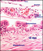
The visceral yolk sac placenta, the thin membrane that lies opposite the placental disk and apposes the peripheral endometrium, is much more simply constructed. A cylindrical epithelium faces the variably cuboidal and cylindrical endometrial epithelium, seemingly without having direct contact; at least this is difficult to ascertain in the sections made after fixation. Those apparent spaces seen in our routine histologic sections are artifact. They are caused by fixation-induced tissue shrinkage. The tissue components are actually contiguous. Important transport of many substances occurs at this interface. They are different, however, than those that reach the embryo through the disk. Brambell (1966) showed that immunoglobulins traverse this barrier from mother to fetus. And since then, many other substances have been shown to pass this epithelial contact zone. This transport, however, was primarily investigated in a variety of other rodents (rat and hamster). While it is true that most of these studies have been done in hamsters and rats, there is reason to believe that the same results hold true for mice. Among the substances studied were viruses, vital dyes, and many other agents administered to the maternal organism. This is well elaborated by Carpenter & Ferm (1969) who traced thorotrast across this membrane in the golden hamster.

This membrane of visceral yolk sac inserts in a ring-like fashion on the surface of the disk (please see below). It has a much more prominent, folded epithelium near the disk, and becomes thinner and thinner as it stretches around the embryo to the dome of the uterine wall.

   
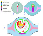
  Diagrams of early placental development (After Mossman, 1983). Figure 1 is at approximately 7-7 1/2 dpc. Figure 2 is at approximately 8 dpc. Figure 3 is at approximately 10-11 dpc.
     
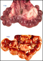
  These two pregnant uteri are in situ. The top is at 13 days and contains 13 embryos with the defective one leaving a space between 3 and 4 at top left. The uterine arterial arcade is well displayed.

The bottom uterus is at 18 days and has 10 normal embryos.
     
  Diagram of mouse implantation at end of gestation (After Mossman, 1983).
     
  Implanted placental disks near end of pregnancy. Note the narrow stalk of the maternal vascular supply and the lateral infolding of the yolks sac membrane.
     
  Implanted placental disks near end of pregnancy. Note the narrow stalk of the maternal vascular supply and the lateral infolding of the yolks sac membrane.
     
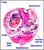
  Cross-section of pregnant uterus with fetus inside at 17 dpc.
     
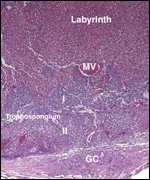
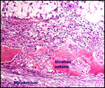
  This 18 dpc placental implantation area shows the two layers (I,II) of the trophospongium to which reference is made in the text. A layer of giant cells (GC) invades the decidua beneath, and a large maternal vessel (MV) penetrates into the labyrinth.
     
 
5) Details of barrier structure

Ramsey (1975) described the complex trophoblast development of the mouse thus:

"The trophoblast forming the ectoplacental cone or träger gives rise to three types of trophoblast. The earliest to appear are wandering giant cells that infiltrate the maternal tissue and eventually come in contact with maternal blood vessels which they open up, thereby creating the hemochorial state. These cells appear to be actively phagocytic for the first 10 days of gestation. The second derivative of the träger is syncytiotrophoblast and the final one the cytotrophoblast. These two compose the walls of the labyrinthine tubules."

(She then refers to an electronmicrophotograph showing this triple layer of trophoblast). Thus, the barrier in the disk is, strictly speaking, a hemotrichorial one. This, of course, is in contrast to the visceral yolk sac placental membrane, which is epithelium to epithelium and, despite this seemingly thick barrier, still provides much exchange function.
   
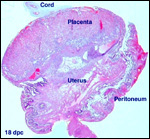
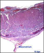
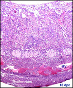
  The pictures (left and below left) show the 18 dpc placenta implanted and mesometrium at bottom (left). Both show the wider labyrinthine placental portion beneath the chorionic plate, in the middle is the layer of trophospongium, and beneath it the second layer. Giant cells follow beneath with invasion of decidua and (minimally) into the myometrium.
     
  The pictures (left and above left) show the 18 dpc placenta implanted and mesometrium at bottom (above left). Both show the wider labyrinthine placental portion beneath the chorionic plate, in the middle is the layer of trophospongium, and beneath it the second layer. Giant cells follow beneath with invasion of decidua and (minimally) into the myometrium.
     
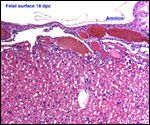
  This picture shows the fetal surface portion of the labyrinth.
     
  Floor of 18dpc mouse placenta with two regions of trophospongium, decidual invasion by giant cells and myometrium below.
     
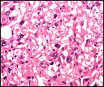
  High magnification of labyrinthine placental portion. Giant trophoblast forms the surface of the fetal villi. Most of the red blood cells present here are in fetal capillaries; the maternal spaces are mostly empty.
     
 
6) Umbilical cord

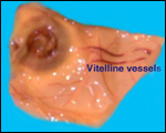
The one umbilical cord that I have measured was 1.5 cm long, on day 18 pc. It was very thin and translucent, and arose from the center of the placental disk. It had no spiral turns. The cord was covered by a very thin amnion and lacked Wharton's jelly. The vitelline cord was also extremely thin. It swung around the embryo to reach the thinnest portion of the "membranes" at the dome of the implantation site, opposite the placental disk. The cord had three very thin blood vessels, an artery and two veins.
   
  Situs at 18 days with placental disk at left and cord remnant. The vitelline vessels course along the membranes to the fundus portion of the inverted yolk sac.
     
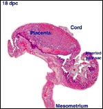
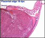
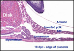
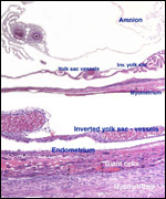
  These two photographs (left, bottom left) show the edge of the 18 dpc placenta with the insertion of the inverted yolk sac placental membrane shown as a fold (seen as a ring in the macroscopic yellow photo). Note that near the insertion of the yolk sac membrane the endodermal epithelium is folded (villus-like) and much thicker than on top. The endometrium is also much thicker here. The amnion is extremely thin.
     
  These two photographs (left, above left) show the edge of the 18 dpc placenta with the insertion of the inverted yolk sac placental membrane shown as a fold (seen as a ring in the macroscopic yellow photo). Note that near the insertion of the yolk sac membrane the endodermal epithelium is folded (villus-like) and much thicker than on top. The endometrium is also much thicker here. The amnion is extremely thin.
     
  The umbilical cord is composed of two portions. At the left side is the vitelline segment with one artery and two veins. At right is the allantoic segment (1A, 1V).
     
  Cross section of umbilical cord near the abdomen. The left portion is the vitelline circulation (1A, 2V), the right is the allantoic portion (1A, 1V).
     
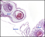
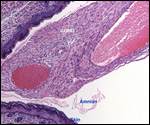
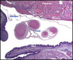
  The picture at left is a section through the main umbilical cord (allantoic). In the picture at the bottom left, the main cord (large vessels) is on the left, while the yolk sac vessels (smaller and here only 2) are on the right.
     
  The picture (above left) is a section through the main umbilical cord (allantoic). In the picture to the left, the main cord (large vessels) is on the left, while the yolk sac vessels (smaller and here only 2) are on the right.
     
 
There are initially three vessels in the vitelline portion of the umbilical cord (one artery and two veins). The vessels split on the way to their distribution across the yolk sac membrane. Thus, this part of the cord leads from the umbilicus to the dome of the dilated sac. It ends on the opposite side of the placental disk. The allantoic portion is thicker and contains one artery and one vein. It also leads to the umbilicus. These vessels have a much thicker wall than those of the omphalomesenteric (vitelline) circulation. There are no vitelline or allantoic ducts.

7) Uteroplacental circulation

The principal maternal vessels enter in the center of the ovoid disk. The arteriolar walls of the maternal vessels are modified by ingrowth of trophoblast. The veins are large and thin-walled, without trophoblast. Large maternal blood channels are situated deep in the labyrinthine tissue whence they divide to make the complex sinusoidal path along the villous surface. They then contact trophoblast.

Cross et al. (2001) have studied the vascularization of the mouse placenta with vascular casts. They suggested that the uterine NK cells are vital in establishing the vascular bed and that much of the vascular development depends on the activity of the giant cells. They found that the uterine artery branches into several spiral arteries. These vessels lack smooth muscle and converge at the junction to the giant cells. Here they open into the trophoblast-lined canals that enter the labyrinth. Mossman (1987) has diagrammed this complex blood flow.

8) Extraplacental membranes

There is no decidua capsularis. The membranous portion of the implanted mouse pregnancy is, sequentially identified from the outside inwards, composed of as follows: Peritoneum, myometrium, thin layer of endometrium, space with some secretion, yolk sac epithelium, connective tissue layer of inverted yolk sac membrane with a few vitelline vessels, amnionic connective tissue, and a very thin amnionic epithelium (which is usually shriveled up in sections). There is no allantoic sac.

   
  Two views of the dome of the implantation sac - the "free membranes" as they are attached to the myometrium - 18 dpc. Note the vitelline vessels in the inverted yolk sac membrane, very thin myometrium, and infiltration of myometrium by trophoblastic giant cells (bottom membrane).
     
  Higher magnification of peripheral membranes on day 18 pc.
     
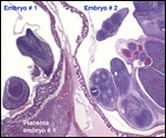
  The membranes of adjacent mouse embryo placentas may be intimately fused, even though their placentas remain separate.
     
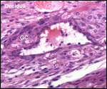
  Maternal blood vessel in the decidua (top left), partially replaced by giant cells (GC) that are also seen below the blood vessel.
     
  Extravillous spongiotrophoblast (giant cells), that opened the maternal blood vessels, is intermixed with decidual cells. This trophoblast, however, does not penetrate deeply into the myometrium. There is some degree of alteration of the maternal arteriolar walls by trophoblastic (giant cell) ingrowth with partial replacement of the muscular wall. This is a common feature of human gestation, but it is much more pronounced in their placentation.

10) Endometrium

There is a marked decidual reaction of the mouse endometrium (which can also be stimulated to develop merely by physical manipulation and also through hormonal manipulation). The decidualization does not include the entire endometrium, however, as will be seen from the photographs of the membranes. At the periphery of the decidual reaction, much glycogen is found in the decidual cells. Capillaries invade this region, and these become subsequently much larger. Finn & Lawn (1967) studied the decidual cells ultrastructurally and identified tight junctions between the cells.

11) Various features

There is no well-developed subplacenta in the mouse but, as can be seen in the center of the discoid placentas here depicted, a suggestion of a subplacenta forms.

12) Endocrinology

The estrous cycle of mice is 3-9 days long. Endocrine aspects of mouse gestation are complex; they have been comprehensively reviewed by Birken et al. (1998). During the early part of gestation, LH stimulates luteal androgen production that is converted to estradiol. This, in turn, stimulates progesterone secretion until midgestation. The placenta then secretes androgens for maintenance of pregnancy, this being referred to as the "luteal shift". When fetectomy is performed, androgen secretion falls dramatically. The placental histologic integrity remains intact, however. It is still unknown if a chorionic gonadotropin exists. Good evidence has been provided by Gardner & Johnson (1972) of hormone production by the trophoblastic giant cells. In paragraphs of "Genetics" below and from Giudice (1999), the reader will find the various genetic factors that are needed for progesterone production and implantation.

13) Genetics

Mice generally have 40 chromosomes, all being acrocentric elements (Committee, 1972). A common finding in mice (as well as some other taxa) is the frequent "Robertsonian fusion" of chromosomes at their centromeres ("whole arm translocation") that reduces the chromosome number, without loss of DNA (Castiglia & Capanna, 1999). There is a huge literature on this topic alone, which commenced with Gropp (review in Gropp et al., 1982; 1983) who identified the low number of chromosomes in Apennine mice (initially the Tobacco mouse, Mus poschiavini 2n=26).

Nesbit (1974) found a 40% banding homology between rat and mouse chromosomes. Although a few reports on mouse hybrids with rats have been issued, they are probably invalid. On the other hand, many different mouse species have hybridized, some with fertility, others with failure. The experiments of hybridization with Mus caroli are especially interesting (Rossant et al., 1983). Only when inner cell mass exchange was done did this lead to the production of heterospecific offspring. Gray (1972) has summarized the extensive literature on mouse hybrids.

Shaver & Martin-DeLeon (1975) reviewed the effect of aging germ cell components. Triploidy, mosaicism and structural anomalies resulted in 2.6% of blastocysts recovered after superovulation. Equally interesting is the study by Yuan et al. (2002) that identifies a synaptonemal complex protein3 gene (SCP3) that functions to allow synapsis of chromosomes at meiosis. In its absence, malsegregation of chromosomes occurs and its absence in mice leads to resorptions, even more so in older mice.

Comprehensive genetic investigations of mice have led to abundant information of their genes. Its genetic structure is now known. There is an abundant literature that cannot be reviewed here. Some of these topics are covered comprehensively by Knobil & Neill (1998). ). How it is possible to localize specific genes and identify the chromosomal location of such a gene by FISH is nicely shown for the X-linked Arhgef6 gene in the contribution by Kutsche & Gal (2001).

The understanding of the major histocompatibility gene complex (MHC) is perhaps most relevant here in view of the problems that occurred in the hybridization experiments of Mus musculus and Mus caroli. The exchange of ICMs succeeded, but the trophoblast was apparently "recognized" and rejected (Rossant et al., 1983). Gill et al. (1983) have reviewed the MHC state of affairs. Hybrids between Mus musculus and Mus caroli were also studied by West et al. (1978) and were found to be sterile. A variety of genetic markers and their expression was explored in this system. The process of imprinting seems to be related to genes located on chromosome 12 and, when the maternal and paternal chromosome are identical, imprinting does not occur, and the mice succumb, with many anomalies and placentomegaly (Georgiades et al., 2001).

Much additional progress has been made in understanding the impact certain genes make on mouse placental development. Thus, the imprinted Esx1 mutational segment of the mouse homeobox complex is imprinted and, when inherited from the mother, has destructive effect on the placental labyrinth (li & Behringer, 1998). Much other work with mice has led to a better understanding of human conditions and the regulation of the placenta. For instance, the work by Sun et al (1997) with the insertion of the transgene for IFG2 leads to Beckwith-Wiedemann-like syndromes. The topic is too extensive, and it is too rapidly moving for a comprehensive discussion here. It has recently been aptly summarized by Rossant & Cross (2001).

Chimerism in mice has been the object of intense study by Tarkowski and later by Mintz (reviewed by Benirschke, 1970). This has have led to much insight into the lineage of cells during development. These topics, however, are beyond the scope of the chapter. Douarin & McLaren (1984) have reviewed the fate of chimeric XX cells in male testes (they vanish). Since mice bear large litters and because numerous corpora lutea can be identified, the question of whether monozygotic ("identical") twins also exist among the litters has often been asked. Wallace & Williams (1965) believed that MZ twins occur as often as 1:100 offspring. They can certainly be produced experimentally by manipulating early stages of development (Sotomaru et al., 1998; Tarkowski et al. 2001). Both these investigative groups of investigators placed single blastomeres into tetraploid carrier embryos and obtained survival under a variety of circumstances, and production of several sets of MZ twins. The investigators successfully made other manipulations that resulted in twins. Kaufman (1982) even produced a set of monoamnionic twins from parthenotes.

McLaren et al. (1995) attempted to identify MZ twins in 200 mice by genetic analysis, but they were unable to find any with certainty. They suggested, therefore, that MZ twinning must be rare. The topic is currently of interest as an increased number of MZ twins accompany the multiples conceived by human infertility therapy. One important question is whether they are the result of embryo handling. Therefore, Cohen & Feldberg (1991) attempted (and succeeded) to show that drilling variable-sized holes into the zona pellucida of mice may lead to premature hatching of blastomeres.

Results of recent studies of genetic mutations have shown convincingly that certain molecules are needed for the development of the labyrinth and for the formation of trophoblastic giant cells (Anson-Cartwright et al., 2000). When a mutation of the Gcm1 locus (encoding the transcription factor glial cells missing-1) is coded, there occur lack of trophoblastic adhesion, lack of villous folding into a labyrinth, lack of giant cell formation, and abortion or early death. Conversely, overgrowth of giant cells and poor development of the diploid trophoblast with fetal death at 10.5 dpc occur when there is a mutational lack of the "orphan nuclear ERR- ß receptor (Luo et al., 1997).

Mice with the chromosomal deletion of one X (39,X) are the genetic equivalent of the human Turner's syndrome. In contrast to the latter, these mice have normal oocytes and may be fertile. Many other chromosomal anomalies (trisomies, XXY, mosaics, chimeras, etc.) have been described. Takagi & Sasaki (1975) showed that the extraembryonic membranes of mice have preferential paternal X-inactivation.

In view of the interest of genomic imprinting (paternal - placenta; maternal - embryo) the study by Ito et al. (1988) is of interest. They were able to follow labeled paternal DNA strands into both, embryo and trophectoderm. Sun et al. (1997) developed a mouse model to study the human Beckwith-Wiedemann syndrome. They introduced the transgene Igf2 (that is responsible for the syndrome's features) with embryonic stem cells. While most of the features of the human syndrome were replicated in the mouse model, some were not (protruding tongue, omphalocele). Nevertheless, this, as well as many similar studies help clarify specific aspects of human maldevelopment that have so far been difficult to access.

14) Immunology

For work on MHC molecules, see Gill et al. (1983). Polyoma virus infection stimulates mouse trophoblastic growth but it does not lead to choriocarcinoma (Koren et al., 1971). One of the most interesting aspects of mouse placentation is the "rejection" of xenotransplanted mouse embryos. When Croy et al. (1982) transplanted blastocysts from Mus caroli into Mus musculus, they initiated a strong inflammatory reaction, hemorrhage and ultimate death of the transplanted embryos. Neighboring Mus musculus embryos and placentas were not affected. It should be pointed out that both species have identical chromosome number with an apparently similar structure. But, when inner cell masses of Mus caroli were placed into the trophectodermal shell of Mus musculus, they developed into normal Mus caroli neonates, supported by the "foreign" placenta. These experiments strongly suggest that, not only is the trophoblast of the foreign genotype immunologically recognized but also, that killer cells of the host destroy the graft. The authors reviewed other interspecific experiments of this kind.

15) Pathological features

Frequently one finds a space in a pregnant mouse uterus that was presumably occupied at an earlier time by an embryo. The reason for its demise are often impossible to discern but with the development of mutant mice and, especially, with the advent of knockout mice and other genetically engineered mice, a better understanding is coming about. Ward & Devor-Henneman (2000) provided for that reason a detailed protocol of how to dissect and assess such fetal deaths. Their chapter is replete with color pictures of the normally developing placenta and membranes and discusses apoptosis in the placenta and various organs and attempts to devise guidelines to differentiate causes of prenatal demise.

One recently identified cause of abortion in mice is the deficient vascular development that results from an absence of a2-adrenoceptors (Philipp et al., 2002). There is a sizeable family of these adreno-receptors; these investigators discriminated among them by knockouts. This particular receptor is beginning to be expressed in the embryo and yolk sac and placenta on day 9.5 and thus, its absence correlated well with the deficient placental vessel development.

   
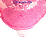
  In one of the uteri dissected, one embryo had died in utero and only remnants of placenta and embryo remained in a mostly empty cavity. These are representative views of the empty cavity, degenerating placenta and embryo. This may well have been due to a chromosomal error.
     
  Monozygotic twins with shared yolk sacs and shared circulation were described by Runner (1984). A plethora of diseases of mice and congenital anomalies have been reported, but there are too many studies to be covered in this context. The reader is referred to two comprehensive reviews (Benirschke et al., 1978; Knobil & Neill, 1998).

16) Physiological data

I have no data or references pertaining to the magnitude of the maternal blood flow, blood volume, or blood pressure of mice.

17) Other resources

Cell lines are available from the Bar Harbor Laboratories and many other agencies. The mouse has been an extraordinary model to study certain genes by the creation of "knockout" mice. There are so many models now available that the topic has become difficult to oversee. For that reason, a "Mouse Knockout & Mutation Database" (MKMD) has been developed in 1995 that now lists over 7000 entries. A sample can be viewed at http://research.bmn.com/mkmd. Ward et al. (2000) published a book on the pathology of genetically engineered mice that may be helpful.

18) Other relevant features

Embryo transfer of mice has been possible since 1935 and is reviewed by Kraemer (1983). This publication also provides references and technical details that may be helpful to the experimenter. Intraspecific chimerae have been produced several times, but the rat x mouse aggregation of blastomeres, while leading to normal appearing blastocysts, did not yield grown chimeric embryos (Stern, 1973). Surani & Barton (1983) produced gynecogenetic embryos, but they failed to go beyond the 25-somite stage. This is presumably due to homozygosity for lethal genes, similar to the androgenetic hydatidiform moles in humans which are lethal to the embryo.


19) Future research needs

There is much information to be gained from the study of mouse placentation. This is especially true for our understanding of the genetic regulation of placentation and trophoblast regulation. The publication by Georgiades et al. (2002) should be consulted for references and directions. In view of the numerous mouse mutations produced, it will be interesting to learn more about the possibly accompanying placental lesions.

References

Anson-Cartwright, L., Dawson, K., Holmyard, D., Fisher, S.J., Lazzarini, R.A. and Cross, J.C.: The glial cells missing-1 protein is essential for branching morphogenesis in the chorioallantoic placenta. Nature Genetics 25:311-314, 2000.

Barlow, P.W. and Sherman, M.I.: The biochemistry of differentiation of mouse trophoblast: Studies on polyploidy. J. Embryol. Exp. Morphol. 27:447-465, 1972.

Barlow, P.W. and Sherman, M.I.: Cytological studies on the organization of DNA in giant trophoblastic nuclei of the mouse and the rat. Chromosoma 47:119-131, 1974.

Benirschke, K.: Spontaneous chimerism in mammals. A critical review. Current Topics in Pathology 51:1-61, 1970. (Springer-Verlag, NY).

Benirschke, K., Garner, F.M. and Jones, T.C.: Pathology of Laboratory Animals. Springer-Verlag, NY 1978.

Birken, S., O'Connor, J., Lobel, L. and Canfield, R.: Chorionic Gonadotropin, Human. Pp. 587-614, in Volume 1 of: Encyclopedia of Reproduction , E. Knobil and J.D. Neill eds.: Academic Press, San Diego, 1998.

Brambell, F.W.R.: The transmission of immunity from mother to young and the catabolism of immunoglobulins. Lancet 2:1087-1093, 1966.

Carpenter, S.J. and Ferm, V.H.: Uptake and storage of thorotrast by the rodent yolk sac placenta: An electronmicroscopic study. Amer. J. Anat. 125:429-456, 1969.

Castiglia, R. and Capanna, E.: Whole-arm translocation (WART) in a feral population of mice. Chromosome Res. 7:493-495, 1999.

Cohen, J. and Feldberg, D.: Effects of the size and number of zona pellucida openings on hatching and trophoblast outgrowth in the mouse embryo. Mol. Reprod. Dev. 30:70-78, 1991.

Committee on standardized genetic nomenclature for mice: Standard karyotype of the mouse. J. Hered. 63:69-72, 1972.

Copp, A.J.: Interaction between inner cell mass and trophectoderm of the mouse blastocyst. I. A study of cellular proliferation. J. Embryol. Exp. Morphol. 48:109-125, 1978.

Copp, A.J.: The development of field vole (Microtus agrestis) and mouse blastocysts in vitro: A study of trophoblast cell migration. Placenta 1:47-60, 1980.

Cross, J.C.: The mouse as a model for investigation of placental function. Chapter 5 (pp.51-60) in, The Placenta: Basic Science and Clinical Practice. J. Kingdom, E. Jauniaux and S. O'Brien, eds. Royal College of Obstetricians and Gynaecologists (RCOG) Press, London, 2000a.

Cross, J.C.: Genetic insights into trophoblast differentiation and placental morphogenesis. Seminars Cell Developm. Biol. 11:105-112, 2000b.

Cross, J.C., Lu, Y., Hemberger, M. and Adamson, L.: Invasion of the mouse uterus by distinct trophoblast subtypes. Placenta 22 (7): abstract P64, p. A24, 2001.

Cross, J.C., Werb, Z. and Fisher, S.J.: Implantation and the placenta: Key pieces of the development puzzle. Science 266:1508-1518, 1994.

Croy, B.A., Rossant, J. and Clark, D.A.: Histological and immunological studies of post implantation death of Mus caroli embryos in the Mus musculus uterus. J. Reprod. Immunol. 4:277-293, 1982.

Dickson, A.D.: Trophoblastic giant cell transformation of mouse blastocysts. J. Reprod. Fertil. 6:465-466, 1963.

Dickson, A.D.: Variations in development of mouse blastocysts. J. Anat. 101:263-267, 1967.

Douarin, N. le and McLaren, A.: Chimeras in Developmental Biology. Academic Press, London, 1984.

Downs, K.M.: Early placental ontogeny in the mouse. Placenta 23:116-131, 2002.

Finn, C.A. and Lawn, A.M.: Specialized junctions between decidual cells in the uterus of the pregnant mouse. J. Ultrastr. Res. 20:321-327, 1967.

Gardner, R.L. and Johnson, M.H.: An investigation of inner cell mass and trophoblast tissues following their isolation from the mouse blastocyst. J. Embryol. Exp. Morphol. 28:279-312, 1972.

Georgiades, P., Watkins, M., Burton, G.J. and Ferguson-Smith, A.C.: Roles for genomic imprinting and the zygotic genome in placental development. Proc. Natl. Acad. Sci. USA 98:4522-4527, 2001.

Georgiades, P., Ferguson-Smith, A.C. and Burton, G.J.: Comparative developmental anatomy of the murine and human definitive placentae. Placenta 23:3-19, 2002.

Gill, T.J., Siew, S. and Kunz, H.W.: Major histocompatibility complex (MHC)-linked genes affecting development. J. Exp. Zool. 228:325-345, 1983.

Giudice, L.C.: Genes associated with embryonic attachment and implantation and the role of progesterone. J. Reprod. Med. 44:165-171, 1999 (Suppl. 2).

Gray, A.P.: Mammalian Hybrids. Second edition. A Check-List with Bibliography. Commonwealth Agricultural Bureaux, Farnham Royal, Slough, UK, 1972.

Gropp, A., Winking, H., Redi, C.A., Capanna, E., Britton-Davidian, J. and Noack, G.: Robertsonian karyotype variation in wild house mice from Rhaeto-Lombardia. Cytogenet. Cell Genet. 34:67-77, 1982.

Gropp, A., Winking, H., Herbst, E.W. and Claussen, C.P.: Murine trisomy: Developmental profiles of the embryo, and isolation of trisomic cellular systems. J. Exp. Zool. 228:253-269, 1983.

Heine, H.: Histoautoradiographie der DNS in Trophoblastriesenzellen der Plazenta der Maus. Acta anat. 95:66-74, 1976.

Heinecke, H. and Grimm, H: Plazentafusionen bei verschiedenen Mausestammen. Z. Versuchstierk. 13:224-231, 1971.

Hemberger, M. and Cross, J.C.: Genes governing placental development. Trends Endocrinol. Metab. 12:162-168, 2001.

Ito, K., McGhee, J.D. and Schultz, G.A.: Paternal DNA strands segregate to both trophectoderm and inner cell mass of the developing mouse embryo. Genes & Development 2:929-936, 1988.

Iwaki, T., Sandoval-Cooper, M.J., Paiva, M., Kobayashi, T., Ploplis, V.A. and Castellino, F.J.: Fibrinogen stabilizes placental-maternal attachment during embryonic development in the mouse. Amer. J. Pathol. 160:1021-1034, 2002.

Kaufman, M.H.: Two examples of monoamniotic monozygotic twinning in parthenogenetic mouse embryos. J. Exp. Zool. 224:277-282, 1982.

Kaufman, M.H.: The Atlas of Mouse Development. Academic Press, 1992.

Kirby, D.R.S. and Bradbury, S.: The hemochorial mouse placenta. Anat. Rec. 152:279-282, 1965.

Knobil, E. and Neill, J.D.: Encyclopedia of Reproduction. Academic Press, San Diego, 1998.

Koren, Z., Damme, O.v., Hirumi, H. and Maramorosch, K.: Growth and stimulation of mouse trophoblastic cells in culture by polyoma virus. Amer. J. Obstetr. Gynecol. 111:846-850, 1971.

Kraemer, D.C.: Intra- and interspecific embryo transfer. J. Exp. Zool. 228:363-371, 1983.

Kunath, T., Strumpf, D., Rossant, J. and Tanaka, S.: Trophoblast stem cells. Chapter 12 (pp. 267-287) in, Stem Cell Biology. D.R. Marshak, R.L. Gardner and D. Gottlieb, eds. Cold Spring Harbor Laboratory Press, Cold Spring Harbor, NY, 2001 (Monograph 40).

Li, Y. and Behringer, R.R.: Essx1 is an X-chromosome-imprinted regulator of placental development and fetal growth. Nature Genetics 20:309-309, 1998.

Kutsche, K. and Gal, A.: The mouse Arhef6gene:cDNA sequence, expression analysis, and chromosome assignment. Cytogenet. Cell Genet. 95:196-201, 2001.

Luo, J., Sladek, R., Bader, J.-A., Matthyssen, A., Rossant, J. and Giguère, V.: Placental abnormalities in mouse embryos lacking the orphan nuclear receptor ERR-ß. Nature 388:778-782, 1997.

McLaren, A., Molland, P. and Signer, E.: Does monozygotic twinning occur in mice? Genet. Res. 66:195-202, 1995.

Mossman, H.W.: Vertebrate Fetal Membranes. MacMillan, Houndmills, 1987.

Nesbitt, M.N.: Evolutionary relationships between rat and mouse chromosomes. Chromosoma 46:217-224, 1974.

Philipp, M., Brede, M.E., Hadamek, K., Gessler, M., Lohse, M.J. and Hein, L.: Placental a2-adrenoceptors control vascular development at the interface between mother and embryo. Nature Genetics 31:311-315, 2002.

Ramsey, E.M.: The Placenta of Laboratory Animals and Man. Holt, Rinehart and Winston, New York, 1975.

Rossant, J. and Cross, J.C.: Placental development: Lessons from mouse mutants. Nature Reviews Genetics 2:538-548, 2001.

Rossant, J., Croy, B.A., Clark, D.A. and Chapman, V.M.: Interspecific hybrids and chimeras in mice. J. Exp. Zool. 228:223-233, 1983.

Rossant, J. and Croy, B.A.: Genetic identification of tissue of origin of cellular population within the mouse placenta. J. Embryol. Exp. Morph. 86:177-189, 1985.

Rossant, J. and Cross, J.C.: Placental development: Lessons from mouse mutants. Nature Reviews Genetics 2:538-548, 2001.

Runner, M.N.: New evidence for monozygotic twins in the mouse: Twinning initiated in the late blastocyst can account for mirror images asymmetries. Anat. Rec. 209:399-406, 1984.

Shaver, E.L. and Martin-DeLeon, P.A.: Effects of aging of sperm in the female and male reproductive tracts before fertilization on the chromosome complement of the blastocysts. In, Aging Gametes. R.J. Blandau, ed. Pp. 151-165. Karger, Basel, 1975.

Snow, M.H.L. and Ansell, J.D.: The chromosomes of giant trophoblast cells of the mouse. Proc. R. Soc. Lond. B.: 187:93-98, 1974.

Sotomaru, Y., Kato, Y. and Tsunoda, Y.: Production of monozygotic twins after freezing and thawing of bisected embryos. Cryobiology 37:139-145, 1998.

Stern, M.S.: Chimaeras obtained by aggregation of mouse eggs with rat eggs. Nature 243:472-473, 1973.

Sun, F.L., Dean, W.L., Kelsey, G., Allen, N.D. and Reik, W.: Transactivation of Igf2 in a mouse model of Beckwith-Wiedemann syndrome. Nature 389:809-815, 1997.

Surani, M.A.H. and Barton, S.C.: Development of gynecogenetic eggs in the mouse: Implications for parthenogenetic embryos. Science 222:1034-1036, 1983.

Takagi, N. and Sasaki, M.: Preferential inactivation of the paternally derived X-chromosome in the extraembryonic membranes of the mouse. Nature 256:640, 1975.

Tarkowski, A.K., Ozdenski, W. and Czolowska, R.: Mouse singletons and twins developed from isolated diploid blastomeres supported with tetraploid blastomeres. Int. J. Dev. Biol. 45:591-596, 2001.

Theiler, K.: The House Mouse. Development and normal stages from fertilization to 4 weeks of age. Springer-Verlag, NY 1972.

Ward, J.M., Mahler, J., Maronpot, R.R., Sundberg, J.P. and Frederickson, R. (eds): Pathology of Genetically Engineered Mice. Iowa State University Press, Ames, 2000.

Ward, J.M. and Devor-Henneman, D.E.: Gestational mortality in genetically engineered mice: Evaluating the extraembryonal embryonic placenta and membranes. Pp. 103-122, in: Ward, J.M., Mahler, J., Maronpot, R.R., Sundberg, J.P. and Frederickson, R. (eds): Pathology of Genetically Engineered Mice. Iowa State University Press, Ames, 2000.

Wallace, M.E. and Williams, D.A.: Monozygotic twinning in mice. J. Med. Genet. 2:26-31, 1965.

West, J.D., Freis, W.I. and Chapman, V.M.: Mus musculus x Mus caroli hybrids: mouse mules. J. Hered. 69:321-326, 1978.

Yuan, L., Liu, J.-G., Hoja, M.-R., Wilbertz, J., Nordqvist, K. and Höög, C.: Female germ cell aneuploidy and embryonic death in mice lacking the meiosis-specific protein SCP3. Science 296:1115-1118, 2002.


back to top

© 2002. All rights reserved.
welcome | home | intro | placentation | glossary | author contact me: kbenirsc@ucsd.edu