(Clicking on the thumbnail images below will launch a new window and a larger version of the thumbnail.)
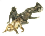
Spider Monkey
Ateles species

Order: Primates
Family: Cebidae

1) General zoological data of species


The term "spider monkey" is used loosely here, as I well appreciate the difficulty in taxonomy. Konstant (1985; 1986) has stated that this designation best, or most properly, applies for Ateles geoffroy,i but that "spider monkey", has now been used for many other, similar-appearing, animals. They are all characterized by their prehensile tail, their body size and black feet. A comprehensive review of these animals was published by Kellogg and Goldman (1944), to be followed by many others. Still, much controversy on taxonomy exists.

This characteristically South American platyrrhine group of species is widely distributed through the Amazon basin and most of Central America. It is composed of many species and/or subspecies that differ greatly in color and some minor other features. Mittermeier (1986) discussed their distribution and status of endangerment in some detail. Despite a lack of publicity, a number of these species/subspecies are critically endangered (Konstant and Mittermeier, 1985). Schneider (2000) compared DNA databases of the major groups of South American primates (Atelidae, Pitheciidae, Cebidae) and further defined their relationships.

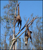
Spider monkeys are extremely nimble, prehensile-tailed animals and are often exhibited in zoological parks. They are very social animals. Often large groups travel in the trees of the rainforest. More often, they form smaller family groups. An extensive literature exists on these species that can be accessed through Nowak's book (1999) and the publication by Mittermeier (1986). A clear differentiation among the great array of phenotypes displayed by these animals has been difficult. A crisper designation of phenotypes was attempted by Konstant & Mittermeier (1985; 1986) when they discussed the need for subspecific recognition because of their chromosomal differences and their many different phenotypes. The importance of this was stressed especially for their captive management. They depicted these different phenotypes in color pamphlets (1985) but the work that was meant to come to a unifying agreement was never completed. The same can be said about the many chromosomal forms that are displayed by spider monkeys from different regions. There are many inversions and translocations, which have not found universal acceptance in their correlation with agreed-upon designations. Nor are they used for a more meaningful management (de Boer, 1974). One reason for the difficulty is that the precise origin of many zoo specimens is unknown, and fieldwork with cytogenetic techniques is problematic. More information on recent phenotype/genotype correlation is found below under the section on genetics. For the understanding of placentation all this is not problematic as the reproductive features of all Ateles species is much the same.

2) General gestational data

Adult spider monkeys weigh about 6-7 kg but, occasionally, twice as much. One of their unusual physical characteristics is the long "clitoris" of females that makes them appear to be males, especially since the testes are small and difficult to see. The function of this pendulous clitoris is unknown, but it is non-erectile. Following mating the male often touches this organ and smells it. The maximal life span recorded for Ateles is 33 years.

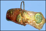
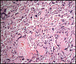
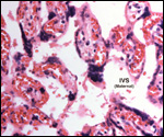
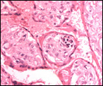
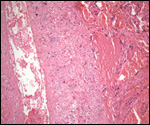
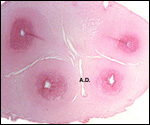
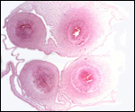
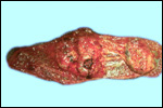
We have had 25 placentas available from term pregnancies, all were bilobed and the average weight without cord and membranes was 105 g (80-135 g). In all but one was the primary lobe (the one with cord insertion) the larger and heavier lobe. It usually weighs 50-75 g and measures 8 cm in diameter and 1-1.5 cm in thickness; the secondary lobe is only slightly smaller. One of my placentas had a tiny third lobe. Blood vessels connect the lobes.

The length of gestation in spider monkeys has been studied in some detail by Eisenberg (1973). He found it to be 7-7½ months (215-226 days). The usually single newborn weighs around 340-480 g (Puschmann, 1989). Sexual maturity is reached between 4-5 years.

   
  Spider monkeys of obviously different types at Children's Zoo of the San Diego Zoo, many years ago. This sort of picture is now only rarely seen.
     
  Spider monkeys with their prehensile tails at the San Diego Wild Animal Park; this is Ateles belzebuth.
     
 
3) Implantation

Few studies have been undertaken on spider monkey implantation. Wislocki (1930) had the best specimens, but they were still insufficient to know details. Implantation in the uterus simplex occurs similar to that in macacs, with one lobe presumably located anterior, the other posterior in the uterus. The bridging vessels run horizontally from one lobe to the other but only in one segment of membranes, not bilaterally. The larger lobe ("primary") usually has the cord insertion. One of our specimens deviated from that rule.

4) General characteristics of placenta

The spider monkey has a bilobed placenta and the lobes are subdivided into smaller lobules. It is thus cotyledonary and hemochorial in the type of its barrier function.
   
  Delivered placenta from spider monkey pregnancy at term. The placenta is bilobed but many smaller lobular dubdivisions are present in the "primary" lobe. There is meconium staining and the "bridging vessels" course only on one side of the membranes.
     
 
Invasive trophoblast is mostly limited to the decidua and is composed of sheets of extravillous trophoblast that characteristically surround the maternal decidual arteries, rather than invading them. The placental villi are much more trabecular than those of man and cercopithecids, as was already mentioned by Mossman (1987). In that respect, the spider monkey placenta is more similar to that of callithricids (marmosets and tamarins). In some areas, particularly near the maternal surface, the villi have a near filiform appearance. Young placentas have larger villi with a rounder appearance and possess foci of hematopoiesis, "blood islands", in their fetal capillaries, much as the callithricids have. These are not found at term, however.

5) Details of barrier structure

This is a characteristic hemochorial placenta. The maternal blood circulates in the intervillous space (IVS) and surrounds the villi with their syncytial surfaces. Syncytial "buds" are commonly found on the villous surfaces and one would expect that, as in humans, they would often detach and land in the maternal lung, as happens in humans.
   
  The "filiform", trabecular nature of the villi is more pronounced near the placental floor (left) than higher up in the placenta (below left).
     
  The "filiform", trabecular nature of the villi is more pronounced near the placental floor (above left) than higher up in the placenta (left).
     
  Term spider monkey villous tissue with little blood in the intervillous space (common). The red blood cells are all contained in the fetal circulation. Multinucleated syncytiotrophoblast covers all villi and forms "knots" (center) that often detach. Cytotrophoblast cannot be discerned at term in paraffin material.
     
  Immature placenta of stillborn fetus with 39 g placenta. Villi are larger than at term and contain foci of hematopoiesis (dark elements). Rare cytotrophoblastic cells can be identified.
     
  Filiform villi on right attach to a large deposit of "X-cells" which has a cystically degenerated center. Such X-cell cysts are common in human placentas and contain major basic protein which is the secretory product of X-cells.
     
 
6) Umbilical cord

There were four umbilical vessels in addition to a large allantoic duct in all but two of our placentas. These had only single arteries, thus altogether only three blood vessels. They were associated with normal fetuses. The allantoic duct may have a good urothelial lining or it may be atrophied. In one cord the duct was apparently occluded. The duct was usually central but in some sections the large fetal vessels "hang" into the sac on broad strands of Wharton's jelly.

The cords measured between13 and 30 cm in length (average 23 cm), had occasional right twists, but generally were not twisted much.
   
  Sections of two different umbilical cords to show the slit-like allantoic duct (left) and the large duct with "hanging" blood vessels in the ductal space.
     
  Sections of two different umbilical cords to show the slit-like allantoic duct and the large duct with "hanging" blood vessels in the ductal space.
     
 
7) Uteroplacental circulation

The general vasculature of the placental/uterine system has not been described in detail in this species. Gruenwald (1972) had one specimen and believed the spider monkey placenta to resemble so much that of the squirrel monkey that appropriate inference can be drawn from their study. He made reference to the fact that several maternal arteries enter the fetal lobules. The decidua basalis has, however, large maternal spiral arterioles that are typically surrounded and only slightly invaded by extravillous trophoblast.
   
  Placental "floor" with large maternal decidual blood vessel at left, surrounded by trophoblast. At "T" is the basal layer of extravillous trophoblast, next to the villous attachment (right). Note the invasion of the decidua by darker trophoblastic cells.
     
 
8) Extraplacental membranes

An unusual feature of spider monkey placentas is their large allantoic sac, an exceptional feature in higher primate placentas (Miller & Benirschke, 1982; 1985). It was found in all of the placentas that we have had available. In contrast to artiodactyla, there was not the universal vascularization of the allantoic membrane. Fluid was expressed from the filled allantoic sac through the umbilical duct, and it was thus assumed to be fetal urine. In a specimen of howler monkey, neither allantoic sac nor a duct was found.
   
  At left is a section from the margin of the placenta with the attachment of the membranes. Note the dark cells (extravillous trophoblast) that surround the maternal blood vessel.
     
  At left is a section of membranes to show the amnion (left) and allantoic sac (center).
     
 
The decidua shows frequently much degeneration, frank yellow necrosis and hemorrhage. This is true for the decidua basalis as well as for the decidua capsularis. In the membranes, of course, travel the thick-walled fetal blood vessels that connect the lobes.

The amnion is cuboidal to slightly columnar, especially when there had been meconium discharge. In that case there is some degeneration of the amnionic epithelium and the columnar appearance becomes more striking. Meconium-laden macrophages can be found as in the human membranes after the discharge of meconium.

9) Trophoblast external to barrier

There is a significant amount of extravillous trophoblast, despite Mossman's (1987) statement that little invasion occurs. A broad band of extravillous trophoblast (the so-called X-cells) is present at the implantation site, surrounded by fibrinoid. These cells are especially clustered around the larger maternal blood vessels in the decidua basalis and on the membranes. There is little infiltration by "giant cells", such as the syncytium. How deeply the trophoblast invades into the uterus is not known, but there is some vascular invasion.

10) Endometrium

Decidualization occurs during pregnancy; the normal cycle is followed by menstrual bleeding. Spider monkeys have a simplex uterus.

11) Various features

There is no subplacenta.

12) Endocrinology

By extracting placental tissues of one spider monkey, Hobson & Wide (1981) were able to demonstrate the presence of chorionic gonadotropin. Hernandez-Lopez et al. (1998) were able to establish the menstrual cycle by means of vaginal swabs and blood estrogen/progesterone measurements. They found a 24 day cycle, with menstrual bleeding. Campbell et al. (2001) used fecal and urinary steroid analysis, confirming the cycle (20-23 days), identifying bleeding, and being also able to determine abortion from progesterone measurements.

As most other simian primates, spider monkeys possess a typical fetal zone of the fetal adrenal gland.

13) Genetics

A recent discovery of a retrovirus envelope gene sequence localizes to the syncytiotrophoblast of human and simian placentas, but it fails to do so in platyrrhine species' placentas, nor does it localize in other, non-primate species (Voisset et al., 1999). Its gene product causes fusion of cells in vitro and this may be related to the non-rejection of the trophoblast (presumably by abolishing surface antigens). It is the first fusion-promoting gene and is thought to have been present for at least 25 Million years (Blond et al., 2000). Whether another gene that encodes for "syncytin" and causes similar cell fusion and expression in trophoblast is related or was separately evolved awaits further study (Mi et al., 2000).

Numerous hybrids among the poorly delineated "species" of Ateles have occurred in zoos where the (sub)species often are housed together (see also Rossan & Baerg, 1977). Gray (1972) has enumerated some of these but many more go unreported. Many, perhaps most, such "hybrids" are also fertile, as the inversions of chromosomal segments produce little of a hybrid barrier at meiosis. Konstant & Mittermeier (1985) have presented what information of genotypes was available at the time. More recently, Collins & Dubach (2001) showed from studies with nuclear DNA study that a species/subspecies separation by pelage phenotype is inconsistent with DNA findings. Much more work is needed before resolution of the relation among some of the more endangered primate species is available.

All spider monkeys studied to date have had 34 chromosomes. There have been many studies of individual species (see Benirschke, 1975; Kunkel et al., 1980; de Boer & de Bruijn, 1990) and a few more comprehensive one, as de Boer (1974). All showed the same chromosome number (2n=34) but many inversions and translocations that may characterize individual "good" species have been demonstrated.

From studies of tranferrins and albumins, as well as from other data, Cronin & Sarich provided cladograms with suggestions of divergence times for Platyrrhini (1975). Histocompatibility complex molecules have been studied by Kriener et al. (2000).

14) Immunology

MHC molecules have been studied by Kriener et al. (2000), which has relevance to an understanding of the phylogeny of the South American primates.

15) Pathological features

Kaplan (1979) has described chorioamnionitis and funisitis due to gram-positive and gram-negative organisms in a term 100 g placenta whose neonate died in ten days from meningitis. We saw placental sac infection in 3 of our 25 cases. Scott (1992) described some inflammatory lesions, especially in the colon, with herniation of the mucosa through the muscular layers , perhaps representing early diverticulosis.
Many of our placentas had meconium-staining, without there being "fetal distress" and with normal, surviving infants. Absence of fetal stress is a common finding in human pregnancies complicated by meconium discharge. It relates more precisely to a prolonged length of gestation (Kaufmann & Benirschke, 2000). Chorioamnionitis and funisitis were found in three cases, one of which was followed by maternal endometritis.

We had two cases of typical abruptio placentae (partial) in our material that was accompanied by excessive decidual necrosis at the base. This was found in this and some other placentas. There also was much calcification in some basal layers, but we do not consider this to be pathological. Infarcts were not seen, but intervillous thromboses occurred several times.

   
  Term placenta with "amnionic sac infection", i.e. chorioamnionitis and funisitis. This shows the fetal surface with emigration of polymorphonuclear leukocytes from the mother in the intervillous space (IVS) and from the fetus' surface vessel. The migration is always towards the surface where the chemotactic agent (bacterium etc.) resides.
     
  Maternal surface of spider monkey placenta showing two central areas of retroplacental hemorrhage (abruptios) in the larger lobe.
     
 
That abortion occurs in wild animals has been shown with fecal steroid determination in the study by Campbell et al. (2001).

16) Physiological data

We know no relevant studies.

17) Other resources

Cell strains of many spider monkeys (poorly identified by proper origin, I believe) are available from CRES at the San Diego Zoo.

18) Other relevant features and need for future studies

Too little is yet known about the species or subspecies designations. They need clarification. Hybrids should have chromosomal and meiotic studies. Cord length is to be better defined and the unusual presence of so large an allantoic sac needs verification and extension to related species.

References

Benirschke, K.: Biomedical research. In, Research in Zoos and Aquariums, Institute Laboratory Animal Resources, eds. Ntl. Acad. Sci. Washington, D.C. pp. 3-11, 1975.

Benirschke, K. and Kaufmann, P.: The Pathology of the Human Placenta, 4th ed. Springer-Verlag, New York, 2000.

Blond, J.L., Lavillette, D., Cheynet, V., Bouton, O., Oriol, G., Chapel-Fernandes, S., Mandrand, B., Mallet, F. and Cosset, F.L.: An envelope glycoprotein of the human endogenous retrovirus HERV-W is expressed in the human placenta and fuses cells expressing the type D mammalian retrovirus receptor. J. Virol. 74:3321-3329, 2000.

Campbell, C.J., Shideler, S.E., Todd, H.E. and Lasley, B.L.: Fecal analysis of ovarian cycles in female black-handed spider monkeys (Ateles geoffroyi). Amer. J. Primatol. 54:79-89, 2001.

Cell lines from: http://www.sandiegozoo.org/conservation/cres_home.html. Please direct your inquiries to Dr. Oliver Ryder (oryder@ucsd.edu).

Collins, A.C. and Dubach, J.M.: Nuclear dna variation in spider monkeys (Ateles). Mol. Phylogenet. Evol. 19:67-75, 2001.

Cronin, J.E. and Sarich, V.M.: Molecular systematics of the new world monkeys. J. Human Evol. 4:357-375, 1975.

de Boer, L.E.M.: Cytotaxonomic Studies in the Primate Suborders Prosimii and Platyrrhini. Drukkerij Bronder-Offset B.V., Rotterdam, 1972.

de Boer, L.E.M. and Bruin, M. de: Chromosomal distinction between the red-faced and black-faced spider monkeys (Ateles paniscus paniscus and A. p. chamek). Zoo Biol. 9:307-316, 1990.

Ateles. Intern. J. Primatol. 1:223-232, 1980.

Eisenberg, J. F.: Reproduction in two species of spider monkeys, Ateles fusciceps and Ateles geoffroyi. J. Mammal. 54:955-957, 1973.

Gray, A.P.: Mammalian Hybrids. A Check-List with Bibliography. Commonwealth Agricultural Bureaux, Farnham Royal, Slough, England, 1972.

Gruenwald, P.: Expansion of placental site and maternal blood supply of primate placentas. Anat. Rec. 173:189-204, 1972

Hernandez-Lopez, L., Mayagoitia, L., Esquivel-Lacroix, C., Rojas-Maya, S. and Mondragon-Ceballos, R.: The menstrual cycle of spider monkey (Ateles geoffroyi). Amer. J. Primatol. 44:183-195, 1998.

Hobson, B.M. and Wide, L.: The similarity of chorionic gonadotrophin and its subunits in term placentae from man, apes, old and new world monkeys and a prosimians. Folia Primatol. 35:51-64, 1981.

Kaplan, C.G.: Intrauterine infections in nonhuman primates. J. med. Primatol. 8:233-243, 1979.

Kellogg, R. and Goldman, E.A.: Review of the spider monkeys. Proc. US National Museum 96:1-45, 1944.

Konstant, W.R. and MittermeierR.A.: Spider monkeys in captivity and in the wild. Primate Conservation #5 (January) pp.82-109, 1985.

Konstant, W.R.: Considering subspecies in the captive management of Ateles. Chapter 61 (pp. 911-920) In, Primates. The Road to Self-Sustaining Populations. K. Benirschke, ed. Springer-Verlag, New York, 1986.

Kriener, K., O'hUigin, C., Tichy, H. and Klein, J.: Convergent evolution of major histocompatibility complex molecules in humans and New World monkeys. Immunogenetics 51:169-178, 2000.

Kunkel, L.M., Heltne, P.G. and Borgaonkar, D.S.: Chromosomal variation and zoography in Ateles. Intern. J. Primatol. 1:223-232, 1980.

Mi, S., Lee, X., Li, X.P., Veldman, G.M., Finnerty, H., Racie, L., LaValle, E., Tang, X.Y., Edouard, P., Howes, S., Keith, J.C. and McCoy, J.M.: Syncytin is a captive retroviral envelope protein involved in human placental morphogenesis. Nature 403:785-789, 2000.

Miller, P.W. and Benirschke, K.: Anatomical and functional differences in the placenta of primates. Biol. Reprod. 26:29-53, 1982.

Miller, P.W. and Benirschke, K.: A large allantoic sac in the placenta of the spider monkey, Ateles geoffroyi. Placenta 6:423-426, 1985.

Mittermeier, R.A.: Primate conservation priorities in the neotropical region. Chapter 16 (pp. 221-240) In, Primates. The Road to Self-Sustaining Populations. K. Benirschke, ed. Springer-Verlag, New York, 1986.

Mossman, H.W.: Vertebrate Fetal Membranes. MacMillan, Houndmills, 1987.

Nowak, R.M.: Walker's Mammals of the World, Vol. II. 6th edition. The Johns Hopkins University Press, Baltimore, 1983.

Puschmann, W.: Zootierhaltung. Vol. 2, Säugetiere. VEB Deutscher Landwirtschaftsverlag, Berlin, 1989.

Rossan, R.N. and Baerg, D.C.: Laboratory and feral hybridization of Ateles geoffroyi panamensis Kellogg and Goldman 1944 and A. fusciceps robustus. Primates 18:235-237, 1977.

Schneider, H.: The current status of the New World monkey phylogeny. An. Acad. Bras. Cienc. 72:165-172, 2000.

Scott, G.B.D.: Comparative Primate Pathology. Oxford University Press, Oxford, 1992.

Voisset, C., Blancher, A., Perron, H., Mandrand, B., Mallet, F. and Paranhos-Baccalà, G.: Phylogeny of a novel family of human endogenous retrovirus sequences. HERV-W, in humans and different primates. AIDS Res. Hum. Retrovir. 15:1529-1533, 1999.

Wislocki, G.B.: Remarks on the placentation of a platyrrhine monkey (Ateles geoffroyi). Amer. J. Anat. 36:465-487, 1926.

Wislocki, G.B.: On a series of placental stages of a platyrrhine monkey (Ateles geoffroyi) with some remarks upon age, sex and breeding period in platyrrhines. Contrib. Embryol. 22:173-192, 1930.


back to top

© 2002. All rights reserved.
welcome | home | intro | placentation | glossary | author contact me: kbenirsc@ucsd.edu