Rhesus Monkey, and some other Cercopithecidae
Macaca mulatta

Order: Primates
Family: Cercopithecidae


1) General Zoological Data


The rhesus monkey is the most widely used laboratory primate. An enormous amount of information has been accumulated on its anatomy, physiology, and on pathologic features afflicting this species. While specimens of rhesus monkeys are not commonly carried in zoos, some placental specimens of rhesus placenta were available to me. But I have had considerably more access to specimens from the lion-tailed macac, Macaca silenus. This endangered Indian species has been bred for many years at the San Diego Zoo. In addition, a few related species with very similar placentation are considered here. The term "macaque" is Portuguese for monkey.

Macacs are members of the large group of Cercopithecidae ("apes with tail"). This genus comprises at least 20 species and some additional subspecies. One species, the Kolb's monkey, has been discussed in a separate chapter. Taxonomic revisions are frequently made in this genus (Nowak, 1999). Many of these species hybridize fertilely (Fa, 1989; Gray, 1972). Hybridization is not only true for the macac species but it occurs with related genera, irrespective of chromosome numbers. Thus, assignment of species rank is somewhat problematic in some animals. A complete review of the genus comes from Lindburg (1971, 1980). It needs to be consulted if more data are desired than what is reviewed by Nowak (1999).

The macacs are largely Asiatic species and are related to other cercopithecid members, such as baboon, mandrill, langurs, etc. Many cercopithecids have very similar or even an identical placentation. Macacs are diurnal animals with considerable arboreal ability. They forage on the ground for variable times and, depending on the species, they are hardy animals, good swimmers, can withstand snow and heat, and macacs generally produce a single young. Twins occur, but this is uncommon. Indeed, Geissmann (1990) determined that the estimates often published suffer from a small sample size. If this is taken into consideration, then twinning frequency in macacs is slightly lower than in human populations (0.19-0.35% vs. 0.8%). Estrous cycles are between 28 and 40 days in length, depending on species and authority, and estrus may last for 9 days. Gestation lasts 160-170 days in rhesus monkeys, slightly longer in some other species. There is, however, considerable variation in rhesus monkeys as well. Specifically, prolonged gestation is not uncommon. Newborns weigh between 400 and 500 g and mothers usually nurse for a year. Sexual maturity occurs between two and three years of age. Longevity for some animals in zoos is over 30 years, and rhesus females are said to go into menopause at age 25 years.

Ramsey has made numerous contributions to our understanding of placentation and reproduction in rhesus monkeys. Her contribution in 1972 evaluates the usefulness of the rhesus monkey for studies to understand human reproductive problems. Despite finding differences in depth of implantation, the basal decidua and vasculature, she nevertheless concluded the animal to be useful, so long as its physiology is better understood. Myers (1979) reviewed the history of the establishment of the primate colony in Puerto Rico and advocated the rhesus for studies of abnormal placentation.

   
  Macaca nigra (Celebes macaque or "Black ape") with neonate.
     
  Liontailed macac, Macaca silenus.


2) General Gestational Data

Gestational length in rhesus monkeys is about 160 days. Newborns weigh then between 450 and 600 g and the corresponding placentas are around 140 and 160 g. There is a general correspondence of placental and fetal weights (Bunton, 1986). Singletons are the rule. The primary disk measures around 9 x 0.75 cm, the secondary disk around 6.5 x 1 cm.

The growth of fetus and placenta was also plotted by v. Wagenen et al. (1965). In their experience, the average length of gestation was 168 (+/- 7) days, at which time the neonate weighed around 470 g and had a crown-rump (CR) length of 19 cm. The average weight of the placenta then was 121 g and their cord measured 20 cm in length. I am impressed with the frequency of longer gestations in this extensive study that reported the data in numerous tables. Placentophagy was observed in this study as well.


3) Implantation

Enders & Schlafke (1981) flushed the uteri of cycling, sexually active female rhesus monkeys and collected various preimplantation conceptuses between days 2 and 8 of the estrogen peak. Five of their specimens had blastocysts without zona pellucida (days 7-8). They showed the trophectoderm to possess microvilli and, near the inner cell mass, there are larger protrusion that presumably will subsequently interact with endometrium at implantation. These authors were impressed with the frequency of cell death in the recovered blastocysts and depicted them in a later publication (Enders et al., 1982). They also saw the earliest differentiation of endoderm in late stages of blastocysts.

The implantation of the rhesus blastocyst was first studied by Wislocki & Streeter (1938) and further elaborated upon by Heuser & Streeter (1941). Its superficial similarity has given rise to hopes that its placentation may thus constitute a useful model for human reproductive studies. Ramsey (1972), while agreeing in general with this, is also adamant to point out significant differences that need to be considered. These include the shallow implantation, the formation of a secondary disk as a result, the development of an endometrial plaque, and differences in maternal vascular supply.

Heuser & Streeter (1941) stated that the blastocyst remains in the uterus for about 5 days, that it is sticky and thus adheres readily to the endometrium. Thereafter the epithelial endometrial plaque forms. They likened the plaque to a deciduoma. One half of the implantations they observed were anterior and one half was posterior in the middle of the uterus. A few exceptional blastocysts implanted laterally in the uterus. Implantation occurred on days 8 to 9. Beautiful photographs and diagrams accompany this publication of data that extend to term gestation.


Ramsey et al. (1976) beautifully illustrated the implanting blastocysts in a comparison between human, rhesus and baboon placentations. The rhesus blastocyst does not implant "interstitially" as the human, but remains only superficially attached and thus lacks the usual decidua capsularis of human placentas. A dome of blastocyst protrudes from the endometrial surface into the endometrial cavity (which is then only a slit) and becomes attached to the other side of the uterus. Also the endometrial reaction is much different. In humans, a pronounced decidua is hormonally initiated. In rhesus monkeys, on the other hand, there is a profound proliferation of surface epithelium, the so-called epithelial plaque.


4) General Characterization of the Placenta

The placenta of rhesus monkeys is similar to that of human placentas, except for the predominance of two disks in most rhesus monkey placentas. The rhesus monkey placenta has two disks in about 80% of term gestations; the remainder, however, has a single disk, as is the normal finding in baboons (Myers, 1972; Chez et al., 1972).

Whether there are two disks or only one, this does not appear to have an impact on fetal growth and survival. The actual cause of the development of single disks in about 20% of rhesus gestations, however, is unknown. Perhaps it is the result of slightly deeper implantation. Chez et al. (1972) found no differences in prenatal bleeding or other parameters that might explain this difference in placental forms. They suggested that this is the result of some autosomal Mendelian trait.

This is a cotyledonary, villous placenta. There are between 4 and 24 lobules of the rhesus placenta according to Myers who also found that wild-born animal placentas had more placental lobules and heavier infants than those born in captivity. In contrast to human placentas, fewer septa are found between the individual cotyledons. As in human placentas, these septa are rarely complete and span only for a short distance into the placenta (Ramsey & Harris, 1966). It is believed that some of the septation is due to the contraction of the uterus during expulsion of the human placenta.

Another feature mentioned by Myers and others is the frequency (around 15%) of circumvallation of the placenta. Circumvallation is a condition in which a wall of degenerated fibrin encircles the placenta with a concomitant narrower insertion of the peripheral membranes. While it seems to be a placental "abnormality", I believe its impact on fetal development to be small.

   
  Term placenta of rhesus monkey with two lobes. Interconnecting vessels are visible.


The study of Wislocki & Streeter (1938) laid the foundation for our knowledge of placentation in this species. At that time, however, the mechanism of trophoblastic growth was not understood. The studies by Galton (1962) on DNA content, by Richart (1961) on thymidine incorporation, and later by Pierce et al. (1964) on gonadotropin-producing cells, laid the foundations for the current understanding. The cytotrophoblast (the "Langhans' cell layer") of the villous surface proliferates and, continuously throughout pregnancy, adds nuclei and cellular material to the syncytiotrophoblast that borders the entire intervillous space. The cytotrophoblast of very young placentas is a continuous sheet of cells that is easily identified. Later in gestation, these cells are less apparent. They are, however, readily seen electronmicroscopically and identified by cell markers (vide infra). The syncytial nuclei have lost their replicative ability. Thus, the syncytium is a single, uninterrupted sheet of cytoplasm within which the nuclei are "loose". Protrusions often form (so-called "knots"), and many of these knots detach. They are then swept away by the intervillous circulation. They are transported to the lung and there they deteriorate. The syncytium is the outermost cell layer of villi. It is studded with numerous microvilli that effects transport in and out of the placenta, produces hormones and has other enzymatic activity. None of these functions are available from the villous cytotrophoblast.

Prior to the knowledge of these details, investigators also considered that the trophoblast may produce the connective tissue, even the vasculature of the villi. I consider this now to be erroneous for many reasons. Importantly, none of the choriocarcinomas that are composed of these two trophoblastic cell types ever produces connective tissue or vessels. This has been studied extensively in human neoplasms and is also true of the few tumors of rhesus monkeys. The mesenchymal elements of the placenta arise from mesodermal precursors (see section of membranes below).

Another topic of concern has been an understanding of the villous structure, especially when the villi were compared with those of other primate placentas. Considerations of this kind have been used to understand the developmental relationships among primates and especially, to understand the possible relationship between platyrrhine and catarrhine primates. Did they have a common ancestor, and when did they separate; and can the placental structure be useful in understanding this evolutionary complexity? These are the principal reasons for such considerations.

It is true, the appearance of the platyrrhine (South American) monkeys' villi differs; they have a trabecular structure (please see chapter on Tamarins). Although cercopithecids have in their final form some similarities with those villi, their villi form quite differently as was pointed out by Starck (1959). In the development of platyrrhines there is much more initial trophoblastic growth, while implantation of cercopithecids is much "thinner" and the growing connective tissue pushes the trophoblast ahead, thus producing the villous outgrowths. Starck also pointed out that the typical longitudinal connecting villi of platyrrhines (that go from chorionic plate to the floor) ar
e very uncommon in cercopithecids, as for instance in the rhesus monkey.


5) Details of fetal/maternal barrier

A superior electronmicroscopic study of rhesus monkey placenta villi was published by Luckett (1970). This animal's placental details are similar to those of human trophoblast. The syncytium contains large amounts of rough endoplasmic reticulum, a Golgi complex, and numerous transport vesicles. The syncytial surfaces are studded with microvilli and at their bases that connect them to the villous mesenchyme, they are complex and infolded. The cytotrophoblast beneath the syncytium is discontinuous. The villi have large, active-appearing macrophages, so-called Hofbauer cells. The trophoblast layer is exposed to the intervillous space within which maternal blood circulates, and the syncytium has the appearance of being the most active cellular component.

   
  Immature villus stained with "Ki-67", a cellular proliferation marker. Note that the brown-stained nuclei are beneath the syncytium. This is the cytotrophoblast, the Langhans' cells.
     
  Cross-section of umbilical cord of rhesus macac at left, and of rolled membrane at right.


6) Umbilical cord

The umbilical cord of macacs, and so far as I know of all cercopithecids, has two arteries, one vein and perhaps an occasional remnant of allantoic duct. The blood vessels have no elastica. The cord is generally inserted on one of the two lobes, usually the larger lobe. The fetal surface vessels thence distribute over the placenta and pursue a membranous course to the secondary lobe. A central insertion upon the lobe is common, but it may also insert more laterally, even upon the membranes. A thin layer of amnionic epithelium covers the cord; there are no caruncles. Myers (1972) found only two cases (0.2%) of single umbilical artery in rhesus monkeys. This anomaly has an incidence of around 1% in human placentas. He also found occasional membranous (velamentous) insertion of the cord. Bunton (1986) described endothelial proliferations in umbilical vessels, which I have not observed. It is perhaps important to point out that the placental surface blood vessels often have an irregular configuration. That is to say, the upper (near amnion) muscular layer is thinner than the lower (toward villi), thicker wall. This eccentric appearance is solely due to "herniation" of the blood-filled lumen (under fetal cardiac load) once the amnionic fluid has been released, and thereby the counter-pressure that keeps the vessel round in utero is lost.

Human and baboon umbilical cords possess an anastomosis between the two umbilical arteries. Whether this is so in rhesus placentas has not been reported, but it is likely. This so-called "Hyrtl anastomosis" (after the Viennese obstetrician who wrote a monograph on the cord) occurs in about 90% of human umbilical cords. It is located near the cord insertion on the placental surface. Houston & Hendrickx (1968) examined the umbilical cords of baboon fetuses and term placentas. They found around 85% of anastomoses in early gestations, and 70% at term. They then related the presence or absence of anastomoses to the manner of vascular distribution on the placental surface, without finding significant correlations. The "Hyrtl anastmosis" has been of interest in human gestations. Raio et al. (2001) have studied the blood flow through a variety of anastomotic types. They found that fusion (rather than an anastomosis) occurs more often in marginally inserted cords.

It is our practice to make a cross section of the umbilical cord and a roll of the free membranes as shown above. The membrane roll is useful for the inspection of decidual blood vessels (when there is a decidua capsularis) and to assure patency of the vessels that connect the two lobes. In addition, inflammatory changes are easiest to identify in this roll. Because Bouin's solution makes the fixed tissue harder and less slippery, its use for the membrane roll is especially useful.

Spatz (1968) gave the length of umbilical cords as measuring around 15-20 cm in rhesus monkeys, which corresponds to the measurements by v. Wagenen et al. (1965) of 20 cm. The longest umbilical cord I have measured is 30 cm in a liontailed macac. Knots had not been reported until Myers (1979) found one loose knot. In another case with tight knot this led to fetal demise. Naaktgeboren & Wagtendonk (1966) hypothesized that the encircling shown in the next pictures may lead to knots of the cord. I should point out though that this is a common feature in human gestations and that knots are rarely the result. Myers (1979) reported cord entanglement in rhesus gestation as well as cord compression during labor with fetal CNS injury. No information is available on the spiraling of cords. The next picture shows no spiraling.
 
   
  Liontailed macac fetus in utero with cord centrally inserted on a single disk and wound about the fetal neck.
     
  Liontailed macac fetus in utero with cord centrally inserted on a single disk and wound about the fetal neck.


7) Uteroplacental circulation

Perhaps the greatest effort has been expended on understanding the circulation of the rhesus monkey placenta. This was done primarily in an effort to better comprehend the circulation of the human placenta that is less accessible for in situ investigations. Several investigators, notably Elizabeth Ramsey in Washington, and Maurice Panigel in France have been the foremost students of this topic.

Ramsey & Harris (1966) comprehensively reviewed all findings on vascular supply and circulation of rhesus and human placentas made prior to this date. When implantation of the rhesus blastocyst has been achieved, the first reaction is the formation of the endometrial epithelial plaque. Next, the capillary endometrial network is opened, to be followed by veins and then the endometrial arteries. Trophoblast invasion is less deep than in humans and "wandering cells" as seen in human decidual floors do not occur in monkeys. Next, the arterial lumens are filled by cytotrophoblastic plug that reach to the myometrial borders. This is followed by invasion of the arteriolar walls and their subsequent modification. The arteriolar lumens subsequently enlarge greatly Ramsey et al., 1979). While veins may contain some trophoblast as well, even some villi have been found within them, the walls are not modified in a manner of the change occurring in arterioles. The veins ultimately form a network around the arterioles. In rhesus monkeys there are approximately 20 arteries and 40 veins that connect the final intervillous space with the maternal circulation. In human placentas the numbers are substantially higher as was also pointed out by Gruenwald (1973). He emphasized that the endometrial arteries are initially convoluted before they enter the final straight portion that injects the blood into the intervillous space. For an understanding of the pathology that occurs in placentation, especially the development of infarcts, it is important to be cognizant that it is principally the arterial injection that is responsible for the well-being of the corresponding villous tree. When it is occluded or when uterine contraction limits the flow through these vessels, the villous tissue becomes hypoxic and may die, depending on the length of the occlusive state. I should point out that many other investigators have made similar observations (e.g., Borell, Richart, Panigel, to name a few) whose publications can be accessed from this review.

Radioangiographic studies with serial x-rays and using contrast media have shown that the arterioles inject "spurts" into the intervillous space. This injection does not occur simultaneously in all vessels, rather, it is irregular and followed subsequently by unlabeled blood. This then drives the previously contrasted blood toward the chorionic plate. Here, it is dissipated peripherally and the appearance of "smoke rings" (also referred to as doughnuts) is produced in the angiographs. These studies also indicated that the vascular jets are partially controlled by uterine contractions and areas of narrowing in the arterioles. Vasomotor agents may also interact in controlling flow. Veins then diffusely drain the blood, and this is subject to uterine contractions as well (Ramsey et al., 1966). The well-known diagram that was produced by Crosby accompanies the large review of the placental circulation by Ramsey & Harris. It portrays the villous organization and blood flow and is shown next.
 
   
  Diagram of primate placenta, with permission from E. Ramsey.
     
 
The fetal circulation has attracted equal attention to that of the maternal side. A single fetal arterial branch supplies a cotyledon, ramifies into a complex network and the blood is then drained through a single vein that usually emerges near the point of arterial entry. Corrosion casts of this cotyledonary vasculature have been made by Panigel (1968, Arts & Lohmann 1974, and others). Arts & Lohman (1974) used dyed polymer injections from both maternal and fetal sides under anesthesia of the rhesus monkeys. They thus showed that maternal blood (blue) enters through single arteriole in the centers of cotyledons. It then fills the intervillous space and reaches the subchorionic plate. After filling the central portion of the cotyledonary intervillous space, the blood is pushed laterally by subsequent injection mass. The arrangement of arteries and veins on the chorionic plate was the same as in human placenta - arteries crossed over the veins. They also made an important observation that is often overlooked when abnormal or twin human placentas are discussed. They found no anastomoses between the capillaries adjacent villi, let alone any of the 10-14 rhesus monkey placental cotyledons. Moreover, they were firm about their conclusion that one maternal artery corresponds to one fetal cotyledon. This artery enters the center of the cotyledon. Ramsey et al. (1979) later showed that occasionally two endometrial arterioles fuse to one and this then injects the cotyledonary blood. They also found that both uterine arteries equally share in blood supply to the placenta with slightly more perfusion to the primary placental lobe. Whether one endometrial arteriole corresponds to the perfusion of one cotyledon in human placentas has been debated extensively and is discussed by these authors. 
   
  Dual colored injection cast of rhesus monkey placenta. Blue=maternal spiral arterioles, entering centers of cotyledons. Red=fetal vasculature of cotyledon. From Arts & Lohman, 1974, with permission.
     
 
Finally I might point out that "holes" in the placenta have caused much discussion. When freshly delivered placentas are examined, the villous tissue at the base often appears to have an empty space. It is assumed that this is filled with blood in vivo and corresponds to the site of initial blood injection, the "spurt," that pushes adjacent villi aside. Arts & Lohman (1974) make extensive reference to this feature. While such holes ("small clefts" by these authors) were found in rhesus placenta by other authors, Arts & Lohman did not observe such spaces.

Martin et al. (1966) did cineoradioangiography of the fetal placental perfusion and added some details of physiologic nature. Panigel (1968) produced elaborate apparatus with which to perfuse maternal and fetal vessels of single cotyledons. He followed this by electronmicroscopy and showed responsiveness of the placenta to hypoxia and pharmacologic agents. The result of many of his studies was that he pleaded for a change of the concept of a "placental barrier".

All of these structures and physiologic considerations have been summarized by Ramsey and Donner (1980). This monograph also provides a wealth of literature citations.


8) Extraplacental membranes

The rhesus monkey placental membranes are very similar to those of apes and humans with the exception of having virtually no decidua and no atrophied villi. The inner surface of the sac in lined by a flat, single-layered amnionic epithelium that is placed on an avascular connective tissue layer. The amnion is pressed passively against the chorionic connective tissue membrane. The fetal vessels that connect the fetal circulation of the two lobes are carried in this membrane. These two layers also contain numerous macrophages and connective tissue cells. Peripheral to the chorionic connective membrane is a layer of trophoblast that is composed principally of extravillous trophoblast ("X-cells"). It often makes small cysts. Peripheral to this trophoblast may be some decidua capsularis. This is minimal in most bilobed placentas and also absent in the baboon (Hendrickx, 1971). Only when there is some adhesion of the membranous dome to the parietal decidua exists, some decidua is delivered with the placenta. When decidua should be present, it is vascularized by maternal blood vessels. This is then a good location in which to find maternal vascular disease, either secondary to hypertension or from pre-eclampsia (especially so-called atherosis). Contrary to the observations of human membranes, there are no atrophic villi in the membrane rolls that I have examined, nor are any present in the baboon (Hendrickx, 1971). This, however, is contrary to the statements by Torpin (1969). He alleged to have found presence of atrophic villi and drew significant conclusions from this without adequately depicting or documenting these structures. In a detailed study of the structure of the rhesus monkey placental chorion, King (1981) did not mention villous remnants either, nor did he include them in his drawing of the placenta. He pointed out that the adjacent trophoblast ("X-cells") is angular and often vacuolated with glycogen content.

King (1980) published a detailed study of the developing amnion in rhesus monkeys. It shows the development of microvillous surface, basal folds and an apparent absorptive capacity. Mitoses are virtually never seen in amnionic epithelium but, when special stains (Ki-67) are used to demonstrate replicative cellular activity, this is found in occasional amnionic epithelial cells. The amnion also contains numerous macrophages.

Enders & King (1988) studied the origin of the mesoderm of rhesus monkey placentas. They were also of the opinion that it does not derive from trophoblast. The earliest differentiation of the extraembryonic mesoderm (that ultimately gives rise to the "membranes" and villous cores) occurs between the primitive endoderm and trophoblast. It can be seen even earlier than the formation of the embryonic disk. These authors indicated that the mesenchyme is likely to commence as a reticulum from the endodermal cells and that, a few days later, this differentiates into proper mesodermal cells. Later still, the extraembryonic mesoderm initiates the formation of villous capillaries. Trophoblast and embryonic disk do not participate in the derivation of mesoderm.

   
  Cross-section of the "free" membranes with connecting fetal blood vessel in lion-tailed macac.


9) Trophoblast external to barrier

Extravillous trophoblast infiltrates diffusely into the basal endometrium and its arterioles (not the veins). In retrospect it is interesting to read about the confusion that existed initially regarding the origin of the large cells that appear to completely plug the endometrial arterioles and subsequently alter the walls of the endometrial vessels. At one time these cells were thought to be endothelial proliferations. This conclusion was rectified when Beck & Beck (1967) studied the distribution of Barr bodies (the expression of the inactivated second X-chromosome of females) in these cells. They found them to correspond to the fetal component and concluded that they are indeed trophoblast. Later investigators supported this conclusion (see Ramsey et al., 1976).

This is now the accepted concept. Extravillous trophoblast (the "X-cells") infiltrates the maternal arterioles early in gestation, appears to completely plug these channels and, later, alters the musculature of the vessels in such a manner as to create a non-contractile blood vessel. This trophoblast may even infiltrate in the blood vessels somewhat beyond the endometrium, the superficial myometrial vessels. This modification of the blood vessels is thought to be essential for normal blood flow to ensue. It has been discussed extensively for human placentation and the reader is referred to our text for further details (Benirschke & Kaufmann, 2000). In that publication we discuss the evidence that through this trophoblastic plugging of vessels, the circulation of the primitive intervillous space is effected by a serum filtrate. It thus also reduces the pressure gradient of the maternal circulation. There is no infiltration of trophoblast into myometrium or beyond the endometrial implantation site except within some blood vessels.

The extravillous trophoblast, the "X-cells", plays an important role in human and simian placentation. For a long time it was unknown whether they were fetal or maternal cells, hence they were referred to as X-cells. They are now known to be a trophoblastic element that differs substantially from the villous trophoblast. It initiates implantation and infiltration into the arterioles. It produces islands within the villous portion of the placenta and lines the membranes. In the X-cell islands of the placenta, the cells produce occasional cysts with proteinaceous content. These cells are also responsible for the production of fibrinoid of the placenta; this is not the coagulation product fibrin but a distinct entity. The regulation of these cells is completely unknown. Indeed, it was only recently that Maddox et al. (1984) and Wasmoen et al. (1989) identified this trophoblast to produce a specific protein, "MBP" (major basic protein), that had been believed to be specific for eosinophilic granule protein. Blood levels during pregnancy are elevated and fall immediately after delivery. Interestingly, this protein from eosinophils is toxic to parasites. In subsequent studies, Wasmoen et al. (1987) identified the same features to exist for simian placentas.

   
  Villous structures and extravillous trophoblastic islands in mature rhesus monkey placenta.
     
  Foci of calcification are common in late pregnancy. It is dystrophic calcification that occurs primarily in deposits of fibrinoid and "X-cells". It has no pathologic significance.
     
  Foci of calcification are common in late pregnancy. It is dystrophic calcification that occurs primarily in deposits of fibrinoid and "X-cells". It has no pathologic significance.
     
  Implantation site of mature rhesus monkey placenta. There is a large profusion of extravillous trophoblast and fibrinoid in the sparse decidua basalis.
     
  High power view of mature rhesus monkey placental villi and trophoblast.


10) Endometrium

The rhesus monkey and other cercopithecids make a fairly good decidua during early pregnancy that it is only somewhat similar to that of human gestations. Although it is readily possible to induce decidualization that becomes similar to deciduomata by the administration of provera (see below), the intense modification of the stroma seen in human decidua differs in rhesus gestations. In general, the development of decidua of rhesus uteri during pregnancy is less prominent than in humans. That is readily apparent when looking at the illustrations of Bartelmez' paper (1951) on the normal rhesus endometrium. There is much less swelling of the stromal cells and the glands do not atrophy as much as is seen in human gestations. Moreover, these illustrations indicate the earlier reduction of the endometrial response in rhesus monkey gestations and the occurrence of focal decidual bleeding, the "decidual sign". Despite these findings, the report by Ramsey (1972) suggested that the animal was still a useful model for human placentation. This difference in decidualization has also been studied by King (1981) who also found some similarities with human placental "membranes".

   
  Deciduoma development after application of "Provera" for fertility control in lion-tailed macac.
     
 
Following pregnancy, macacs and related simians retain the endometrial implantation site for many months as hyalinized scars. On occasion, these plaques or scars calcify. Sections of these areas are shown below. It has not been reported how this voluminous scar is ultimately resolved. Indeed, Bronson et al. (1972) identified these scars for over a year after pregnancy. 
   
  Post partum uterus of rhesus monkey five months after normal birth.
     
  The same placental site scar with regenerating endometrium beneath. Note the continued presence of "X-cells" (extravillous trophoblast) on top.


11) Various features

There is no subplacenta in higher primates. The base is composed of some decidua basalis with extensive infiltrating trophoblast and fibrinoid deposits. Endometrial glands are generally less atrophied than in humans. Some retain thickened secretion.


12) Endocrinology

The endocrine performance and regulation of fetal growth, and the differentiation of these regulatory events are very complex. They have been studied in considerable detail, especially in rhesus monkeys and baboons. Numerous hormones are produced in the placenta and fetus, as well as in the mother, and these have interactions that undergo changes as pregnancy proceeds. Moreover, numerous specific enzymes interact and alter these hormonal profiles. Good evidence also exists to show that hormones modulate vascularization of the placenta and regulate blood flow. The complexity is exceedingly well reviewed by Pepe & Albrecht (1995).

The placenta of rhesus monkeys and related simians produces chorionic gonadotropin. Since this hormone is deemed necessary for the initial maintenance of the corpus luteum and thence its progesterone support of pregnancy, it has attracted many studies. One such study was undertaken to ascertain the need for the presence of a fetus in order to assure pregnancy continuation. Initially, studies showed that fetectomy leads to the delivery, many months later, of the normal placenta. Renewed investigations by Lewis and Hertz (1966), however, clearly demonstrated that this is not the case. The villi atrophy or degenerate after fetectomy and progesterone support did nothing to enhance survival. Contrary to their expectations, however, no hydatidiform moles developed either. Now it is recognized that hydatidiform moles of human gestations have an androgenetic origin (two male sets of chromosomes only) and such change is not expected any longer from fetectomy. No hydatidiform moles have been reported in rhesus monkeys. Other investigators have subsequently made similar observations on the sequelae of fetectomy, namely that the living fetus is needed for normal parturition to occur (the most recent paper summarizes others - Nathanielsz et al., 1992).

In a related study by Novy et al. (1981) it was found that fetal hypohysectomy or interruption of the inter-disk blood vessels did not change the monkey chorionic somatomammotropin (sMC) levels. [sMC is also called placental lactogen]. This vascular ligation merely led to atrophy of the secondary disk and to hypertrophy of the primary placental disk. Placental endocrine function was not changed, and the normal mCS rise continued throughout pregnancy.

All simians produce some chorionic gonadotropin during pregnancy but its length of secretion and the nature of the glycoprotein structure are incompletely known for most species. Macacs produce mCG during the first 35 days of gestation and the amounts then fall to immeasurable levels (Atkinson et al., 1975). Estrogens and progesterone are also produced during pregnancy and the levels for various gestational ages can be found in Hobson (1971), and Hodgen et al. (1972). An exhaustive review of all endocrine functions of placenta and fetus of primate pregnancies is found in Pepe & Albrecht (1995).

In efforts to understand the macac chorionic gonadotropin (mCG) production, Wilken et al. (in press) investigated the expression of mCG genes in cynomolgus monkeys (Macaca fascicularis). They demonstrated the existence of a single gene for the alpha-subunit, but multiple genes for the beta-subunit and put this into an evolutionary context.


13) Genetics

Rhesus monkeys have 42 chromosomes. Numerous cytogenetic studies have been undertaken, including all staining methods. Stock & Hsu (1973) have discussed the possible rearrangements that are the basis for such marked differences in the karyotypes of cercopithecids, specifically between rhesus (2n=42) and African green monkey (2n=60). Indeed, there is a great deal of variability of chromosome number and structure among cercopithecine monkeys. Therefore, hybridization while perhaps possible among many forms, it may lead to fertile offspring. Ruppenthal et al. (2004) reported trisomy 16 (homologous to trisomy 13 of humans) in a pigtailed macac (M. nemestrina) that had many anomalies but lived to age 3 years.

At least one chromosomal error has been recorded, a 41,X animal, monosomy-X. This is the human Turner-syndrome equivalent (Weiss et al., 1973). Perhaps some of the abnormal blastocysts that have been studied in the past were chromosomally abnormal. This warrants further study.

The rhesus monkey, of course, has been a vital model for the detection of the "rhesus factor" (Landsteiner & Wiener, 1940). Through this study and the large number of subsequent developments in immunogenetics, erythroblastosis or "Rh-incompatibility" in gestations has been made a harmless entity in human pregnancies. Socha (1986) and Wiener et al. (1964) provided further insight into blood groups of rhesus and other simians.


14) Immunology

The expression of major histocompatibility loci (MHC complex) in the placentas of rhesus monkeys is beyond the scope of this chapter. Nevertheless, many studies have been done to understand the possible antigenicity of this "graft" and its possible role in selection of fitness. The reader is referred to recent publications by Knapp et al. (1997, 1998), and Boyson et al. (1997). Insight into cytotoxic lymphocyte population in rhesus pregnancies can be obtained from the publication by Kuroda et al. (1998).

The fact that the syncytium is apparently non-antigenic has evoked much interest. The recent identification of the incorporation of a viral envelope gene into the simian genome and its effect of cell fusion ("syncytin") are the most important advances made in understanding primate placentation. The reader is referred to the discussion on Genetics in the chapter on Ateles (Spider monkey).


15) Pathological features

Many pathologic features have been described regarding the reproductive physiopathology of rhesus monkeys, baboons and related species. Many of the common lesions have been detailed by Scott (1992) or reference to them can there be found. For instance, the reports on ectopic pregnancy, or cervical cancer, ovarian teratoma, and infections are given. The macac and baboon have been long-term research animals for a variety of diseases. For instance, initially the cause of cerebral palsy was attacked with this "animal model". Infectious diseases, transplacental chemical teratogenesis, and so on have been studied. Effects of drugs on placental blood perfusion have been a specific target, and transplacental transfer of nutrients and chemicals have been studied.

In the Japanese macac (Macaca fuscata) best known for its ability to endure ice and snow, a terrible series of outbreaks of phocomelia occurred all over Japan. The animals shown below are from a troop of these animals in Awaji Island where 30% of newborns suffered phocomelia, some of much greater degree than those shown. This was traced to insecticides. Likewise, Wilson (1972) showed that a single dose of 19 mg thalidomide given on day 27 of pregnancy led to phocomelia. This single finding convinced Society of the culpability of this agent in causing human phocomelia.

   
  Two Japanese macacs on Awaji island (1987) with phocomelia.
     
 
Rhesus monkeys also are one of the few species with documented choriocarcinoma following pregnancy (Lindsey et al., 1969). In contrast to human gestations, however, hydatidiform moles have not been identified. The next photographs show a post partum uterus and lung with metastatic choriocarcinoma. It had the typical hemorrhagic tendency and was composed of trophoblast only. 
   
  Post partum choriocarcinoma in uterus and metastasis to lung in rhesus monkey.
     
  Post partum choriocarcinoma in uterus and metastasis to lung in rhesus monkey.
     
 
I have found abscesses in occasional placentas when the mother was febrile. Such a lesion is shown next. In the center of the abscess were gram-positive bacilli. While this specimen was not cultured, it is consistent with listeriosis. This placenta came from a pregnancy with intrauterine demise, and was associated with severe chorioamnionitis and deciduitis. In human pregnancies, such lesions would always be expected to result from maternal sepsis due to Listeria monocytogenes. Despite this uncommon observation, the most frequent finding of ascending infections that leads to chorioamnionitis and premature delivery in human gestations, is rarely described in rhesus monkeys (Myers, 1972). 
   
  Slightly immature placenta of macerated stillborn of liontailed macac pregnancy with abscess. Presumably this is due to listeriosis.
     
 
In an experimental rhesus monkey I saw a post partum "tumor" that developed between uterus and colon. It occurred three weeks following Cesarean section. While it had initially been thought to be a sarcoma, histologically trichomonads were identified to cause this large mass (Migaki et al., 1978). Its ultimate origin has remained obscure. 
   
  Post partum trichomonas "tumor" (reactive inflammatory lesion) in rhesus monkey.
     
  Post partum trichomonas "tumor" (reactive inflammatory lesion) in rhesus monkey.
     
  In the H&E section at left and below, the poorly stained round bodies are the organisms. Their EM and scanning features are at right.
     
  In the H&E section above and at left, the poorly stained round bodies are the organisms. Their EM and scanning features are at right.
     
  Their EM and scanning features are at left.
     
 
Infarcts and intervillous thromboses are relatively common in the placentas of cercopithecids. As has been detailed in the chapter on Patas monkeys, that species seems to be especially vulnerable to develop placental infarcts. Nevertheless, I have seen several placentas of rhesus monkeys, liontailed macacs, baboons, orangutans and langurs to have infarcts accompanied by the thrombotic lesions in the decidual blood vessels that caused the villous infarcts. They are very similar to human placental infarcts. It should be pointed out again that infarcts of the placenta do not truly "organize". No connective tissue or neovascularization occurs. The tissue merely atrophies and may occasionally calcify. 
   
  Implanted rhesus placenta with an infarcted lobe at left. MV=maternal vein in decidua basalis; MA=maternal arteriole.
     
  Infarcted placenta of Liontailed macac with thrombosis of decidual blood vessels.
     
 
We have had one liontailed macac neonate suffer for many years from what was clearly cerebral palsy. It was blind and had uligyria at autopsy. But the cause of the lesion remained obscure. 
   
 
Sections of brain of Liontailed macac with uligyria.
     
 
Sections of brain of Liontailed macac with uligyria.


Spontaneous abortions occur but are perhaps not so common as seen in human gestations. This is particularly so in breeding colonies of Primate Research Centers. Wilson (1972) reviewed this topic extensively. Around 10-15% of gestations that occurred in Primate Research Centers abort spontaneously or end in premature delivery. Many more do so when the females were freshly imported. Similarly, Hertig et al. (1971) reported that nearly one half of newly imported pregnant animals had either abortions or premature deliveries. Most were due to common infections and/or measles infection. In contrast to human studies of abortions, a majority of which are due to trisomies or other chromosomal errors, such investigations have apparently not been done in rhesus monkey colonies.

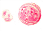
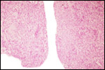
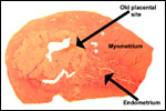
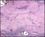
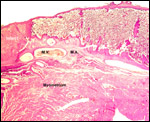
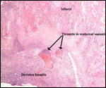
Placenta previa, abruptio placentae, infarcts and stillbirths with fetus papyraceus all have been recorded in rhesus monkeys (Myers, 1972). Indeed, Myers asserted that most of the anomalies or pathologic conditions seen in human placentas may be observed in rhesus monkeys. Endometriosis, adenomyosis and endometrial anomalies are some other pathologic features studied in rhesus monkeys.

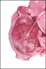
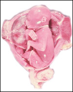
Structural abnormalities of neonatal primates have been studied by Wilson (1972). A variety of congenital anomalies have been identified, but apparently fewer than found in humans. Importantly, the same author clearly identified the cause of phocomelia following a single dose of thalidomide. Several of those animals are depicted in his report.


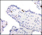
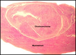
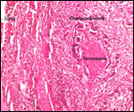
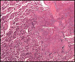
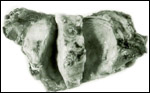
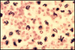
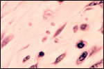
Several extrauterine choriocarcinomas have been reported, even in juvenile catarrhine monkeys. They may have arisen from germ cells or stem cells; alternatively they are associated with a teratoma. Their apparent relative frequency is nevertheless surprising and they can be quite destructive. The last-cited authors have used very modern tools for their exploration. (Farman et al. 2005; Giusti, et al. 2005; Marbaix et al., 2008; Moore et al., 2003; Toyosawa et al. 2000; Yamamoto et al. 2007).


16) Physiologic data

Physiologic data on blood pressure and pulse rate of fetus and mother in pregnant rhesus monkeys under anesthesia were reported by Misenhimer & Ramsey (1970. In addition to that report, a wealth of information has been accumulated in fetal and placental physiology of rhesus gestations. Access to this literature can be achieved through the comprehensive book of Ramsey and Donner (1980). Some information on the vasculature is summarized above.


17) Other resources

The Regional Primate Research Centers of the USA, several foreign research centers, and industrial companies have large holdings of rhesus monkeys. Most are now bred in captivity, very few are being imported. And the zoological parks of the world, of course, house large numbers of these animals and more commonly some of the related species. There is much expertise in breeding and pathology in these agencies.


18) Other remarks - What additional Information is needed?

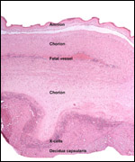
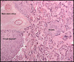
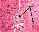
One of the more interesting future studies should be on the regulation of the "X-cells", the extravillous trophoblast. Not only does it achieve the implantation but also, it modifies the blood vessels of the endometrium and produces a specific protein whose function is unknown so far. The means by which MHC is modulated so as to prevent rejection, the exploration of syncytin and syncytium formation will be of interest. Observations on intrauterine mobility and its effect on spiraling (not described in rhesus) of the cord should be made. Do MZ twins occur and how is their placenta structured.


Acknowledgement

Most of the animal photographs in these chapters come from the Zoological Society of San Diego. I appreciate also very much the help of the pathologists at the San Diego Zoo.


References

Arts, N.F.Th. and Lohman, A.H.M.: An injection=corrosion study of the fetal and maternal vascular systems in the placenta of the rhesus monkey. Europ. J. Obstet. Gynecol. Reprod. Biol. 4:133-141, 1974.

Atkinson, L.E., Hotchkiss, J., Fritz, G.R., Surve, A.H., Neill, J.D. and Knobil, E.: Circulating levels of steroids and chorionic gonadotropin during pregnancy in the rhesus monkey, with special attention to the rescue of the corpus luteum in early pregnancy. Biol. Reprod. 12:335-345, 1975.

Bartelmez, G.W.: Cyclic changes in the endometrium of the rhesus monkey (Macaca mulatta). Contrib. Embryol. Carnegie Institution # 227. 34:101-144, 1951.

Beck, A.J. and Beck, F.: The origin of intra-arterial cells in the pregnant uterus of the macaque (Macaca mulatta). Anat. Rec. 158:111-114, 1967.

Benirschke, K. and Kaufmann, P.: The Pathology of the Human Placenta. 4th ed. Springer-Verlag, NY, 2000.

Boyson, J.E., Iwanaga, K.K., Golos, T.G. and Watkins, D.I.: Identification of a novel MHC class I gene, Mamu-AG, expressed in the placenta of a primate with an inactivated G locus. J. Immunol. 159:3311-3321, 1997.

Bronson, R., Volk, T.L. and Ruebner, B.H.: Involution of placental site and corpus luteum in the monkey. Amer. J. Obstet. Gynecol. 113:70-75, 1972.

Bunton, T.E.: Incidental lesions in nonhuman primate placentae. Vet. Pathol. 23:431-438, 1986.

Chez, R.A., Schlesselman, J.J., Salazar, H. and Fox, R.: Single placentas in the Rhesus Monkey. J. med. Primatol. 1:230-240, 1972.

Enders, A.C. and King, B.F.: Formation and differentiation of extraembryonic mesoderm in the rhesus monkey. Amer. J. Anat. 181:327-340, 1988.

Enders, A.C. and Schlafke, S.: Differentiation of the blastocyst of the rhesus monkey. Amer. J. Anat. 162:1-21, 1981.

Enders, A.C., Hendrickx, A.G. and Binkerd, P.E.: Abnormal development of blastocysts and blastomeres in the rhesus monkey. Biol. Reprod. 26:353-366, 1982.

Fa, J.E.: The genus Macaca: a review of taxonomy and evolution. Mamm. Rev. 19:45-81, 1989.

Farman, , C.A.: Benirschke, K., Horner, M. and Lappin, P: Ovarian choriocarcinoma in a rhesus monkey associated with elevated serum chorionic gonadotropin levels. Vet. Pathol. 42:226-229, 2005.

Fitch, H., Coen, R., Lindburg, D., Robinson, P., Hajek, P. Hesselink, J. and Crutchfield, S.: Cerebral palsy in a macaque monkey. Amer. J. Primatol. 14:181-187, 1988.

Galton, M.: DNA content of placental nuclei. J. Cell. Biol. 13:183-191, 1962.

Geissmann, T.: Twinning frequency in catarrhine primates. Human Evolution 5:387-396, 1990.

Giusti, A.M.F., Terron, A., Belluco, S., Scanziani, E. and Carccangiu, M.L.: Ovarian epithelioid trophoblastic tumor in a cynomolgus monkey. Vet. Pathol. 42:223-226, 2005.

Gray, A.P.: Mammalian Hybrids. A Check-list with Bibliography. 2nd edition. Commonwealth Agricultural Bureaux Farnham Royal, Slough, England, 1972.

Gruenwald, P.: Lobular structure of hemochorial primate placentas, and its relation to maternal vessels. Amer. J. Anat. 136:133-152, 1973.

Hendrickx, A.G.: Embryology of the Baboon. University of Chicago Press, Chicago, 1971.

Hertig, A.T., King, N.W. and MacKey, J.: Spontaneous abortion in wild-caught rhesus monkeys, Macaca mulatta. Labor. Animal Sci. 21:510-519, 1971.

Heuser, C.H. and Streeter, G.L.: Development of the macaque embryo. Contrib. Embryol. Carnegie Institution # 181. 29:9-55, 1941.

Hobson, B.M.: Production of gonadotropin, oestrogens and progesterone by the primate placenta. Adv. Reprod. Physiol. 5:67-102, 1971.

Hodgen, G.D., Dufau, M.L., Catt, K.J. and Tullner, W.W.: Estrogens, progesterone and chorionic gonadotropin in pregnant rhesus monkeys. Endocrinology 91:896-900, 1972.

Houston, M.L. and Hendrickx, A.G.: Observations on the vasculature of the baboon placenta (Papio sp.) with special reference to the transverse communicating artery. Folia primatol. 9:68-77, 1968.

King, B.F.: Developmental changes in the fine structure of rhesus monkey amnion. Amer. J. Anat. 157:285-307, 1980.

King, B.J.: Developmental changes in the fine structure of the chorion leave (smooth chorion) of the rhesus monkey placenta. Anat. Rec. 200:163-175, 1981.

Knapp, L.A., Lehmann, E., Piekarczyk, M.S., Urvater, J.A. and Watkins, D.I.: A high frequency of Mamu-A*01 in the rhesus macaque detected by polymerase chain reaction with sequence-specific primers and direct sequencing. Tissue Antigens 50:657-661, 1997.

Knapp, L.A., Cadavid, L.F. and Watkins, D.I.: The MHC-E locus in the most ancient and well-conserved of all known primate class I histocompatibility genes. J. Immunol. 160:180-196, 1998.

Kuroda, M.C., Schmitz, J.E., Barouch, D.H., Craiu, A., Allen, T.M., Sette, A., Watkins, D.I., Forman, M.A. and Letvin, N.L.: Analysis of gag-specific cytotoxic T lymphocytes in SIVmac-infected rhesus monkeys by cell staining with tetrameric MHC Class I/peptide complex. J. Exp. Med. 187:1373-1381, 1998.

Landsteiner, K. and Wiener, A.S.: An agglutinable factor in human blood recognized by immune sera for rhesus blood. Proc. Soc. Exp. Biol. Med. 43:223-224, 1940.

Lewis, J. and Hertz, R.: Effects of early embryectomy and hormonal therapy on the fate of the placenta in pregnant rhesus monkeys. Proc. Soc. Exp. Biol. Med. 123:805-809. 1966.

Lindburg, D.G.: The rhesus monkey in north India: an ecological and behavioral study. Primate Behav. 2:1-106, 1971.

Lindburg, D.G., ed.: The macaques: studies in ecology, behavior, and evolution. Van Nostrand Reinhold, NY, 1980.

Lindsey, J.R., Wharton, L.R., Woodruff, J.D. and Baker, H.J.: Intrauterine choriocarcinoma in a rhesus monkey. Pathol. Vet, 6:378-384, 1969.

Luckett, W.P.: The fine structure of the placental villi of the rhesus monkey (Macaca mulatta). Anat. Rec. 167:141-164, 1970.

Maddox, D.E., Kephart, G.M., Coulam, C.B., Butterfield, J.H., Benirschke, K. and Gleich, G.J.: Localization of a molecule immunochemically similar to eosinophil major basic protein in human placenta. J. Exp. Med.. 160:29-41, 1984.

Marbaix, E., Defrere, S., Ho Minh Duc, K., Lousse J.-C. and Dehoux, J.-P.: Non-gestational malignant placental site trophoblastic tumor of the ovary in a 4-year-old rhesus monkey. Vet. Pathol. 45:375-378, 2008.
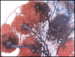
Martin, C.B., Ramsey, E.M. and Donner, M.W.: The fetal placental circulation in rhesus monkeys demonstrated by radioangiography. Amer. J. Obstet. Gynecol. 95:943-947, 1966.

Migaki, G., Benirschke, K., McKee, A.E. and Casey, H.W.: Trichomonal granuloma of the pelvic cavity in a rhesus monkey. Vet. Path. 15:679?681, 1978.

Misenhimer, H.R. and Ramsey, E.M.: The effect of anesthesia and surgery in pregnant rhesus monkeys. Gynec. Invest. 1:105-114, 1970.

Moore, C.M., Hubbard, G.B., Leland, M.M., Dunn, B.G. and Best, R.G.: Spontaneous ovarian tumors in twelve baboons: a review of ovarian neoplasms in nonhuman primates. J. Med. Primatol. 32:48-56, 2003.

Myers, R.E.: The gross pathology of the Rhesus Monkey placenta. J. Reprod. Med. 9:171-198, 1972.

Myers, R.E.: The pathology of the rhesus monkey placenta. Pp. 221-260. In, Symposium on the Use of Non-human Primates for Research on Problems of Human Reproduction. Sukhumi, 1971. WHO Research & Training Centre on Human Reproduction, Karolinska, Sweden, 1972.

Myers, R.E.: Placental pathology. Chapter 85 in, Spontaneous Animal Models of Human Diseases, Vol. 1, E.J. Andrews, B.C. Ward, and N.H. Altman eds., Academic Press, NY, 1979. pp. 208-209.

Naaktgeboren, C. and Wagtendonk, A.M.v.: Wahre Knoten in der Nabelschnur nebst Bemerkungen über Plazentophagie bei Menschenaffen. Z. Säugetierk. 31:376-382, 1966.

Nathanielsz, P.W., Figueroa, J.P. and Honnebier, M.B.O.M.: In the rhesus monkey placental retention after fetectomy at 121 to 130 days' gestation outlasts the normal duration of pregnancy. Amer. J. Obstet. Gynecol. 166:1529-1535, 1992.

Novy, M.J., Aubert, M.L., Kaplan, S.L. and Grumbach, M.M.: Regulation of placental growth and chorionic somatomammotropin in the rhesus monkey: Effects of protein deprivation, fetal anencephaly, and placental vessel ligation. Amer. J. Obstet. Gynecol. 140:552-562, 1981.

Nowak, R.M.: Walker's Mammals of the World. 6th ed. The Johns Hopkins Press, Baltimore, 1999.

Panigel, M.: Comparative anatomical, physiological and pharmacological aspects of placental permeability and haemodynamics in the non-human primate placenta and in the isolated perfused human placenta. In, The Foeto-Placental Unit. Pp.279-295, Excerpta Medica Internat. Congress Series # 183, Milan 1968.

Pepe, G.J. and Albrecht, E.D.: Actions of placental and fetal adrenal steroid hormones in primate pregnancy. Endocrine Reviews 16:608-648, 1995.

Pierce, G.B., Midgley, A.R. and Beals, T.F.: An ultrastructural study of differentiation and maturation of trophoblast of the monkey. Laboratory Invest. 13:451-464, 1964.

Raio, L., Ghezzi, F., DiNaro, M. Franchi, M., Balestreri, D., Durig, P. and Schneider, H.: In-utero characterization of the blood flow in the Hyrtl anastomosis. Placenta 22:597-601, 2001.
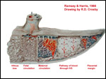
Ramsey, E.M. and Harris, J.W.S.: Comparison of Uteroplacental vasculature and circulation in the rhesus monkey and man. Contrib. To Embryol. # 261. Carnegie Inst. Washington Publ. # 625. Vol. 38:59-70, 1966.

Ramsey, E.M., Martin, C.B., McGaughey, H.S., Kaiser, I.H. and Donner, M.W.: Venous drainage of the placenta in rhesus monkeys: Radiographic studies. Amer. J. Obstet. Gynecol. 95:948-955, 1966.

Ramsey, E.M.: Evaluation of Macaca mulatta as an experimental model for studies of primate reproduction. In, Medical Primatology, part 1, pp. 308-316, 1972. Karger, Basel.

Ramsey, E.M., Houston, M.L. and Harris, J.W.S.: Interactions of the trophoblast and maternal tissues in three closely related primate species. Amer. J. Obstet. Gynecol. 124-647-652, 1976.

Ramsey, E.M., Chez, R.A. and Doppman, J.L.: Radioangiographic measurement of the internal diameters of the Uteroplacental arteries in rhesus monkeys. Amer. J. Obstet. Gynecol. 135:247-251, 1979.

Ramsey, E.M. and Donner, M.W.: Placental Vasculature and Circulation. Anatomy, Physiology, Radiology, Clinical Aspects. Atlas and Textbook. W.B. Saunders Company, Philadelphia, 1980.

Richart, R.: Studies of placental morphogenesis. I. Radioautographic studies of human placenta utilizing tritiated thymidine. Proc. Soc. Exp. Biol. Med. 106:829-831, 1961.

Ruppenthal, G.C., Moore, C.M., Best, R.G., Walker-Gelatt, C.G., Delio, P.J. and Sackett, G.P.: Trisomy 16 in a pigtailed macaque (M. nemestrina) with multiple anomalies and developmental delays. Amer. J. Ment. Retard. 109:9-20, 2004.

Scott, G.B.D.: Comparative Primate Pathology. Oxford University Press, 1992.

Socha, W.W.: Blood groups of nonhuman primates. In, Comparative Biology. Vol. 1: Systematics, Evolution, and Anatomy. Pp. 299-333, A. R. Liss, 1986.

Spatz, W.B.: Nabelschnur-Längen bei Insektivoren und Primaten. Z. Säugetierk. 33:226-239, 1968.

Starck, D.: Vergleichende Anatomie und Evolution der Placenta. Verh. Anat. Gesellsch. 56th Versammlung, Zurich, 1959. Gustav Fischer Verlag, Jena, 1959. Pp.5-26.

Stock, A.D. and Hsu, T.C.: Evolutionary conservatism in arrangement of genetic material. A comparative analysis of chromosome banding between the rhesus macaque (2=42, 84 arms) and the African green monkey (2n=60, 120 arms). Chromosoma 43:211, 1973.

Torpin, R.: Placentation in the rhesus monkey (Macaca mulatta). Obstet. Gynecol. 34:410-413, 1969.

Toyosawa, K., Okimoto, K., Koujitani, T. and Kikawa, E.: Choriocarcinoma and teratoma in the ovary of a cynomolgus monkey. Vet. Pathol. 37:186-188, 2000.

Wagenen, G.v., Catchpole, H.R., Negri, J. and Butzko, D.: Growth of the fetus and placenta of the monkey (Macaca mulatta). 23:23-34, 1965.

Wasmoen, T.L., Benirschke, K. and Gleich, G.J.: Demonstration of immunoreactive eosinophil granule protein major basic protein in the placenta and placentae of non-human primates. Placenta 8:283-292, 1987.

Wasmoen, T.L., McKean, D.J., Benirschke, K., Coulam, C.B. and Gleich, G.J.: Evidence of eosinophil granule major basic protein in human placenta. J. Exp. Med. 170:2051-2063, 1989.

Weiss, G., Weick, F., Knobil, E., Wolman, S.R. and Gorstein, F.: An X-O anomaly and ovarian dysgenesis in a rhesus monkey. Folia primatol. 19:24, 1973.

Wiener, A.S., Moor-Jankowski, J. and Gordon, E.B.: Blood groups of apes and monkeys. V. Studies on the human blood group factors A,B,H and Le in old and new world monkeys. Amer. J. Phys. Anthropol. 22:175-188, 1964.

Wilken, J.A., Matsumoto, K., Laughlin, L.S., Lasley, B.L. and Bedows, E.: A comparison of chorionic gonadotropin expression in human and macaque (Macaca fascicularis). In press, 2001.

Wilson, J. G.: Abnormalities of intrauterine development in non-human primates. Pp. 261-292. In, Symposium on the Use of Non-human Primates for Research on Problems of Human Reproduction. Sukhumi, 1971. WHO Research & Training Centre on Human Reproduction, Karolinska, Sweden, 1972.

Wislocki, G.B. and Streeter, G.L.: On the placentation of the macaque (Macaca mulatta) from the time of implantation until the formation of the definitive placenta. Contrib. Embryol. Carnegie Inst. 27:1-66, 1938.

Yamamoto, E., Ino, K. Yamamoto, T., Sumigama, S., Nawa, A., Nomura, S. and
Kikkawa, F: A pure nongestational choriocarcinoma of the ovary diagnosed with short tandem repeat analysis: case report and review of the literature. Int. J. Gynecol. Cancer 17:254-258, 2007.


back to top

© 2010. All rights reserved.
welcome | home | intro | placentation | glossary | author contact me: kbenirsc@ucsd.edu