(Clicking on the thumbnail images below will launch a new window and a larger version of the thumbnail.)
Kolb's Monkey and other Guenons
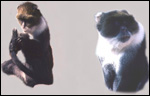
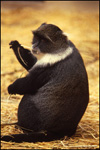
Cercopithecus mitis (kolbi)

Order: Primates
Family: Cercopithecidae

1) General Zoological Data


Of catarrhine African monkeys, the guenons represent a very varied group of primates. They differ much in size, coloration, distribution and cytogenetic characters. Several groups are formally segregated, and the phylogenetic relations are incompletely worked out. Moreover, the taxonomic classification is confusing and has often been revised. The Kolb's monkey is one of these. It is a rarely discussed representative of this group. The Eastern African primate has, therefore, many synonyms. One of these is the "blue monkey", a commonly used designation. The scientific literature now groups Kolb's monkey under Sykes' monkey, or C. mitis (kolbi), which includes, among other less familiar names, the following: C. kolbi, C. albogularis, C. diadematus, C. m. stuhlmanni (Wilson & Reeder, 1992). Others, like Nowak (1999), separate some of these animals into different species with their own denominations, but they may not even list Kolb's monkey. A preliminary, modern phylogeny of guenons was presented only in abstract form by Raaum (2000).

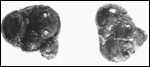
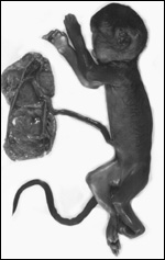
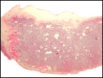
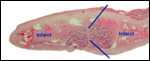
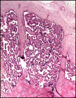
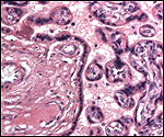
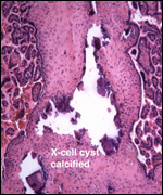
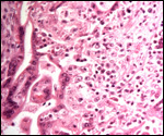
The animals whose placentas are shown here come from the collection of the Zoological Society of San Diego, where reproduction was achieved in the past with Kolb's monkeys. This chapter is based on those pregnancies but also makes reference to some related forms. In addition, I draw on three other placentas of related animal species: the Talapoin money (Miopithecus talapoin), Diana monkey (Cercopithecus diana), Mustached monkey (Cercopithecus cephus), and Mandrill (Mandrillus sphinx).

It should be noted, however, that a separate chapter is devoted to the lesser spot-nosed guenon (Cercopithecus petauritus).

2) General Gestational Data

"Blue monkeys" live in troops in Eastern Africa, have many color variations and a wide distribution throughout those forests. They also vary considerably in size. Guenons breed throughout the year and have birth intervals of about 6 months. Animals observed in the wild by Cords & Rowell (1987), however, had intervals of between 24 and 54 months. Gestation is about 5.6 months long, and females produce their first young when they are about 4-5 years old. Longevity is around 30+ years in captivity. Many species of guenons (in German: "Meerkatzen") are widely represented in zoological gardens. The origin of the designation "guenon" is unknown. Puschmann (1989) has provided much detail concerning their management in zoos, and provided this specifically for a variety of named species. Cycles average 30 days, gestational length is about 160-170 days, and newborns weigh around 400-500 g, depending on the species. In a review of the twinning frequencies, Geissmann (1990) concluded that the sample size studied for most species is too small for definitive conclusions to be reached. He reviewed all reports and concluded that the twinning frequency is probably lower in Old World Monkeys that is that of humans (which is around 1:80 births).

   
  Kolb's monkey (guenon) at San Diego Zoo, 1981.
     
  Other Kolb's monkeys (guenons) at San Diego Zoo.
     
 
3) Implantation

No reports on early implantational stages have been available to me. They are probably similar to those of rhesus monkeys and baboons. These will thus be dealt with in separate chapters.

4) General Characterization of the Placenta

Cercopithecus mitis kolbi:
One of my placentas, shown here, was a bilobed, hemochorial placenta with similarity to that of rhesus monkeys. It weighed 65 g (50 and 15 g per lobe, respectively) and measured 8 x 11 x 1 and 5 x 5 x 1 cm. The cord was marginally inserted but absent in the specimen obtained. The membranes were also absent. Much retroplacental decidua was present. There were marginal infarcts accompanied by fresh hemorrhage. The infant and mother both lived.

Another placenta had a stillborn premature female infant attached, which weighed 167 g. The placenta was bilobed (both lobes were of the same dimension), and had its 13 cm umbilical cord attached at the margin of one lobe. There were large infarcts and much calcification was present in the maternal floor.

A third placenta came from a term pregnancy with a surviving 368 g female neonate. The placenta weighed 35 g and had four cotyledons of different sizes. Again, the umbilical cord and membranes were absent.

Soma (1978) depicted a "guenon" placenta in which the lesser lobe was composed of several smaller, distinct lobules.

   
  Multilobed placenta of Kolb's monkey (maternal aspect on right).
     
  Third trimester fetal demise with bilobed placenta attached. The fetus died in utero. This was perhaps related to the chronic villitis found in the placenta.
     
  Margin of mature placenta, fetal side.
     
  Margin of immature placenta with infarcts.
     
 
The structure of the placenta is very similar to that of other primates, including the human placentas. A significant difference to the latter, however, is the presence of some long stem villous connections between chorion and the base of the placenta, as is shown in the low power view above and indicated by blue arrows and also in the next photographs. The stem villous connections between chorion and maternal floor are shown under higher magnification next.
   
  Two long stem villi connect the chorion with the floor. The left one is artifactually disrupted.
     
 
As is true of many catarrhine monkeys, many placentas have infarcts of the villous tissue. Because of the similarity to the human condition known as pre-eclampsia (toxemia of pregnancy), they have therefore been recommended as potential "animal models" for this common human disease. All of the blue monkey placentas (for species) I have had an opportunity to study, had placental infarcts. Perhaps this is a reason for the high stillbirth rate (24%) referred to above. The placental disease is especially common in the Patas monkey> This is the reason for writing a separate chapter on that species. Further details on toxemia and placental structure can be found in the text by Benirschke & Kaufmann (2000).

5) Details of fetal/maternal barrier

The cercopithecid monkeys all have a typical discoid (or bidiscoid), hemochorial, villous placenta. Maternal blood circulates around the villi, following "injections" of blood from basal decidual arterioles. The exchange of nutrients, water, gases, etc. takes place through the syncytiotrophoblast with its finely microvillous "brush" border. The syncytium is post-mitotic and is replenished continuously from underlying cytotrophoblast that is difficult to identify in H&E preparations. The syncytium is continuously added to until term. In later gestation, the syncytium has foci of apoptosis, and syncytial "knots" flake off into the intervillous space. Large numbers of these syncytial knots are transported into the maternal lung during pregnancy; at least this is so in human gestations where this has been amply documented. Such studies are desirable for other hemochorial placentations.

   
  Mature placental villous tissue of Kolb's monkey. Stem villus on the left. Note the syncytial cover of the villi with protrusion into the intervillous space.
     
 
Miopithecus talapoin, the Talapoin monkey:

I had one term placenta available of a term delivery of a healthy newborn. This was a bilobed placenta weighing 65 g. The eccentric cord was attached to the major disk and measured 16 cm. It had a pronounced right twist. The histological study showed essentially identical features as those described above. There was a minute remnant of allantoic duct epithelium in the cord and a small infarct was present in the villous tissue.

Mandrillus sphinx, the Mandrill:

This placenta had a single disk and weighed 250 g, with an 825 g newborn. It had a centrally inserted 27 cm long umbilical cord, had numerous infarcts of various ages and an abruptio placentae that caused the stillbirth. The mother of this fetus succumbed from tuberculosis.

Cercopithecus cephus, the Moustached guenon:

No details of this gestation are now available, but the histologic preparations show a mature placenta with large infarcts and attachment of the uterus. Representative photos are shown here to illuminate the structure of the placental floor and its myometrial relationship. This placenta also shows the collections of "X-cells" nicely. They form larger aggregates, make small cysts, and are focally markedly calcified. X-cells are now usually referred to as extravillous trophoblast and represent a different line of cells than the villous trophoblast. Calcification of the floor of primate placentas is common. It betrays maturity, does not interfere with transport function, and has no other consequences. It usually takes place in fibrinoid (produced by the X-cells) or in degenerating regions. It should be pointed out that the placenta does not have the capacity to truly "organize" dead tissue (i.e. have ingrowing fibrous tissue to replace infarcts). Infarcts merely shrink and become firma. The low power view of implantation shown above reveals the thin layer of basal decidua remaining.
   
  Implantation site of guenon placenta. At left is an accumulation of "X-cells" with central degeneration and calcification.
     
  One sees the columns of "X-cells" mingle with the decidua basalis and surround a large maternal arteriole.
     
  Decidua basalis of guenon placenta. There is a nearly occlusive thrombus in the maternal spiral artery. It is the reason for villous infarction above. Note the presence of numerous lymphocytes.
     
 
Cercopithecus diana, the Diana monkey:

No details are now available of the pregnancy from this placenta. Histology it is mature and has many infarcts.

6) Umbilical cord

Few reports give adequate information of the umbilical cord length. Spatz (1968), who tabulated much information, found it to be around 12-14 cm long. One of ours, however, was 27 cm long. The cercopithecids have three blood vessels in their umbilical cord, 2 arteries and 1 vein. There are no ducts. The surface is covered by a thin squamous amnion. A moderate amount of Wharton's jelly is present.

7) Uteroplacental circulation

I have to assume that the circulation is similar to that of rhesus monkeys, where Ramsey (1982) and others have studied this is considerable detail. "Spurts" of blood from the maternal arterioles enter, in irregular fashion, into the intervillous space> here, the blood is displaced radially ("smoke rings" in radiography) by the next jet of blood, and it is thus driven back to the maternal floor after having been turned around under the chorionic plate. The arterial walls of the spiral arteries are "modified" in a fashion, which is similar to that described for the human placenta. Perhaps it is even more pronounced, as the vessels are much thicker and larger than the comparable arterioles in human gestations. This vascular alteration is believed to afford certain stiffness to these vessels and render them non-reactive to maternal pressure agents during pregnancy.

8) Extraplacental membranes

The membranes insert at the margin, are composed of amnion, chorion and decidua capsularis. Beneath the chorionic plate is a layer of cytotrophoblast that is commonly markedly vacuolated. I have previously commented upon the absence of atrophied villi in these membranes of catarrhine primates (Benirschke & Miller, 1982). Decidual necrosis is common in the periphery.

   
  Cross-section of rolled membranes. The amnion is artifactually absent (a common feature). Beneath the chorion is a layer of vacuolated normal trophoblast abutting the decidua capsularis.
     
 
9) Trophoblast external to barrier

When the trophoblast columns (largely extravillous cytotrophoblast) attach the decidua basalis, some cells invade the decidua, and specifically also the walls of the maternal arteries. This trophoblast is a specific type. It is now commonly referred to as "extravillous" trophoblast and has formerly been called the "X-cells" (because the origin of the cells was disputed for a long time). These cells make up a large proportion of the trophoblastic shell and have other specific functions. The X-cells produce "MBP" (major basic protein) that is similar to the protein contained in the eosinophilic granulocytes. It occurs in cysts that are often formed by these cells, and is also secreted into the intervillous space. Considerable maternal serum levels are achieved during pregnancy (Wasmoen et al., 1987). In addition, the X-cells produce "fibrinoid". This eosinophilic substance is widely scattered throughout primate placentas and may be mistaken for fibrin - it is not a coagulation product, however.
   
  The implantation site shows anchoring fetal villi and infiltration of the decidua basalis with trophoblast (dark nuclei). Large maternal vein at bottom.
     
  Decidua basalis with a large maternal spiral artery that shows the modification of its wall by trophoblastic infiltration.


10) Endometrium

Normal decidua forms in the second half of the cycle and is present throughout gestation. There is a broad decidua basalis with altered arterioles and trophoblastic ingrowth. There are many lymphocytes and macrophages in the decidua basalis (see below) and a surprising amount of distension of uterine glands (with secretion) can be found.
   
  Guenon placenta attached to myometrium. Note: 1) the large stem villous connection to the floor in the left view (faint arrows); 2) the large, distended endometrial glands; 3) the location of the large maternal artery (A) in the left view. FV=fetal vessel in chorion. D.b.=decidua basalis.
     
  Guenon placenta attached to myometrium. Note: 1) the large stem villous connection to the floor in the left view (faint arrows); 2) the large, distended endometrial glands; 3) the marginal infarct of the placenta shown at left;
FV=fetal vessel in chorion. D.b.=decidua basalis.
     
 
11) Various features

Fairgrieve (1995) described infanticide and infant eating for wild Sykes' monkeys.

12) Endocrinology

The reproductive cycle has been studied by Else et al. (1985). They observed 35 gestations in blue monkeys and determined the cycle length for Kolb's monkey to be 32 days and provided serum levels of estrogen and progesterone during the menstrual cycle. They commented on the absence of seasonality in reproduction (in Kenya) and especially on the frequency of stillbirths (24%).

13) Genetics

A modern study with Giemsa banding of chromosomes was performed by Sineo (1990). He reported C. albogularis and C. mitis to have 72 chromosomes, and C. nictitans had 70 elements. This is in partial agreement with older reports he mentioned. His study indicates that there is considerable inversion polymorphism of chromosomes in the guenons.

Numerous other cytogenetic studies have been done, on many species and subspecies of the Cercopithecidae. Their chromosomal number varies widely. Here is a very brief overview: Allenopithecus nigroviridis 2n=48; Cercopithecus torquatus 2n=42; Cercopithecus aethiops 2n=60; Cercopithecus ascanius 2n=66; Cercopithecus cephus 2n=66; Cercopithecus hamlyni 2n=64; Erythrocebus patas 2n=54; Macaca sp. 2n=42; Cercopithecus talapoin 2n=54; Papio sp. 2n=42 (see Hsu & Benirschke, 1967-1977; Chiarelli & Vaccarino, 1963; Chiarelli, 1973). Bramblett (1992) described several hybrids bred in a colony composed of vervets and Sykes' monkeys. Regrettably, their fertility is not commented upon.

Serum protein polymorphism was studied in vervets and blue monkeys by Turner & Mott (1984). They found relatively little serologic protein polymorphism in vervets; in C. mitis, however, there was a considerable amount.

14) Immunology

No relevant studies are known to me. In general, however, there is always a population of lymphocytes and macrophages in the decidua basalis. This has given rise to much investigation and speculation. Generally speaking, the invading trophoblast, having a different genome, should be recognized immunologically by the mother. Theoretically, this should lead to placental rejection. The reason why this is not the case (and why this may be different in toxemia of pregnancy) has been explained for human pregnancy by the lack of MHC surface antigen on the trophoblast. Haig (1993) has beautifully covered this topic of "conflict", and it is also discussed in Benirschke & Kaufmann (2000). The lymphocytes found in human decidual floors are mostly "Large Granular Lymphocytes".

15) Pathological features

One outstanding feature of many Cercopithecidae is the presence of placental infarcts. This is most pronounced in the Patas monkeys (Please see that chapter). Infarcts of placental tissue differ greatly in appearance. When they are recent, their villi appear to be fused, the intervillous space is absent. The nuclear detail diminishes and karyorrhexis becomes prominent. After several days, infarcts become macroscopically white/yellow and histologically pale and indistinct. Infarcts are usually the result of obstruction of maternal spiral arterioles. One such occluded blood vessel is shown here. It has "atherosis", a term borrowed from the human literature, where the lesion is characteristic of pre-eclampsia ("toxemia of pregnancy"), and is most commonly seen in primiparae.
   
  Spiral arteriole in Kolb's monkey decidua basalis showing partial thrombosis and an alteration of the wall referred to as "atherosis". This is an accumulation of cholesterol-laden macrophages in endothelial and muscular layers.
     
  The lesions shown in the previous picture are responsible for the recent infarct (left in this photograph). This attachment site also shows the collection of trophoblastic "X-cells" and syncytial "knots" on the right side in the intervillous space (IVS).
     
 
Another lesion found in a Kolb's monkey placenta is shown next. In the human placental literature, this is called chronic villitis of unknown etiology (VUE). It is comprised of focal ( or occasionally widespread) infiltration of villi with chronic inflammatory cells. While this was found in one of our animals (Kaplan, 1979), its etiology is obscure. In this case, the fetus was a macerated stillborn and the placenta had additionally numerous hemosiderin-laden macrophages in the chorionic membrane. These commonly arise when there is retroplacental hemorrhage or hemolysis for other reasons.

Hirsch et al. (1993) reported finding a distinct lentivirus in Sykes' monkeys. Munene et al. (1998) reported on the parasites found in catarrhine monkeys. Otsyula et al. (1996) studied antibodies to SIV and STLV in Kenyan primates.

Griner (1983) found a biliary carcinoma in a Diana guenon and referred to a few cases of infectious and parasitic diseases in the animals from the San Diego Zoo.
   
  Chronic villitis (infiltration of the villous core with lymphocytes and macrophages).
     
  Hemosiderin-laden macrophages within the chorionic surface of the same Kolb's monkey placenta.
     
 
16) Physiologic data

I know of no relevant data on guenons. Many exist in rhesus monkeys and will be discussed in the chapter on macacs.

17) Other resources

The Kolb's monkey is not commonly seen in zoological gardens, however, many other species of guenons are exhibited. Cell strains of one male Kolb's monkey are available from CRES.

18) Other remarks - What additional Information is needed?

More data on cord length are desirable. Is there more pathology than we found in our colony? And are the decidual vascular lesions and infarcts due to a condition that is similar to human pre-eclampsia?

Acknowledgement

Most of the animal photographs in these chapters come from the Zoological Society of San Diego. I appreciate also very much the help of the pathologists at the San Diego Zoo.

References

Benirschke, K. and Kaufmann, P.: The Pathology of the Human Placenta. 4th ed. Springer-Verlag, NY, 2000.

Benirschke, K. and Miller, C.J.: Anatomical and functional differences in the placenta of primates. Biol. Reprod. 26:29-53, 1092.

Bramblett, C.: Physical and behavioral phenotype of a vervet/Sykes' hybrid. Amer. J. Physical Anthropol. 27:19 (abstr.), 1992.

CRES: Cell strains from Kolb's monkey and other guenons available through: http://www.sandiegozoo.org/conservation/cres_home.html. Please direct your inquiries to Dr. Oliver Ryder (oryder@ucsd.edu).

Chiarelli, B.: Numerical data on the chromosomes of the Old World primates. J. Hum. Evol. 2:297-300, 1973.

Chiarelli, B. and Vaccarino, C.: Cariologia ed evoluzione nel genere Cercopithecus. Atti Ass. Genet. Ital. 9:329-339, 1963.

Cords, M. and Rowell, T.E.: Birth intervals of cercopithecus monkeys of the Kakamega forest, Kenya. Primates 28:277-281, 1987.

Else, J.G., Eley, R.M., Suleman, M.A. and Lequin, R.M.: Reproductive biology of Sykes and blue monkeys (Cercopithecus mitis). Amer. J. Primatol. 9:189-196, 1985.

Fairgrieve, C.: Infanticide and infant eating in the blue monkey (Cercopithecus mitis stuhlmanni) in the Budongo Forest Reserve, Uganda. Folia Primatol. 64:69-72, 1995.

Geissmann, T.: Twinning frequency in catarrhine primates. Human Evolution 5:387-396, 1990.

Gray, A.P.: Mammalian Hybrids. A Check-List with Bibliography. 2nd edit. Commonwealth Agricultural Bureaux Farnham Royal, Slough, England, 1972.

Griner, L.A.: Pathology of Zoo Animals. Zoological Society of San Diego. San Diego, California, 1983.

Haig, D.: Genetic conflicts in human pregnancy. Quarterly Rev. Biol. 68:495-532, 1993.

Hirsch, V.M., Dapolito, G.A., Goldstein, S., McClure, H., Emau, P., Fultz, P.N., Isahakia, M., Lenroot, R., Myers, G. and Johnson, P.R.: A distinct African lentivirus from Sykes' monkeys. J. Virol. 67:1517-1528, 1993.

Hsu, T.C. and Benirschke, K.: Mammalian Chromosomes Atlas. Springer-Verlag, NY, 10 Volumes, 1967-1977.

Kaplan, C.G.: Intrauterine infection in nonhuman primates. J. Med. Primatol. 8:233-243, 1979.

Munene, E., Otsyula, M., Mbaabu, D.A.N., Mutahi, W.T., Muriuki, S.M.K and Muchemi, G.M.: Helminth and protozoan gastrointestinal tract parasites in captive and wild-trapped African non-human primates. Vet. Parasitol. 78:195-201, 1998.

Nowak, R.M.: Walker's Mammals of the World. 6th ed. The Johns Hopkins Press, Baltimore, 1999.

Otsyula, M., Yee, J., Jennings, M., Suleman, M., Gettie, A., Tarara, R., Isahakia, M., Marx, P. and Lerche, N.: Prevalence of antibodies against simian immunodeficiency virus (SIV) and simian T-lymphotropic virus (STLV) in a colony of non-human primates in Kenya, East Africa. Ann. Tropic. Med. Parasitol. 90:65-70, 1996.

Puschmann, Zootierhaltung. Vol. 2 Säugetiere, VEB Deutscher Landwirtschaftsverlag, Berlin, 1989.

Raaum, R.L., Disotell, T.R., Jolly, C.J., Wildman, D.W., Telfer, P.T. and Teaford, M.: A preliminary molecular phylogeny of the guenons. Amer. J. Phys. Anthropol. Suppl. 30:256-257, 2000.

Ramsey, E.M.: The Placenta. Human and Animal. Praeger Publ. NY, 1982.

Sineo, L.: The banded karyotype of Cercopithecus mitis maesi compared with the karyotype of C. albogularis samango and C. nictitans stampflii. Intern. J. Primatol. 11:541-552, 1990.

Soma, H.: Comparative placentology. Chapter VII. Modern Obstet. Gynecol. Series A. 123-159, 1978 (In Japanese).

Spatz, W.B.: Nabelschnur-Längen bei Insektivoren und Primaten. Z. Säugetierk. 33:226-239, 1968.

Turner, T.R. and Mott, C.S.: Genetic and morphological studies on two species of Kenyan monkeys, Cercopithecus aethiops and Cercopithecus mitis. Intern. J. Primatol. 5:388 (abstr.), 1984.

Wasmoen, T.L., Benirschke, K. and Gleich, G.J.: Demonstration of immunoreactive eosinophil granule major basic protein in the plasma and placentae of non-human primates. Placenta 8:283-292, 1987.

Wilson, D.E. and Reeder, D.A. M., Eds.: Mammal Species of the World. Second Ed. Smithsonian Instit. Press, Washington, D.C., 1992.


back to top

© 2002. All rights reserved.
welcome | home | intro | placentation | glossary | author contact me: kbenirsc@ucsd.edu