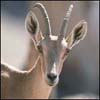
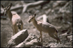
Typical Nubian ibex at San Diego Zoo | ||
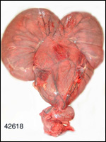
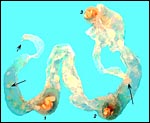
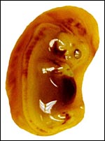
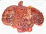
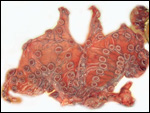
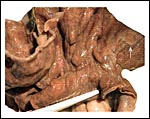
3) Implantation This cotyledonary placenta is epitheliochorial; it is implanted on the rows of caruncles of the typically bicornuate uterus. Mossman (1987) referred to contact of the spherical blastocyst and the mesometrial endometrium at about 15 days and also, that in some bovidae, perhaps the ibex, there is some interdigitation of trophoblast with intercaruncular endometrial glands. We have had the opportunity to study the intact uterus of a female Nubian ibex that had died traumatically in early to mid-pregnancy (San Diego Zoo autopsy # 42618). There were female twins, one occupying each horn but having different directions, head down and up. The whole uterus weighed 3,300 g; after removal of fetuses and placentas it weighed 650 g. The fetuses weighed 300 g each, with a 17 cm Crown-to-Rump length. The placentas weighed 250 g each when all fluid was drained and without umbilical cords. Two corpora lutea were present in the right ovary. The cotyledons were rather flat and arranged in rows. Each horn had 45 cotyledons and corresponding endometrial caruncles. They measured from 5 x 4 cm to a few very tiny cotyledons of 0.5 cm. When they were detached they had an 0.5 cm thickness. There was no associated hemorrhage with the cotyledons. The "partition" of the bicornuate uterus was employed for placentation of both sides. Newborn twins of an additional observations weighed 1,350 g (male) and 550 g (female). A pregnant Nubian ibex died following trauma and had very young triplets in utero. The large allantoic sacs connected to each other but had no vascular connections. They were filled with a jelly-like clear fluid and contained the much smaller amnionic sac that also contained a jelly. At the free ends of the allantoic sac, the tissues were degenerated as expected. The embryos were 2 cm long, but the cotyledons had already detached at autopsy. Interestingly, the left ovary had two, the right ovary one corpus luteum, while two embryos were located in the right uterine horn. The allantoic sacs connected through the uterine connection; the cervix was filled with thick mucus. An additional placenta we saw came from a term gestation, had 35 cotyledons, weighed 132 g, had hippomanes and a 21 cm umbilical cord The East Caucasian Tur (Capra cylindricornis) placenta weighed 400 g, had 38 cotyledons and a 19 cm long umbilical cord. Another, more recently obtained twin placenta of tur weighed 400 g (both together), each measuring 50x20 cm in dimensions and one possessing 39, the other 35 cotyledons. They are shown below. The umbilical cords were 9 and 11 cm long. |
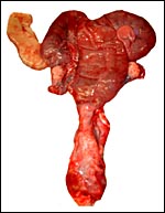
Uterus of pregnant Nubian ibex. | ||
4) General characteristics of placenta This cotyledonary organ had 45 cotyledons, arranged in rows, a large allantoic sac, an epitheliochorial barrier with interdigitation of villi, and a caruncular endometrium. The fetal surface of the cotyledons was finely yellow stippled. The reason for this is unknown. There is no invasive trophoblast. 5) Details of barrier structure As is true for most artiodactyl mammals, this placenta has a typical epitheliochorial relation between trophoblast and endometrium, as is true for most artiodactyl mammals. |
|||||||
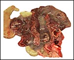
Opened uterus of pregnant Nubian ibex with immature twin fetuses, and placenta still attached. | ||
Uterus of same ibex with placenta and fetuses removed. Note the rows of maternal caruncles. | ||
6) Umbilical cord The cord has a midmesometrial location (Mossman, 1987), possesses four blood vessels and a large allantoic duct. It was not spiraled and measured 9 x 1.5 cm at this gestational age. There were no surface callosities. No hippomanes were present in this specimen and there was no vitelline duct. 7)
Uteroplacental circulation 8)
Extraplacental membranes 9)
Trophoblast external to barrier
10) Endometrium
12)
Endocrinology 13)
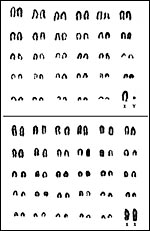
Genetics
15)
Pathological features 16)
Physiological data 17)
Other resources
Corbet, G.B.: The Mammals of the Palaearctic Region: A Taxonomic Review. British Museum (Nat. Hist.), London, 1978. Courrier,
R.: Endocrinologie de la Gestation. Paris, 1945. Griner, L.A.: Pathology of Zoo Animals. Zoological Society of San Diego, 1983. Hsu, T.C. and Benirschke, K.: An Atlas of Mammalian Chromosomes. Vol. 3, Folio, 140. Springer-Verlag, NY, 1969. Mossman, H.W.: Vertebrate Fetal Membranes. MacMillan, Houndmills, 1987. Nowak, R.M. and Paradiso, J.L.: Walker's Mammals of the World, Vol. II. 4th edition. The Johns Hopkins University Press, Baltimore, 1983. Porter, D.G., Heap, R.B. and Flint, A.P.F.: Endocrinology of the placenta and the evolution of viviparity. J. Reprod. Fertil., Suppl. 31:113-138, 1982. Puschmann, W.: Zootierhaltung. Vol. 2, Säugetiere. VEB Deutscher Landwirtschaftsverlag, Berlin, 1989. Stüwe M. and Grodinsky, C.: Reproductive biology of captive Alpine ibex (Capra i. ibex). Zoo Biol. 6:331-339, 1987. |
|||||||||||||||
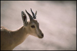
Other views of Nubian Ibex at the San Diego Zoo. | ||
back
to top © 2005. welcome | home | intro | placentation | glossary | author | contact |