(Clicking on the thumbnail images below will launch a new window and a larger version of the thumbnail.)
 

Rock hyrax - Rock dassie (rabbit) - Cony
Procavia capensis

Order: Hyracoidea
Family: Procaviidae


1) General Zoological Data

There are three hyrax genera: Procavia, Heterohyrax, and Dendrohyrax. Varied opinions have been expressed regarding their validity. The three genera, however, were affirmed when Hoeck (1978) studied various anatomical features, importantly the morphology of the penile structure. The rock hyrax (Procavia capensis) that is considered in this chapter extends from Israel and Syria to most of Africa. Taxonomists distinguish four species, as well as several subspecies (Nowak, 1999). South Africans refer to the commonest animal, the Cape hyrax, as the "Klippdachs"; in German it is known as "Schliefer". The name "hyrax" originates from the Greek "hurax", for mouse. Despite their similarity to rodents, they form a distinct, separate Order of mammals. They have a complex developmental history, with many much larger forms representing their ancestors. Now the hyraxes are small, diurnal, browsing animals that live in groups of up to 60 individuals, with a watchful male dominating the group. Dassies, as they are often called, have a dark rump patch. Beneath this patch is a sebaceous gland that is activated in states of alarm and reproduction. The animals weigh up to 4 kg and may live at least to an age of 12+ years in captivity (Fourie, 1978). Since hyraxes are relatively easily maintained in captivity, many zoos have colonies. The hooves of dassies are remarkable in addition to their suction pads that characterize the soft part of the sole. This animal has neither sweat glands nor a gall bladder. Griner (1968) suggested that this species would be a suitable laboratory animal.

Hyraxes also have a specialized tooth morphology, an expanded frontal sinus, short snouts, and two intestinal appendices (ceca). Some of these features, as well as the abdominal location of their testes, have made the animal known to be the closest living relative of the elephant. A closer affinity to Sirenia, however, may be more likely.

Kleinschmidt et al. (1983, 1986) examined the hemoglobin sequences of several mammalian species and thereby identified a monophyletic clade of Paenungulata. This contains Proboscidea, Sirenia and Hyracoidea. Stanhope et al. (1998) examined their nuclear protein coding sequences and mitochondrial DNA. Ozawa et al. (1997) reported on the mitochondrial cytochrome b gene sequences of the extinct mammuth, sea cow, and the extant elephants, hyrax and manatees.

   
  Rock hyrax at San Diego Zoo.
     
  Rock hyrax female with young at San Diego Zoo.


2) General Gestational Data

Hyraxes have the onset of sexual maturity at 16-17 months of age (Glover & Sale, 1968; Murray, 1942). The length of gestation is 205-245 days, with 1-6 young born. Kingdon (1971) suggested a gestational length of 214-225 days. The long gestational period of these animals was thought to be related to their having had much larger ancestors. The average litter size is 3.2, according to Nowak (1999), and the average weight of newborns was 240 g, according to Griner (1968). Soma et al. (1976) deduced from a litter of four offspring that two of these macerated fetuses were monozygotic because there were only three corpora lutea. Sale (1965) also provided data on the length of gestation and neonatal weights.

No placental weights are available. The placenta of the quadruplets shown below measured 9.5 cm in length and 2.5-3 cm in width, with a thickness of 0.5 cm (Soma et al., 1976). The near-term fetuses weighed 107 and 93 g, respectively; the dam had died from gastric ulcers due to nematode infection. Another gestation of a 4 kg female had two fetuses, one of which was macerated. The dam died from nematode gastritis.

Since this original description two placentas have become available and they were weighed. The first comes from a stillborn female fetus (317 g) in which the placenta encircled the mature fetus as shown next. The placenta weighed 19 g and measured 16 x 3 cm; it was 0.3 cm thick, uniformly so. The second placenta was from a term live born and weighed 25.6 g, measuring5.5 x 5.5 x 0.3 cm. The umbilical cords were short and split into their major vessels.

Photograph of the new placenta from a liveborn fetus.


3) Implantation

The earliest stages of implantation have not been described, but the publication by Wislocki & v.d. Westhuysen (1940) provides much insight from their description of eight uteri with fetuses ranging from early limb bud stages to near term. The uterus of the rock hyrax is bicornuate, assuming an Y-shape. Implantations were not necessarily on the side of the corpus luteum and they were occasionally in the body of the uterus, in addition to their location in the uterine horns.

In discussing the structure of the hyrax placenta, these authors drew analogies to the placenta of the manatee and elephant, which they considered to be very similar in structure.

   
  Hyrax placenta of quadruplets, two (bottom) with maceration. Note the ring-shaped (zonary) type of the disks. At the red arrows are the cord insertions.


4) General Characterization of the Placenta

Wislocki & v.d. Westhuysen (1940) have studied the placenta of the rock hyrax especially well. Their contribution needs to be read for detailed information. They dissected various developmental stages and illustrated the placental development extremely well. They also referred to previous reports, and discussed some of their different interpretations of the microscopic findings. The hyrax has a hemochorial, villous, zonary placenta with a maximal width of 2.5 - 3 cm at term. The initially diffuse trophoblastic growth over the entire chorionic sac regresses at the poles during the course of gestation, so that eventually a zonary organ develops.

In early stages of development then, the entire chorion is covered with a thick layer of trophoblast that as yet lacks villous formation. Beneath the chorionic mesoderm of what these authors described as zone I, there is a thin layer of syncytium in which maternal sinusoids are present. Zone II is the broadest region at this stage and consists largely of cytotrophoblast that forms numerous fine channels that carry maternal blood. Zone III is the thin layer of basal, columnar trophoblast, interrupted by maternal venous blood channels. This region shows trophoblastic phagocytic activity, with debris contained within the cells.

Next in the course of development, fetal mesenchymal cells proliferate from the chorionic plate into the trophoblast; they form the initial villi. These are separated from the maternal blood by syncytium and cells with iron content, according to these authors. Thursby-Pelham (1924) interpreted this region differently, as is discussed in this detailed contribution. It is at this stage also, that the trophoblast at the "poles" of the placenta (as opposed to the "girdle") atrophies; villous transformation never takes place here. Degenerating, fusing decidual cells are also present at this location that simulate giant cells.

The final stage of placental transformation is reached when a true zonary shape is attained. The chorionic plate now has the usual fetal vasculature and, beneath the chorion, large maternal blood-filled channels are seen. The main portion of the placenta (representing the original zone I) has now increased in thickness remarkably and makes up 5/6th of the placental thickness. It is composed of parallel, thin villi (trabeculae) that are covered with trophoblast and bathed in sinusoids of maternal blood. While thus zone I expands in width remarkably, zones II and III become much thinner.

   
  The three layers of a nearly mature hyrax placenta, as described by Wislocki & v.d. Westhuysen (1940).
     
  The two stillborn hyrax fetuses (107 and 93 g) of the placenta shown first. The macerated fetuses (two of quadruplets) are shown in the section on Pathology.


5) Details of fetal/maternal barrier

The fine structure of the barrier was primarily studied in Heterohyrax brucei (Oduor-Okelo et al. (1983). These authors found the "barrier" to be hemomonochorial. Only trophoblast and fetal capillary endothelium separated fetal from maternal blood. This contrasted with earlier literature cited by the authors. Since then, however, Dempsey (1969) has also clarified the structure of Procavia capensis as being hemochorial with cellular, rather than syncytial trophoblast next to maternal blood, as was published by the histopathologic study of Wislocki & v.d. Westhuysen (1940). As I have indicated above, these authors divided the full term rock hyrax placenta into three zones: 1) trophoblast vascularized by fetal mesenchyme and arranged in tall columns (trabeculae) - the labyrinth; 2) trophoblast forming vascular channels of maternal blood, without fetal mesenchyme; 3) columnar basal trophoblast contacting decidua.

   
  Border of zones I (above) and II (below left)
     
  Surface portion of zone I with large maternal blood sinuses beneath the chorionic plate and mesenchyme streaming from the chorion into the villi. These are covered with dark trophoblast, largely empty spaces are maternal sinusoids.
     
  Section through the trabecular labyrinth with mesenchyme of villi (F) and maternal blood (M). The cellular trophoblastic surface is somewhat autolyzed in this post mortem specimen. It has been variably interpreted as syncytium or cellular trophoblast.
     
  Another view of the villi (trabeculae) and maternal blood in between.
     
  Zone III with large maternal artery (Wislocki & v.d. Westhuysen considered this to be a vein) infiltrated by trophoblast. Beneath is the degenerating decidua.


6) Umbilical cord

Wislocki & v.d. Westhuysen (1940) stated that the umbilical cord of dassies is "excessively short". They found that the earlier mesenchymal and epithelial caruncles that are present on the amnionic surface diminish with maturation, although squamous plaques remain. The cord contains 2 arteries, 1 vein and the allantoic duct, in addition to many small "vasa propria". There are no spirals. The cords of the two near-term fetuses shown above were 3 cm long, and possessed two arteries, one vein and allantoic duct.


7) Uteroplacental circulation

There are no specific studies.


8) Extraplacental membranes

The initially large yolk sac shrinks early in development and, with advancing gestation, it disappears completely, as does the vitelline duct. The allantoic sac is complex and multi-chambered ("sacculated"), as the illustrations by Wislocki & v.d. Westhuysen (1940) demonstrated. It is vascularized in the portion that is fused with the chorion, not elsewhere. Ultimately it surrounds the amnion completely. The amnion develops initially round caruncles (mesenchymal proliferations), which may later disappear nearly completely. In our specimen of quadruplets shown above, the amnion had numerous plaques. The amnion is not vascularized. There is no decidua capsularis. The decidua has atrophied completely in the second half of gestation.

   
  Mesenchymal nodule on amnionic surface.


9) Trophoblast external to barrier

Trophoblast infiltrates the decidua basalis and surrounds the maternal arteries (not the veins), "almost down to the layer of uterine muscle" (Wislocki & v.d. Westhuysen (1940). There is a proliferation of large, prominent epithelioid cells around these blood vessels whose cellular origin is debated. No myometrial trophoblastic infiltration occurs.


10) Endometrium

The endometrium of early stages of implantation shows frequent pyknotic nuclei and edematous transformation. Glands are inconspicuous, and they do not appear to be secretory. Later in the development, the endometrial cells enlarge and have the appearance of decidual cells as in human gestation.

   
  Floor of the placenta with maternal artery that is surrounded by the large "epithelioid cells" described in the text (? trophoblast). Note the foci of dark blue calcification and, at bottom right, the compacted, degenerating decidua.


11) Various features

In the final stages of placental development, the basal portion of the placenta is composed of multinucleated trophoblast and decidua, with some leukocytic infiltration. There is much debris at this site. The giant cells located here were interpreted to be decidual in origin. Eventually, the decidua basalis becomes fibrous.


12) Endocrinology

Heap et al. (1975) have studied the progesterone metabolism during the hyrax pregnancy. They found low serum levels, not unlike those of elephants. The erythrocytes were found to metabolize progesterone. Kirkman et al. (2001) concluded from their detailed analysis that the progesterone from the corpus luteum was reduced to 5alpha-pregnanes which bind to uterine progesterone receptors and aid in the maintenance of gestation, similar to that observed in the ("related") elephant. This progesterone reduction in the blood of hyraxes had previously also been examined by Makawiti et al. (1991).

The structure of the hyrax ovary is unique, according to Mossman & Duke (1973; see also Kayanja & Sale, 1973) in that it has a very sharp border between cortex and medulla. The medulla has a massively developed interstitial gland that these authors interpreted as being of adrenal origin. They suggested that a close similarity to the ovarian structure of elephants exists. The ovaries of the hyrax are macroscopically also unusual in that they are deeply furrowed and, thus, they are also somewhat similar in appearance to those of elephants. Wislocki & v.d. Westhuysen (1940) showed them in Procavia; and O'Donoghue (1963) depicted the ovaries and uterus of Dendrohyrax arborea ruwenzorii, and described their structure in detail.

It has been suggested that chorionic gonadotropin had been isolated from hyrax placental tissue (Oduor-Okelo et al., 1983 who cited unpublished observations by Bambra & Gombe). No further confirmation exists of that finding. Millar & Aehnelt (1977) developed an antibody to measure, with RIA, the serum and pituitary LH of a variety of mammals. They found it sensitive for rock hyrax studies.


13) Genetics

Hungerford & Snyder (1969), and other authors, have studied the chromosomes of Procavia capensis. The animal has 54 chromosomes, and much of the karyotype is very similar to that of the elephants (2n=56).

Studies of mitochondrial DNA restriction fragment length polymorphism (RFLP) were employed to assess the geographical variation in rock hyraxes (Prinsloo & Robinson, 1992). Two major clades of this species were identified. They were estimated to have separated 2 MYA. They, therefore, suggested that these major differences found between Northern and Southern rock hyraxes are indicative of speciation in what is currently considered to be only one species, Procavia capensis.

Hyraxes are known to live in small rock colonies ("kopjes") that are often separated by larger stretches of savannah. Therefore, a study of their population dynamics was of interest to Gerlach & Hoeck (2001). They studied two species, Heterohyrax brucei and Procavia johnstoni. In the latter species, these investigators found a "surprisingly low allelic diversity". They interpreted the results as due to colonization of a small number of (female) individuals in Heterohyrax. In Procavia johnstoni, there was an excess of homozygotes, suggesting a high rate of inbreeding.

Hybrids of hyraxes have not been reported.


14) Immunology

I know of no studies.


15) Pathological features

The rock hyrax serves as a "stable focus" for Leishmania aethiopica, according to studies by Morsy et al. (1997a,b). Horak & Fourie (1986) showed that dassies in South Africa also commonly harbor ixodid ticks and fleas. Kingdon (1971) stated that large numbers of dassies have died in the wild from bubonic plague.
Tuberculosis was identified in an imported rock hyrax by Cousins et al. (1994). Hyraxes are very susceptible to toxoplasma infection; vegetables, which were infected by cat feces, decimated a colony of different hyrax species in Switzerland.

Bilateral ovariectomy in pregnant hyraxes results in abortion within 48-72 hours (Gombe et al., 1977).

Rehg et al. (1980) identified hemosiderosis ("hemochromatosis") in ten hyraxes and suggested a probably dietary origin of this iron storage (see also Frye, 1982). Only mild hepatic fibrosis resulted. They, as Griner (1968), suggested that this species may be a good laboratory animal. In his book on zoo animal pathology, Griner (1983) reviewed the causes of deaths of 76 dassies. Of these, 41% were related to postnatal problems, and there were eleven stillbirths. Grassenema procavia was a common organism in the stomach and caused gastritis and ulcers. Despite their tractability, long-term survival of colonies has been problematic in dassies even though numerous colonies have existed in various zoos. One case of granulosa cell tumor of the ovary was mentioned. Müller et al. (1976) found the gastric parasite in all three genera of hyraxes.

The twins whose placenta and fetuses are shown in this chapter come from a female that died from gastric ulcers at the end of pregnancy (Soma et al., 1976). The two macerated embryos shown below where enclosed in separate membranes and were assumed to be monozygotic. They weighed 4 g each, and were 1 and 1.6 cm long. Their placentas were 4 cm each and were infarcted.

   
  Two macerated fetuses of quadruplets discussed in text. These were presumed to be monozygotic twins because of the presence of only three corpora lutea in a quadruplet gestation.
     
  Histologic appearance of the placenta shown above, from the macerated fetuses showing massively dilated maternal blood channels and collapsed fetal surface vessels.


16) Physiologic data

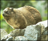
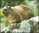
A comprehensive review on physiologic data in rock hyraxes was presented by Rubsamen et al. (1982). McNairn & Fairall (1984) described temperature adaptations of dassies with respect to climate adaptation. The gastrointestinal 
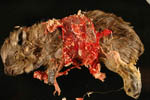
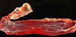
tract of hyraxes, known to be unusual because of the presence of two appendices, was studied by Bjornhag et al. (1994, 1995). The intestinal tract, including their double appendices (ceca), was depicted by Griner (1968).

Hyraxes have nondescended testes; they are "testicond". Their testes are located at the lower renal poles. This anatomic feature was studied by v.d. Schoot (1996) in the fetal development of rock hyraxes. The findings suggested that there was only minimal, partial development of the gubernaculum and the primordia of the cremaster sac were absent.

The study of hyrax brains has disclosed the presence of an unusual, small structure in the aqueduct (Quay, 1971). This is a small ependymal proliferation whose function is unknown.


17) Other resources

The "Frozen Zoo" of CRES at the San Diego Zoo has numerous cell lines of both sexes from rock hyraxes. They may be made available by contacting Dr. O. Ryder at oryder@ucsd.edu.


18) Other remarks - What additional information is needed?

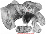
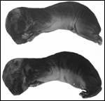
The multiple gestations of hyraxes have always been believed to be multizygotic. The report of Soma et al. (1976) suggested that MZ littermates may occur. More correlation with the number of corpora lutea or RFLP study of DNA differences is warranted. The secretory product of the large "interstitial gland" of the ovaries needs exploration. There are few good studies of umbilical cord (length, structure), and placental weights are lacking.


Acknowledgement

I appreciate very much the help of the pathologists at the San Diego Zoo.


References

Assheton, R.: The morphology of the ungulate placenta, particularly the development of that organ in sheep, and notes upon the placenta of the elephant and hyrax. Phil. Trans. Roy.Soc. B 198:143-220, 1906.

Bjornhag, G., Becker, G., Buchholz, C. and von Engelhardt, W.: The gastrointestinal tract of the rock hyrax (Procavia habessinica). 1. Morphology and motility patterns of the tract. Comp. Biochem. Physiol. A Physiol. 109:649-653, 1994.

Bjornhag, G., Becker, G., Heller, R.. and von Engelhardt, W.: The gastrointestinal tract of the rock hyrax (Procavia habessinica). 2. Fluid flow, production of short chain fatty acids and absorption of water and e electrolytes. Comp. Biochem. Physiol. A Physiol. 111:433-438, 1995.

Cousins, D.V., Peet, R.I., Gaynor, W.T., Williams, S.N. and Gow, B.L.: Tuberculosis in imported hyrax (Procavia capensis) caused by an unusual variant belonging to the Mycobacterium tuberculosis complex. Vet. Microbiol. 42:135-145, 1994.

Dempsey, E.W.: Comparative aspects of the placenta of certain African mammals. J. Reprod. Fertil. Suppl. 6:189-192, 1969.

Fourie, P.B.: The life-span of mammals: estimates for the dassie (Procavia capensis). J. S. Afr. Vet. Assoc. 49:143-145, 1978.

Frye, F.L.: Iron storage disease (hemosiderosis) in an African rock hyrax (). J. Zoo Anim. Med. 13:152-156, 1982.

Gerlach, G. and Hoeck, H.N.: Islands on the plains: metapopulation dynamics and female biased dispersal in hyraxes (Hyracoidea) in the Serengeti National Park. Mol. Ecol. 10:2307-2317, 2001.

Glover, T.D. and Sale, J.B.: The reproductive system of male rock hyrax (Procavia and Heterohyrax). J. Zool. 156:351, 1968.

Gombe, S., Oduor-Okelo, D. and Amoroso, E.C.: Role of progesterone in pregnancy of the hyrax. E. Afr. J. Med. Res. 4:133-134, 1977.

Griner, L.A.: The rock hyrax (Procavia capensis): a potential laboratory animal. Lab. Anim. Care 18:144-150, 1968.

Griner, L.A.: Pathology of Zoo Animals. Zoological Society of San Diego, San Diego, California, 1983.

Heap, R.B., Gombe, S. and Sale, J.B.: Pregnancy in the hyrax and erythrocyte metabolism of progesterone. Nature 257:809-811, 1975.

Horak, I.G. and Fourie, I.J.: Parasites of domestic and wild animals in South Africa. XIX. Ixodid ticks and fleas on rock dassies Procavia capensis () in the Mountain-Zebra National Park. Onderstepoort J. Vet. Res. 53:123-126, 1986.

Hungerford, D.A. and Snyder, R.L.: Chromosomes of the rock hyrax, Procavia capensis (Pallas), 1767. Experientia 25:870, 1969.

Kayanja, F.I. and Sale, J.B.: The ovary of rock hyrax of the genus Procavia. J. Reprod. Fertil. 33:223-230, 1973.

Kingdon, J.: East African Mammals. Vol. I, pp. 329-339. Academic Press, London, 1971.

Kirkman, S., Wallace, E.D., van Aarde, R.J. and Potgieter, H.C.: Steroidogenic correlates of pregnancy in the rock hyrax (Procavia capensis). Life Sci. 68:2061-2072, 2001.

Kleinschmidt, T. and Braunitzer, G.: The primary structure of hemoglobins of the rock hyrax Procavia habessinica, Hyracoidea): insertion of glutamine in the alpha chains. Hoppe Seylers Z. Physiol. Chem. 364:1303-1313, 1983.

Kleinschmidt, T., Czelusniak, J., Goodman, M. and Braunitzer, G.: Paenungulata: a comparison of the hemoglobin sequences from elephant, hyrax, and manatee. Mol. Biol. Evol. 3:427-435, 1986.

Makawiti, D.W., Osaso, J. and Gombe, S.: In vitro metabolism of progesterone by peripheral blood of rock hyrax (Procavia capensis). Gen. Comp. Endocrinol. 83:159-163, 1991.

McNairn, I.S. and Fairall, N.: Metabolic rate and body temperature of adult and juvenile hyrax Procavia capensis. Comp. Biochem. Physiol. A 79:539-545, 1984.

Millar, R.P. and Aehnelt, C.: Application of ovine luteinizing hormone (LH) radioimmunoassay in the quantitation of LH in different mammalian species. Endocrinology 101:760-768, 1977.

Morsy, T.A., al Dakhil, M.A. and el Bahrawy, A.F.: Characterization of Leishmania aethiopica from rock hyrax, Procavia capensis trapped in Najran, Saudi Arabia. J. Egypt. Soc. Parasitol. 27:349-353, 1997a.

Morsy, T.A., al Dakhil, M.A. and el Bahrawy, A.F.: Natural leishmania infection in rock hyrax, Procavia capensis (Pallas, 1766) order: Hyracoidea, trapped in Najran, Saudi Arabia. J. Egypt. Soc. Parasitol. 27:75-81, 1997b.

Mossman, H.W. and Duke, K.L.: Comparative Morphology of the Mammalian Ovary. University of Wisconsin Press, Madison, 1973.

Müller, R., Rüedi, D. and Hörning, B.: Magenparasiten bei Schliefern. In, L.A. Page: Wildlife Diseases. Pp. 69-71. Plenum Press, NY, 1976.

Nowak, R.M.: Walker's Mammals of the World. 6th ed. The Johns Hopkins Press, Baltimore, 1999.

Murray, G.N.: The gestation period of Procavia capensis (dassie). J. S. Afr. Vet. Med. Assoc. 13:27, 1942.

O'Donoghue, P.N.: Reproduction in the female hyrax (Dendrohyrax arborea ruwenzorii). Proc. Zool. Soc. London 141:207-237, 1963.

Oduor-Okelo, D., Musewe, V.O. and Gombe, S.: Electron microscopic study of the chorioallantoic placenta of the rock hyrax (Heterohyrax brucei). J. Reprod. Fertil. 68:311-316, 1983.

Ozawa, T., Hayashi, S. and Mikkelson, V.M.: Phylogenetic position of mammoth and Steller's sea cow within Tethytheria demonstrated by mitochondrial DNA sequences. J. Mol. Evol. 44:406-413, 1997.

Prinsloo, P. and Robinson, T.J.: Geographic mitochondrial DNA variation in the rock hyrax, Procavia capensis. Mol. Biol. Evol. 9:447-456, 1992.

Quay, W.B.: A mid-aqueductal ependymal organ in the brain of the hyrax (Procavia capensis). J. Comp. Neurol. 142:249-253, 1971.

Rehg, J.E., Burek, J.D., Strandberg, J.D. and Montali, R.J.: Hemochromatosis in the rock hyrax. In, R.J. Montali & G. Migaki, eds.: The Comparative Pathology of Zoo Animals. Pp. 113-120, Smithsonian Institution Press, Washington, DC, 1980.

Rubsamen, K., Hume, I.D. and von Engelhardt, W.: Physiology of rock hyrax. Comp. Biochem. Physiol. A 72:271-277, 1982.

Sale, J.B.: Gestation period and neonatal weight of the hyrax. Nature 205:1240-1241, 1965.

Schoot, van der, P.: Foetal genital development in Hyrax capensis, a species with primary testicondia: proposal for evolution of Hunter's gubernaculums. Anat. Rec. 244:386-401, 1996.

Soma, H., Spraker, T. and Benirschke, K.: Macerated monozygotic twins in quadruplet pregnancy of the Cape hyrax (Procavia capensis). J. Mammal. Soc. Japan 6:210-213, 1976.

Stanhope, M.J., Madsen, O., Waddell, V.G., Cleven, G.C., de Jong, W.W. and Springer, M.S.: Highly congruent molecular support for a diverse superordinal clade of endemic African mammals. Mol. Phylogenet. Evol. 9:501-508, 1998.

Thursby-Pelham, D,.: The placentation of Hyrax capensis. Phil. Trans. Roy. Soc. London, Series B. 213:1-20, 1924.

Wislocki, G.B.: On an unusual placental form in the Hyracoidea: its bearing on the theory of the phylogeny of the placenta. Contrib. Embryol. 21:83-95, 1930.

Wislocki, G.B. and van der Westhuysen, O.P.: The placentation of Procavia capensis, with a discussion of the placental affinities of the hyracoidea. Contrib. Embryol. Carnegie Inst. 28:(171)67-88, 1940.


back to top

© 2008. All rights reserved.
welcome | home | intro | placentation | glossary | author contact me: kbenirsc@ucsd.edu