(Clicking on the thumbnail images below will launch a new window and a larger version of the thumbnail.)
 
Sept. 21, 2011.
Lowland Gorilla
Gorilla gorilla

Order: Primates
Family: Pongidae

1) General Zoological Data


Lowland gorillas are an African species. There are local differences in phenotype and genetic makeup that subdivide Eastern and Western gorillas into subspecies, with perhaps additional subspecies. The larger Mountain gorilla placenta has not been studied and none of this endangered species are in captivity. Numerous gorillas have bred in captivity, in various zoos (Cousins, 1976). The largest colony exists in Aspinall's Howletts and Port Lympne Parks, Kent, UK (Aspinall, 1982). In Africa, the principal predator of gorillas is the leopard. Osteomyelitis from such an apparent attack has been reported by Tutin & Benirschke (1991).
   
  Typical term gorilla placenta, maternal surface at left.
Note the long umbilical cord with few twists. Note also the circummarginate insertion of the membranes on the right.
     
 
2) General Gestational Data

Adult females weigh from 70-140 kg (Nowak & Paradiso, 1983). Newborns weigh around 2 kg and have closed skull sutures with thick skull bones. The estrous cycle is 31 days; usually one young is born after 251-295 days of gestation. Twins are uncommon and are usually stillborn or aborted (Rosen, 1972). The newborn weight is about 1.6 to 2 kg at term. Sexual maturity of females is reached at 8 years, and at 11 years in males. In captivity, the lifespan is over 50 years. The placental weight, at term (excluding membranes and cord), is around 350 g. The average placental disk measures approximately 15 x 13 x 2 cm. The membranes (chorion laeve) attach marginally or in a circummarginate fashion. The organ bears great similarity to human placentas, except for its smaller size and the longer umbilical cord. The most recently studied placenta from the "Safari Center" at San Diego Zoo Global was 15 cm in diameter and weighed 225 g. It had the cord knot to be described below.

Wislocki (1932) has described the anatomy and histology of the female reproductive tract, including placentation, in great detail.
   
  Gorilla group at the Wild Animal Park of the Zoological Society of San Diego.
     
  Mother and neonate gorilla at San Diego's Wild Animal Park.
     
 
3) Implantation

As is true of the human placenta, the gorilla placenta implants "interstitially", with decidua surrounding the entire implanted blastocyst. Once the membranes "herniate" into the uterine cavity, the peripheral villi, those on the membrane surface, atrophy. Implantation of the blastocyst onto the other endometrial surface does not occur. Thus, a single discoid organ forms with membranes like those in humans. The placenta is anterior or posterior in its location. A wide margin of membranes separates it from the endocervical canal.

4) General Characterization of the Placenta

Other than for their smaller size, the discoid shape and appearance of the gorilla placenta are very similar to those of human placentas. It is cotyledonary with few, relatively indistinct cotyledonary subdivisions seen on the maternal surface. There are much basal calcification and a considerable amount of fibrinoid material. The barrier is identical to that of humans, it is hemochorial and there is moderately extensive trophoblastic invasion of the endometrium and probably also invasion of the superficial myometrium. Vascular trophoblastic invasion occurs in the decidua basalis. There are large deposits of extravillous trophoblast ("X-cells") and some have cystic centers. The same cells infiltrate into the decidua basalis.
   
  Cyst of extravillous trophoblast ("X-cells")
     
  Term gorilla placental villus. It is much like the human.
     
 
5) Details of fetal/maternal barrier

The surface area of villous structures was determined by Baur (1970). It is similar to that of other ape placentas and humans. The surface is composed of syncytiotrophoblast with underlying cytotrophoblast. The syncytium has brush (microvillous) borders.
   
  Placental floor, villi below, much fibrinoid and mild calcification
     
  More term villi of gorilla placenta.


6) Umbilical cord

The umbilical cord of the gorilla placenta has two arteries and one vein. There are normally no ducts, but remnants of the former allantoic duct occasionally occur in between the two arteries. Compared to human umbilical cords (55 cm), that of the gorilla is unusually long, often as much as 100 cm. The reason for this extraordinary length is unknown. This aspect has been further discussed by Spatz (1968) who related the length of cords to fetal length. The most recently studied placenta had a 185 cm cord length with a true umbilical knot and the neonate did well.

The cord shown in our photograph was 70 cm long and there was an estimated 20 cm still attached to the newborn. The umbilical cord had apparently ruptured spontaneously or it was bitten through by the mother. This is not often observed in apes, nor is placentophagy, as was discussed by Naaktgeboren & Wagtendonk (1966). In our experience, the cord is most frequently marginally inserted in gorillas, but this was not so in the placentas shown here. Ludwig (1961a,b) also found a marginal insertion of a 65 cm long cord and in other umbilical cords that he observed. He considered the specimen depicted to be a "usual" gorilla cord, even though it had a true knot. The female offspring of this gestation did well.

Last placenta seen with true knot and 185 cm long cord.
   
This is the true knot in the cord.
   
This is a section through the knot in the umbilical cord.
   
Another section through the cord knot.
   
Section through the cord away from the knot.
   
The mother with offspring from the mother with long cord and knot.
   
Remnant of allantoic duct in cord.


7) Uteroplacental circulation


This is essentially the same as in human gestations, except that the maternal decidual arterioles usually have thicker walls.
   
  Membranes with chorion at left and decidua at right. Note the thick, hyalinized arterioles.
     
 
8) Extraplacental membranes

Wislocki (1932) and others have shown the presence of a decidua capsularis with remnants of villi in the chorion laeve. The amnion is identical to the human placental amnion, and there is no allantoic membrane.
   
  Membranes with atrophied villus in between decidua (right) and chorion (left), within trophoblast layer.
     
 
9) Trophoblast external to barrier


There are extensive islands of "X-cells" and there is invasion by extravillous trophoblast into the decidua basalis and into the maternal vessels. Whether it also extends into the myometrium, as is true in humans, is unknown.

10) Endometrium

There is decidualization, much like that of human placentation with wandering trophoblast, which invades the decidua basalis.

11) Various features

In all respects this placenta is morphologically like human placentas.

12) Endocrinology

A complete review of hormones and their actions in primate pregnancies has been made by Pepe & Albrecht (1995). Much of this applies to gorilla pregnancies for which only few specific studies have been reported, e.g. Czekala et al. (1983), Watson (1984), Mitchell et al. (1982), and Martin et al. (1977).

Successful birth after embryo transfer was reported by Pope et al. (1997). These authors have provided much endocrine information as well and a comprehensive bibliography.

Chorionic gonadotropin is present during pregnancy, but the total urinary estrogen of gorilla pregnancies is significantly lower than that found in human or chimpanzee pregnancies. The syncytiotrophoblast of the ape placenta produces corticotropin-releasing hormone (CRH) and the serum of pregnant gorillas contains its binding protein (CRH-BP) (Bowman et al., 2001).

13) Genetics

Gorillas, as all large apes, possess 48 chromosomes with extensive homology to human chromosomes. Hybrids have not been described.

We have observed a growth-retarded, phenotypically normal 5 year-old gorilla that has a large deletion of chromosome 3 (at q30). The parents have normal chromosomes (Lear et al., 2001).

Mitochondrial DNA was used to differentiate among subspecies (Garner & Ryder, 1996); and Arnason et al. (1996) used mtDNA to survey patterns of hominid evolution.

DNA from hair samples has enabled paternity diagnosis (Field et al., 1998). Using FISH methodology for 24 specimens of gorilla, Schempp et al. (1998) localized human ribosomal DNA to chromosomes 22 and 23 (corresponding to 21 and 22 in humans).
The numerous publications on gorilla chromosomes and genetics are partially cited in the references below.

14) Immunology

No truly immunological studies of pregnancy are known to us.

15) Pathological features

Numerous reproductive diseases have been described, and a review of gorilla mortalities has been published (Benirschke & Adams, 1980). Examples include placental abruption with stillbirth and presumed preeclampsia equivalent to human gestations, fatally acute amebic meningoencephalomyelitis, tularemia, coccidioidomycosis, nephritis, ruptured aortic aneurysm, and a multitude of ailments similar to those found in humans. Scott (1992) and Griner (1983) have compiled some of this information in books. Other aspects of pathology are summarized in a book, which evolved from a primate meeting in 1985 (Benirschke, ed., 1986). Trophoblastic tumors and hydatidiform moles have not been described. Spontaneous abortions do occur, but their chromosomes have not been studied. Insufficient numbers of few placentas have been studied to eliminate ascending placental infections as a possible disease, as exists in chimpanzees.

In the placenta of abruptio and a few others we have observed, there occur typical infarcts, very similar to those seen in human placentas. Intervillous thromboses and so-called "Tenney Parker changes" (excessive syncytial knotting) also occur. Eclampsia, with a live-born neonate, was reported by Baird (1981); in my opinion, however, that diagnosis is somewhat dubious.

   
  Abruptio placentae with green and yellow
discoloration at left; at right is the underlying
infarct (From Benirschke, 1980).
     
  Term gorilla placental villi with focal necrosis and hemosiderin deposition in villus. The cause is unknown; in human placentas this would be suspicious of former hemorrhage or CMV infection.
     
 
16) Physiological data

Whereas alkaline phosphatase, (the most abundant enzyme of human placentas), is present in placentas of chimpanzees and orangutans, it is extremely low or absent in gorilla placentas (Doellgast et al., 1979, 1981). Information on blood groups and many other physiological data can be found in Benirschke, 1986.

Sonographic estimation of birth size has been advanced by Yeager et al. (1981).

17) Other resources

Cell strains of numerous gorillas and DNA are available from CRES at the Zoological Society of San Diego.

18) Other features of interest

Here follow a few examples of pathology with similarity to human reproductive pathology. Surely, many more of these conditions exist but that information is unpublished. One may ask, why are the umbilical cords of gorillas and chimpanzee twice as long as those of humans? What is the frequency of chromosomal errors at conception? Many comparative questions of this kind remain to be answered.

   
  Spontaneous abortus with hydatid villi lacking
vessels and degenerating decidua. This is essentially
similar to a human spontaneous abortus
due to chromosomal error.
     
  This is another view of the aborted pregnancy. The villi are significantly enlarged (swollen, "hydatid"), but there is still a floor of decidua basalis. Villi contain no vessels and are comparable to what in human pregnancies might be a "partial mole" due to triploidy. No trophoblastic proliferation.
     
  Another, similar spontaneous abortus.
     
  A very young spontaneous abortus with defective embryo in the center of a blood-filled chorion.
     
  Infarct of mature placenta with live infant
Decidual necrosis is on top.
     
  Histologic appearance of the infarcted placental tissue.
     
  Gorilla placenta with fetal vascular latex injection- note marginal cord and one-to-one correspondence of fetal blood vessels.
     
  Knot in umbilical cord of gorilla placenta, at right.
     
  Five year-old male gorilla with growth retardation, presumably due to a large deletion of a portion of one chromosome # 3.
     
  Stillborn gorilla male. Fetal death because of abruptio placentae.
     
 
References

Anderson, M.P., Oosterhuis, J.E., Kennedy, S. and Benirschke, K.: Pneumonia and meningoencephalitis due to amoeba in a lowland gorilla. J. Zoo Anim. Med. 17:87-91, 1986.

Antonius, J.I., Ferrier, S.A. and Dillingham, L.A.: Pulmonary embolus and testicular atrophy in a gorilla. Folia Primatol. 15:277-292, 1971.

Arnason, U., Gullberg, A., Janke, A. and Xu, X.: Pattern and timing of evolutionary divergences among hominoids based on analysis of complete mtDNAs. J. Molec. Evol. 43:650-661, 1996.

Arnheim, N., Krystal, M., Schmickel, R., Wilson, G., Ryder, O. and Zimmer, E.: Molecular evidence for genetic exchanges among ribosomal genes on nonhomologous chromosomes in man and apes. Proc. Natl. Acad. Sci. 77:7323-7327, 1980.

Aspinall, J.: The husbandry of gorillas in captivity. Help (in-house magazine). Spring 1982, pp. 12-17.

Baird, J.N.: Eclampsia in a lowland gorilla. Amer. J. Obstet. Gynecol. 141:345-346, 1981.

Baur, R.: Über die Relation zwischen Zottenoberfläche der Geburtsplacenta und Gewicht des Neugeborenen bei verschiedenen Säugetieren. Z. Anat. Entw.-Gesch. 131:31-38, 1970.

Benirschke, K. ed.: Primates. The Road to self-sustaining Populations. Springer-Verlag, NY 1986.

Benirschke, K. and Adams, F.D.: Gorilla diseases and causes of death. J. Reprod. Fertil. Suppl. 28:139-148, 1980.

Bernstine, J.J.: An epizootic of hydatid disease in captive apes. J. Zoo Anim. Med. 3:16-20, 1973.

Bowman, M.E., Lopata, A., Jaffe, R.B., Golos, T.G., Wickings, J. and Smith, R.: Corticotropin-releasing hormone-binding protein in primates. Amer. J. Primatol. 53:123-130, 2001.

Carter, A.M.: J.P.: Hill on placentation in primates. Placenta 20:513-317, 1999.

Cell strains available from: http://www.sandiegozoo.org/conservation/frozen.html.

Czekala, N.M., Benirschke, K., McClure, H. and Lasley, B.L.: Urinary estrogen excretion during pregnancy in the lowland gorilla (Gorilla gorilla), orangutan (Pongo pygmaeus) and the human (Homo sapiens) Biol. Reprod. 28:289-294, 1983.

Cousins, D.: The breeding of gorillas, Gorilla gorilla, in Zoological Collections. Zool. Garten 46:215-236, 1976.

Doellgast, G.J. and Benirschke, K.: Placental alkaline phosphatase in Hominidae. Nature 280:601-602, 1979.

Doellgast, J., Wei, S.C., Kennedy, M., Stills, H. and Benirschke, K.: Primate placental alkaline phosphatase. FEBS Letters 135:61-64, 1981.
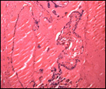
Dutrillaux, B., Rethoré, M-O., Prieur, M. and Lejeune: Term gorilla placenta villus, much like the human, J.: Analyse de la structure fine des chromosomes du gorille (Gorilla gorilla). Comparaison avec Homo sapiens et Pan troglodytes. Humangenetik 20:343-354, 1973.

Dutrillaux, B., Rethoré, M.-O. and Lejeune, J.: Comparaison du caryotype de l'orang-outang (Pongo pygmaeus) a celui de l'homme, du chimpanzé et du gorille. Ann. Génét.: 18:153-161, 1975.

Egozcue, J. and Chiarelli, B.: The idiogram of the lowland gorilla (Gorilla gorilla gorilla). Folia Primatol. 5:237-240, 1967.

Field, D., Chemnick, L., Robbins, M., Garner, K. and Ryder, O.A.: Paternity determination in captive lowland gorillas and orangutans and wild mountain gorillas by microsatellite analysis. Primates 39:199-209, 1998.

Garner, K.J. and Ryder, O.A.: Mitochondrial DNA diversity in gorillas. Molec. Phylogenet. Evol. 6:39-48, 1996.

Griner, L.A.: Pathology of Zoo Animals. Zool. Soc. San Diego, 1983.

Haaf, T. and Schmid, M.: Chromosome heteromorphisms in the gorilla karyotype. Analyses with distamycin A/DAPI, quinacrine and 5-azacytidine. J. Hered. 78:287-292, 1987.

Hamerton, J.L., Fraccaro, M., de Carli, L., Nuzzo, F., Klinger, H.P., Hulliger, L., Taylor, A. and Lang, E.M.: Somatic chromosomes of the gorilla. Nature 192:225-228, 1961.

Hamerton, J.L., Klinger, H.P., Mutton, D.E. and Lang, E.M.: The somatic chromosomes of the Hominoidea. Cytogenetics 2:240-263, 1963.

Henderson, A.S., Atwood, K.C. and Warburton, D.: Chromosomal distribution of rDNA in Pan paniscus, Gorilla gorilla beringei and Symphalangus syndactylus: Comparison to related primates. Chromosoma 59:147-155, 1976.

Lear, T.L., Houck, M.L., Zhang, Y.W., Debnar, L.A., Sutherland-Smith, M.R., Young, L., Jones, K.L., and Benirschke, K.: Trisomy 17 in a bonobo (Pan paniscus) and deletion of 3q in a lowland gorilla (Gorilla gorilla gorilla): comparison with human trisomy 18 and human deletion 4q syndrome. Cytogenet. Cell Genet. 95:228-233, 2001).

Lucas, M. and Wallace, I.: Chromosomes of Gorilla gorilla gorilla. J. Zool. 169:403-407, 1973.

Ludwig, K.S.: Beitrag zum Bau der Gorilla-Placenta. Acta Anat. 45:110-123, 1961a.

Ludwig, K.S.: Ein weiterer Beitrag zum Bau der Gorilla-Placenta. Acta Anat. 46:304-310, 1961.b

Martin, R.D., Kingley, S.R. and Stavy, M. Prospects for coordinated research into breeding of great apes in zoological collections. Dodo#14, pp.45-55, 1977.

Miller, D.A.: Evolution of primate chromosomes. Man's closest relative may be the gorilla, not the chimpanzee. Science 198:1116-1124, 1977.

Miller, D.A., Firschbein, I.L., Dev., V.G., Tantravahi, R. and Miller, O.J.: The gorilla karyotype: chromosome lengths and polymorphisms. Cytogenet. Cell Genet. 13:536-550, 1974.

Mitchell, W.R., Loskutoff, N.M., Czekala, N.M. and Lasley, B.L.: Abnormal menstrual cycles in the female gorilla (Gorilla gorilla). J. Zoo Anim. Med. 13:143-147, 1982.

Morgan, D.G.: Dissecting aneurysm of the aorta in a gorilla. Vet. Rec. 86:502-505, 1970.

Mrasek, K., Heller, A., Rubtsov, N., Triforov, V., Starke, H., Rocchi, M., Claussen, U. and Liehr, T.: Reconstruction of the female Gorilla gorilla karyotype using 25-color FISH and multicolor banding (MCB). Cytogenet Cell Genet. 93:242-248, 2001.

Naaktgeboren, C. and Wagtendonk, A.M.van: Wahre Knoten in der Nabelschnur nebst Bemerkungen uber Plazentophagie bei Menschenaffen. Z. Säugetierk. 31:143-147, 1982.

Nowak, R.M. and Paradiso, J.L.: Walker's Mammals of the World. Vol. I. 4th ed. Johns Hopkins University Press, Baltimore, 1983.

Pepe, G.J. and Albrecht, E.D.: Actions of placental and fetal adrenal steroid hormones in primate pregnancy. Endocrine Reviews 16:608-648, 1995.

Pope, C.E., Dresser, B.L., Chin, N.W., Liu, J.H., Loskutoff, N.M., Behnke, E.J., Brown, C., McRae, M.A., Sinoway, C.E., Campbell, M.K., Cameron, K.N., Owns, O.M., Johnson, C.A., Evans, R.R. and Cedars, M.I.: Birth of a western lowland gorilla (Gorilla gorilla gorilla) following in vitro fertilization and embryo transfer. Amer. J. Primatol. 41:247-260, 1997.

Rideout, B.A., Gardiner, C.H., Stalis, I.H., Zuba, J.R., Hadfield, T. and Visvesvara, G.S.: Fatal infections with Balamuthia mandrillaris (a free-living amoeba) in gorillas and other old world primates. Vet. Path. 34:15-22, 1997.

Robinson, P.T. and Benirschke, K.: Congestive heart failure and nephritis in an adult gorilla. J. Amer. Vet. Assoc. 117:937-938, 1980.

Rosen, S.I.: Twin gorilla fetuses. Folia Primatol. 17:132-141, 1972.

Schempp, W., Zeitler, S. and Rietschel, W.: Chromosomal localization of rDNA in the gorilla. Cytogenet. Cell Genet. 80:185-187, 1998.

Scott, G.B.D.: Comparative Primate Pathology. Oxford University Press, 1992.

Spatz, W.B.: Nabelschnur-Längen bei Insektivoren und Primaten. Z. Säugetierk. 33:226-239, 1968.

Templeton, A.R.: The phylogeny of the hominoid primates: A statistical analysis of the DNA-DNA hybridization data. Mol. Biol. Evol. 2:420-433, 1985.

Turleau C., de Grouchy, J. and Klein, M.: Phylogénie chromosomique de l'homme et des primates hominiens (Pan troglodytes, Gorilla gorilla et Pongo pygmaeus). Essai de reconstruction du caryotype de l'ancêtre commun. Ann. Génét. 15:225-240, 1972.

Tutin, C.E.G. and Benirschke, K.: Possible osteomyelitis of skull causes death of a wild lowland gorilla in the Lopé reserve, Gabon. J. Med. Primatol. 20:357-360, 1991.

Watson, L.M.: Hormone levels and overt social behaviors, including signed output, in a captive lowland gorilla. Zoo Biol. 3:285-306, 1984.

Stanyon, R. and Chiarelli, B. Phylogeny of the Hominoidea: The chromosome evidence. J. Hum. Evol. 11:493-504, 1982,

Wislocki, G.B.: On the female reproductive tract of the gorilla, with a comparison of that of other primates. Carnegie Inst. Contrib. to Embryol. # 135, pp. 163-204, 1932.

Yeager, C.H., O'Grady, J.P., Esra, G., Thomas, W., Kramer, L. and Gardner, H.: Ultrasonic estimation of gestational age in the lowland gorilla: A biparietal diameter growth curve. J.A.V.M.A. 179:1309-1310, 1981.


back to top

© 2002. All rights reserved.
welcome | home | intro | placentation | glossary | author contact me: kbenirsc@ucsd.edu