Last updated:
June 19, 2007.
Giraffe
Giraffa camelopardalis

Order: Artiodactyla
Family: Giraffidae

General Zoological Data


Giraffes are believed to have originated in Africa during the Miocene (Thenius, 1967; Spinage, 1968; Gentry, 2000). This is in contrast to the okapi, the other member of the family Giraffidae. The okapi presumably stems originally from short-necked giraffoid ancestors in Asia. The much lower chromosome number of the giraffes (2n=30, down from 2n=46 in okapis and nilgai) also suggests a later derivation of the giraffes from bovid ancestral stock. Investigation of tooth morphology (Solounias et al., 2000) additionally provides significant differences in the browsing behavior of the giraffe and okapi. There is, however, ongoing discussion of the development of giraffidae in the literature that may ultimately only be resolved by more sophisticated genetic studies (Gatesy & Arctander, 2000).

In the past, giraffes extended over Eurasia but they are now confined to sub-Saharan Africa. They are browsing animals of the open savannah. Many behavioral and structural differences exist between the giraffe and okapi that will also be of interest in the future exploration of their evolution (Gijzen, 1959). Suggestions have, therefore, been made to place these animals into separate subfamilies.

There is only one good species of giraffe, with at least four (some authors suggested eight) subspecies. They have various color and reticulation patterns, largely depending on their location in Africa. Their distinct status as defined subspecies is often challenged; these forms also have often hybridized. Giraffes weigh between 500 and 1,930 kg (Hayssen et al., 1993). Males are larger than females. Neonates are recorded to weigh between 30 and 100 kg.
   
  Masai giraffes at San Diego Zoo.
     
  Masai giraffe at San Diego Zoo.


2) General Gestational Data

Sexual maturity of giraffes is attained at 3 years and 10 months (in captivity), and at 4 years 8 months (in the wild), according to Hall-Martin & Skinner (1978). Calving intervals in captive and wild giraffes was nearly identical, around 20 months. At 24 years of age, female giraffes may still be pregnant. Although Spinage (1968) stated that twins never occurred in this species, this is contradicted by at least two records cited by Hall-Martin & Skinner (1978). Moreover, in June 2003 a set of twins was born at Marwell Zoological Park , UK . One twin did not develop properly and died neonatally. The length of gestation of giraffes was determined to be 433 days in the accurately studied case reported by Deka et al. (1980). Other publications record 400-480 days (Hayssen et al., 1993), and the average is 457 days, according to Skinner & Hall-Martin (1975). Longevity is around 15-20 years in the wild and at least 25 years in zoos.


3) Implantation

Hall-Martin & Skinner (1978) stated that embryos are free, unimplanted, in the uterus for 30 days. The implantation site was always found to be on the side of the corpus luteum. This suggested to these authors that uterine transmigration of the blastocyst does not occur in giraffes. The implanted placenta, however, occupied both uterine horns.


4) General Characterization of the Placenta

The giraffe placenta is polycotyledonary. It is implanted in both uterine horns, with the fetus located in the side of the corpus luteum. Hall-Martin & Skinner (1978) counted the number of cotyledons as 149, 183 and 191 in three pregnancies. The largest cotyledon measured was 25 cm in diameter. Deka et al. (1980) found 178 cotyledons (140 "large", 38 "small"). I found over 100 cotyledons in one placenta with diameters ranging from 8.8 x 8 and 1.7 x 0.8 cm. Another placenta had more than 120 cotyledons. A reticulated giraffe specimen had 140 cotyledons measuring between 2 and 10 cm. Another reticulated giraffe had 155 cotyledons, measuring from 2 to 12 cm. A Baringo giraffe I studied had 125 cotyledons measuring between 2 and 11 cm in diameters. An additional specimen obtained in September, 2003, weighed 5,100 g and had 190 cotyledons. A placenta of a Ugandan giraffe obtained in June, 2004 had 102 cotyledons which were significantly larger in the occupied horn than in the horn that was not occupied by the fetus. Its umbilical cord was 33 cm long. Ludwig (1962), who made the most detailed description of the giraffe placenta, did not enumerate the cotyledons of his two specimens. An additional specimen obtained in March, 2005 weighed 3,220 g and had only 47 large cotyledons. Its male neonate died shortly after birth from unknown causes. Its testes were 4.3 and 4 g. The cord had four vessels and was 20 cm long.

The following weights of giraffe placentas are available:

Deka et al. (1980): 2,850 g
   
My own observations: 4,400 g
4,900 g
5,900 g
3,850 g
4,880 g
3,220 g
   
Reticulated giraffe 3,800 g
Reticulated giraffe 5,100 g
Baringo giraffe 5,450 g
Uganda giraffe 5,100 g
Uganda giraffe 5,375 g

The maximal expansion of the opened sac was 156 cm in the gravid horn and 135 cm in the nongravid horn of the placenta described by Deka et al. (1980); maximum breadth was 56 cm. In the placenta here shown, the diameters were 181 x 35 cm. In another Masai giraffe placenta that I examined, the expansion was 181 x 40 cm. It had 155 cotyledons in four rows, with considerable difference in sizes. The reticulated giraffe specimen measured 160 cm in greatest length, 70 cm across and had a large allantoic sac.

A Uganda giraffe placenta obtained in March 2004 weighed 5,100 g, measured 187 x 48 cm and had 95 cotyledons in four rows.

Ludwig (1962) depicted the (formalin-fixed) placental cotyledons of Owen's paper (1849). These figures suggested that the cotyledons have surface indentations, thus having the appearance of beans. This is not pronounced in fresh placentas of my experience.

In July, 2004 I obtained the pregnant uterus from a Ugandan giraffe that had died from aspiration, following the “bloat”. The 190 g male fetus was in the left horn, the side that also contained the large corpus luteum in its ovary. The placenta extended into the right horn as well, but the cotyledons were remarkably smaller in that horn. The left horn contained 60 cotyledons, the right had 50. The cord was 12 cm long, had a decidedly right spiral and was studded with diminutive foci of squamous metaplasia. There was abundant light yellow allantoic and amnionic fluid present, and the fetal bladder was filled with urine as well.

   
  Fetal surface of a portion of a term, delivered giraffe placenta with cord insertion site.
     
  Maternal surface of a portion of giraffe placenta at term. There are four somewhat irregular rows of cotyledons of great size difference.
     
  Term Masai Giraffe placenta with surviving female newborn - 155 cotyledons in four rows. Insertion of umbilical cord at arrow.
     
  Term Masai Giraffe placenta with surviving female newborn - 155 cotyledons in four rows. Insertion of umbilical cord at arrow.
     
  Giraffe uterus with 190 g conceptus in left horn. Ovaries are at arrows.


Uterus before opening the fetal sacs.

     
  Opened uterus with fetus in left horn; the sac of the right horn is not yet opened.
     
  The 190 g fetus with short but twisted cord.


5) Details of fetal/maternal barrier

The giraffe has an epitheliochorial, villous placenta. Hradecky et al. (1987), in comparing giraffe and okapi placentas, made the point that the number of cotyledons and structure of placental villi are sufficiently different to negate successful interspecific embryo transfer. The giraffe's placental villi are long, thin, and sparsely branched, and the villi are of different lengths according to Ludwig (1962) who dissected individual villi. He showed them to branch at least twice and identified their flat, rather than round shapes. In contrast to other placentas from the bovid lines, there are only very few trophoblastic binucleate cells, a finding also commented upon by Ludwig (1962). They are concentrated at the tips of villi. Mossman (1987) was tentative about their occurrence. The binucleate cells are situated between the more cuboidal, single-layered trophoblast. In addition to the few binucleate cells, occasional trophoblastic cells have markedly enlarged, hyperchromatic nuclei. They simulate binucleate cells in all other features. The fetal capillaries often project in between the trophoblastic epithelium, as was also pointed out by Ludwig. Since no implanted placenta has been available, no information can be provided concerning the relationship to the maternal caruncles. The trophoblast layer is usually single-layered, except beneath the chorionic plate. Here, it is often bilayered and may have hemosiderin inclusions. The external surface has microvilli. Glycogen was found in the amnion, but in no other regions (Ludwig, 1962). Electronmicroscopic studies were reported by Reissig (1977). The placenta he studied had 167 cotyledons, which were of brown color on the maternal side. He described the region of areolae as having small folds. The trophoblast was cuboidal and had microvillous surfaces. These stained for alkaline phosphatase. Binucleated cells were very rarely found, and these had very sparce organellar contents. The trophoblast did possess numerous cytolysosomes and, at their basal surface, the cells had much infolding that created a labyrinth. The protruding capillaries caused the formation of villous surfaces that were devoid of nuclei.

   
  Partial cotyledon of delivered giraffe placenta. The maternal surface is ragged, as it withdrew from the caruncle. Note the straight and minimally branched villi. Fetal surface vessels above, within the chorion.
     
  Edge of giraffe cotyledon. Arrow is at the continuation of the chorion between cotyledons.
     
  Term giraffe placental surface with focal pigment deposition beneath the chorionic plate.
     
  Edge of cotyledon of giraffe placenta. At right top, the chorion and trophoblast extend over the intercotyledonary region and the areolae.
     
  Chorion at right, trophoblast over the intercotyledonary region (left) with areolar proteinaceous fluid (uterine milk).
     
  Binucleate trophoblastic cell at tip of villus. Note superficial location of fetal capillaries.


The placenta of the immature gestation described above (190 g fetus) had 110 cotyledons. They were significantly smaller in the unoccupied sac. The histology is not much different from that of the term gestation but there was absolutely no pigment beneath the chorionic plate. Binucleate cells were very rarely found. The relationship to the maternal endometrial surface is nicely shown in the photographs.

Implanted cotyledon in the young placenta described above. The Endometrium has few glands and very prominent, thick-walled blood vessels.
   
Higher magnification of previous implantation site.
   
Villus (center) between maternal endometrial tufts. Note the huge endometrial nuclei.
   

Reflections on trophoblastic pigment : Although I have stated several times that the trophoblast possesses hemosiderin inclusions, this is far from certain when reviewing other ungulate placentas. I had never stained the giraffe placentas but have now and there is no hemosiderin in the granular inclusions shown next. Thus, are they iron-containing or are we wrong in this general assumption of this route being a way for iron to be absorbed by the fetus? In other sections of this book I have suggested that this may actually be melanin, perhaps liberated from the skin. Granted, it is in big chunks and has the superficial appearance of hemosiderin and not that which we consider to be melanin in melanocytes. But, bleaching with hydrogen peroxide abolishes the pigment (as it does melanin), it stains with silver (as does melanin) and is iron-stain negative. It also does not stain for bilirubin, the only other pigment that one might consider. Thus, at present I think we must consider that it is perhaps melanin that is accumulated from “melanemia” perhaps from rubbing the skin and liberating the pigment. Besides, if this region of pigmentation really did serve as an important organ to transfer iron to the fetus, as is suggested by the term “hemophagous organ”, then one should surely expect it in immature placentas as well. It is not present in this case shown here.

Pigment in cylindrical trophoblast below the chorionic surface. See “reflections on the nature of this pigment” immediately ahead.


6) Umbilical cord

The umbilical cord inserts in the mid-portion of the two horns. Hradecky (1983) described the length of the cord as 140 cm in one specimen. One of my placentas had a 50 cm umbilical cord with four vessels and large allantoic duct. Two other umbilical cords were 35 cm long with a diameter of 3.5 cm, and 28 x 9 cm. Two arteries were fused at midpoint of the longer cord. Numerous small blood vessels are also present, some surround the allantoic duct. The surface has many keratinized plaques that were also referred to by Hradecky. He mentioned that they extend to the adjacent amnion (see figure below). The cord has no spirals and no remnants of vitelline structures.

Ludwig (1962) described the vascular structure more extensively. He depicted the difference found in elastic fiber distribution of artery and vein. Major villous vessels lacked an elastica.

   
  Allantoic duct in center of umbilical cord. Large vessel edge at right. Numerous small vessels are present and the allantoic duct has a muscular coat.
     
  Allantoic duct of the immature placental umbilical cord.
     
  Squamous nodule on the surface on immature umbilical cord.


7) Uteroplacental circulation

This has not been investigated.


8) Extraplacental membranes

Areolae were reported to exist by Mossman (1987). These are located in the intercotyledonary regions and represent discrete areas within the epithelial cover. There is a concentration of fetal chorionic vessels in this region. Areolae were first described and named by Eschricht (1837). They are now considered to be specific regions of protein absorption and transfer from mother to fetus. (See the chapter on Sheep). Ludwig (1962) depicted their macroscopic feature in the giraffe placenta. The giraffe has a large, tubular allantoic sac. No vitelline remnants are present at term. There is no decidua capsularis.

   
  Amnion near the insertion of umbilical cord with squamous pearl (verruca) at right.
     
  Edge of cotyledon with intercotyledonary membrane spanning to the right.
     
  Trophoblast of membrane at left; allantoic epithelium at right.


9) Trophoblast external to barrier

There is no invasive trophoblast.


10) Endometrium

The endometrium has the usual caruncles that are illustrated here in a neonate.

   
  Low power view of neonatal giraffe uterus with four caruncles (C=caruncles)
     
  Fetal uterus with caruncle at top and glandular endometrium below.


11) Various features

No attached giraffe placenta has been described. Thus, it is unknown exactly what the floor of the implantation site might show. It is unlikely, however, that a subplacenta or metrial glands exist.

The cervix of the pregnant giraffe with the 190 g fetus. It was very tough, and the thick cervical mucus can be seen exuding from the central os.


12) Endocrinology

Gombe & Kayanja (1974) determined that the progesterone levels rose during the second half of pregnancy. Hall-Martin & Rowlands (1980) interpreted that finding to support their contention that corpora lutea occurred in fetal life and persisted throughout youth. They suggested that these structures occurred non-cyclically. Their discussion opposed the view of Mossman & Duke (1973). These authors referred to the publication by Kellas et al. (1958) that demonstrated similar findings. Mossman & Duke suggested that, more likely, the stimulated follicles were corpora atretica with theca luteinization rather than true corpora lutea. I am also very skeptical that true corpora lutea can develop in fetal ovaries and also interpret these features as representing luteinized follicles and luteinized corpora atretica. It is highly unlikely that fetuses ovulate in utero; on the other hand, a minor degree of follicular maturation is frequent in many fetuses, albeit without such massive endocrine stimulation, and certainly without ovulation.

Luteinizing hormone (LH) is believed to stimulate the theca/granulosa cells. But the origin of this LH in the fetal giraffe is unknown. In view of the scarcity of placental binucleate cells, and absence of another indication that the placenta is active in an endocrine sense, the origin of LH would be more likely the fetal pituitary gland. The fetal pituitary has no unusual histological appearance, however, nor is it especially large. The persistence of these structures in neonatal life also suggests a fetal, rather placental origin of the stimulating LH. Other endocrine interactions may also be at play. Thus, the unusually large fetal adrenal gland of giraffes needs to be considered.

In addition to these luteinized atretic follicles, the interstitial tissue of the ovaries is also markedly stimulated, consisting of epithelioid cells with much cytoplasm. Since Hall-Martin & Rowlands (1980) provided no pictures of these unusual ovarian structures in fetal giraffes, I attach some photographs that provide examples of the fetal ovarian morphology. Hall-Martin & Rowlands (1980) also discussed the former allegation that the placenta produced gonadotropins; they found them to be absent, however. Neonatal testes have no evidence of interstitial cell stimulation, nor do the testes of the very immature fetus shown above. As mentioned, the adrenal gland appears to be unusually large (weights are not available). It has an unusually wide zona fascicularis.

It may here then be of interest to note that okapi newborns do not share this ovarian stimulation. Moreover, the adrenal fascicularis is considerably thinner than that of giraffes and involutes to some degree at birth.

Loskutoff et al. (1986) have studied the urinary progesterone excretion in pregnant and post partum giraffes. They found marked elevation of progesterone during pregnancies in giraffe and okapi and saw a moderate decline prior to delivery. They speculated that some of this progesterone may come from the luteinized fetal ovaries. Since okapis do not possess these follicles and the steroid hormonal profile is similar to that of giraffes, an origin of progesterone from fetal ovaries seems to be an unlikely event. They also drew attention to the interaction of adrenal steroids, luteinizing hormone and ovarian activity that needs to be considered to explain these unusual features of fetal ovarian activity in giraffes. An earlier unconfirmed report of placental gonadotropins was mentioned in their discussion. More work is needed.

Estrous cycles occur every 14+ days, and last 24 hours (Hall-Martin & Skinner, 1978). The sections of ovary from a female giraffe dam that I had occasion to examine (in pregnancy) had a corpus luteum in addition to several luteinized cystic follicles.

   
  Neonatal giraffe ovary with three adjacent luteinized ovarian follicles (probably atretic follicles). In the very center is a fibrous scar of one.
     
  Low-power view of neonatal giraffe ovary with immature oocyte mantle and stimulated follicles below.
     
  Higher power view of a similar area as the previous photograph. Note the large, luteinized theca/granulosa cells. The deep red cytoplasm of these cells suggests early involution.
     
  Luteinization of the "interstitial cells" of a giraffe neonatal ovary (arrows point to the collections of epithelioid cells.)


Fetal ovary of stillborn giraffe with apparent corpora lutea.


The other ovary of the same stillborn giraffe fetus.

     
  The two ovaries of the pregnant giraffe with 190 g fetus. The left ovary has the very large corpus luteum, the right has a small luteinized old follicle.


13) Genetics

All types of giraffes have 30 chromosomes; 28 elements are metacentric, except the smallest pair that is an acrocentric element (Taylor et al., 1967; Hösli & Lang, 1970; Koulisher et al., 1971). Numerous hybrids have been produced among the various subspecies (Gray, 1972). Most are apparently fertile. Vermeesch et al. (1996) identified the telomeric repetitive DNA by FI SH (fluorescent in situ hybridization) in the interstitial portions of the fusion chromosomes. This finding, as other evidence, supports the notion of the chromosomal evolution in this family by chromosomal fusion.

Karyotype of female giraffe with 30 chromosomes.


14) Immunology

No studies are known to me.


15) Pathological features

Numerous external and internal parasites plague the life of giraffes (Spinage, 1968). Fowler (1978) reviewed the causes of acute deaths in giraffes from many institutions. Unexplained "peracute" deaths may be secondary to poor nutrition combined with "stress". Bovine tuberculosis is a serious threat to this species in zoological parks; other diseases were also reviewed. Focal calcification of the placentas has been reported.

Lang (1955) described a premature delivery, followed by inattention of the dam that fractured the leg of the newborn. The neonate was successfully reared but, because of the non-healing fracture, that calf was euthanized age 6 months. The calf weighed only 30 kg at birth, and was born 11 months after last breeding.

In several placentas that I have been able to observe I found focal villous degenerations. Sometimes there was thrombosis associated with this necrosis, at other times there was calcification and considerable pigment deposition. The pigment, similar to that in the cylindrical subchorionic trophoblast (see above) was iron-stain negative. The placentas appeared otherwise normal and were associated with term, live births. Perhaps this is a “normal” finding.

   
  Villus from normal giraffe placenta with extensive hemosiderin deposit around a thrombosed blood vessel.
     
  Area of old degenerative change in a large villus (arrows) of an otherwise normal term giraffe placenta.


Similar villous degeneration of a reticulated giraffe placenta.


Higher magnification of degenerating villus of term reticulated giraffe with yellow pigment of focal mineralization.


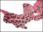
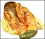
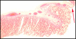
In July, 2003, we had an abortion in a Baringo giraffe. The dam went spontaneously into labor and was known to be ill. The structurally normal male fetus weighed only 19 kg and the small placenta weighed 1,300 g. Most remarkably, it had only 23 cotyledons that were yellow-red and appeared infected. The cord was 34 cm long. Pictures are shown next.

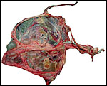
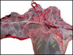
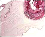
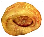
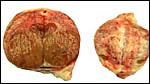
Fetal surface of aborted giraffe fetus' placenta.


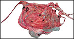
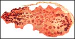
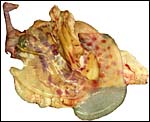
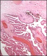
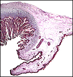
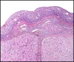
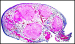
Maternal surface of the same placenta showing the yellow/red cotyledons.


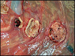
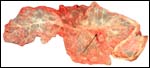
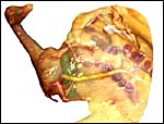
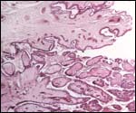
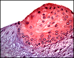
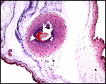
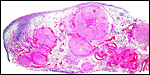
Three apparently degenerating, yellow-discolored cotyledons.


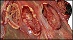
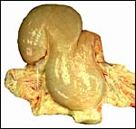
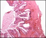
Three additional discolored cotyledons.


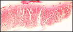
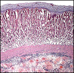
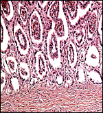
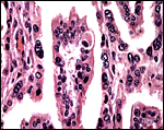
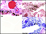
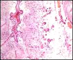
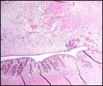
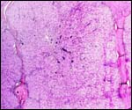
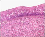
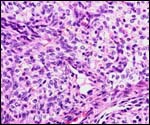
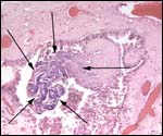
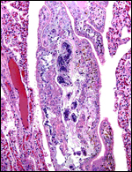
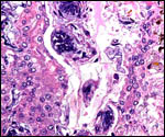
Touch preparations from the cotyledonary surface yielded very long bacilli, with some similarity to actinomyces. Sections of umbilical cord, membranes, fetal lung and testis were all normal and not inflamed or degenerated. The cotyledons, however, had a largely necrotic surface and the same long organisms were identified. Post partum the dam has a vaginal discharge but, while on antibiotics, seems reasonably healthy. Sections are shown next. I speculate that the dam must have had a preexisting endometritis that prevented normal cotyledons to form so that she ended with only 23 and very small cotyledons. That the fetus grew to become 19 kg in size is remarkable. The only similar histology I have encountered is that of a dromedary (see that chapter) in which a stillborn had a largely calcified villous surface that has great similarity to this placenta.

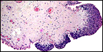
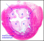
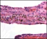
Low power view of cotyledonary/villous surface in the Baringo giraffe abortus from San Diego Zoo's Wild Animal Park . The surface is largely necrotic but there is only minimal inflammation and part of the cotyledonary trophoblast (left) is still intact.


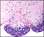
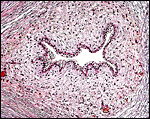
The same cotyledon under higher magnification. There is only minimal inflammation.


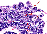
In between cotyledons debris of this kind is found with the red arrows pointing to some of the many long bacilli.


Murai et al. (2007) have described a large (20x36x20 cm) teratoma of the umbilical cord of a reticulated giraffe two months before birth; the dam died during gestation.


16) Physiologic data


Many physiologic data, such as hematologic values, blood pressure, and respiration were compiled by Spinage (1968), and also by Warren (1974). Cerebral blood pressure is of normal proportion, and the mechanism of prevention of foot edema, often discussed in the literature, is probably due to a difference in the structure of blood vessels and connective tissue.


17) Other resources

Cell lines of many subspecies are stored in the Frozen Zoo of CRES at the San Diego Zoo. They can be made accessible by contacting Dr. O. Ryder (oryder@ucsd.edu).


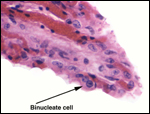
18) Other remarks - What additional Information is needed?

No implanted placenta has been examined, thus not much is known about the fetal/maternal interface. Examination of the fetal/maternal interface would thus be of interest. Since there are so few binucleate trophoblastic cells, it would also be interesting to know if placental lactogen is produced. Estrogen determinations during pregnancy would be helpful. Finally, the status of the endocrine stimulus for the fetal corpora lutea or luteinized fetal atretica follicles needs clarification.


Acknowledgement

I appreciate very much the help of the pathologists at the San Diego Zoo in collecting specimens.


References

Deka, B.C., Nath, K.C. and Borgohain, B.N.: Clinical note on the morphology of the placenta in a giraffe (Giraffa camelopardalis). J. Zoo An. Med. 11:117-118, 1980.

Eschricht, D.F.: De organis, quae respirationi et nutritioni foetus mammalium inserviunt; anniversaria (Schultzianis, Havniae 1837). Quoted by Ludwig, 1962.

Fowler, M.E.: Peracute mortality in captive giraffe. JAVMA 173:1088-1093, 1978.
Gijzen, A.: Das Okapi. (Neue Brehm-Bucherei). A. Ziemsen Verlag. Wittenberg Lutherstadt, 1959.

Gatesy, J. and Arctander, P.: Molecular evidence for the phylogenetic affinities of ruminantia. Chapter 9, pp. 143-170, in Vrba, E.S. and Schaller, G. B., eds. Antelopes, Deer, and Relatives. Yale University Press, New Haven, 2000.

Gentry, A.W.: The ruminant radiation. Chapter 2, pp. 11-25, in Vrba, E.S. and Schaller, G. B., eds. Antelopes, Deer, and Relatives. Yale University Press, New Haven, 2000.

Gombe, S., and Kayanja, F.I.B.: Ovarian progestins in Masai giraffe (Giraffa camelopardalis). J. Reprod. Fert. 40:45-50, 1974.
Gray, A.P.: Mammalian Hybrids. A Check-list with Bibliography. 2nd edition. Common-
wealth Agricultural Bureaux Farnham Royal, Slough, England, 1972.

Hall-Martin, A.J. and Rowlands, I.W.: Observations on ovarian structure and development of the southern giraffe, Giraffa camelopardalis giraffa. S. Afr. J. Zool. 15:217-221, 1980.

Hall-Martin, A.J. and Skinner, J.D.: Observations on puberty and pregnancy in female
giraffe (Giraffa camelopardalis). S. Afr. J. Wildl. Res. 8:91-94, 1978.

Hayssen, V., van Tienhoven, A. and van Tienhoven, A.: Asdell's Patterns of Mammalian Reproduction: a Compendium of Species-specific Data. Comstock/Cornell University Press, Ithaca, 1993.

Hösli, P. and Lang, E.M.: A preliminary note on the chromosomes of the Giraffidae: Giraffa camelopardalis and Okapia johnstoni. Mammalian Chromosomes Newsletter 11:109-110, 1970.

Hradecky, P.: Placental morphology in African antelopes and giraffes. Theriogenology 20:725-734, 1983.

Hradecky, P., Benirschke, K. and Stott, G.G.: Implications of the placental structure compatibility for interspecies embryo transfer. Theriogenology 28:737-746, 1987.

Kellas, L.M., van Lennep, E.W. and Amoroso, E.C.: Ovaries of some foetal and prepuberal giraffes [Giraffa camelopardalis (Linnaeus)]. Nature 181:487-488, 1958.

Koulisher, L., Tijskens, J. and Mortelmans, J.: Mammalian Cytogenetics. V. The chromosomes of a female giraffe. Acta Zool. Pathol. Antverp. 52:93-96, 1971.

Lang, E.M.: Frühgeburt und künstliche Aufzucht einer Giraffe. Schweiz. Arch. Tierheilk. 97:198-205, 1955.

Loskutoff, N.M., Walker, L., Ott-Joslin, J.E., Raphael, B.L. and Lasley, B.L.: Urinary steroid evaluations to monitor ovarian function in exotic ungulates: II. Comparison between the giraffe (Giraffa camelopardalis) and the okapi (Okapia johnstoni). Zoo Biol. 5:331-338, 1986.

Ludwig, K.S.: Beitrag zum Bau der Giraffenplacenta. Acta anat. 48:206-223, 1962.

Mossman, H.W.: Vertebrate Fetal Membranes. MacMillan, Houndmills, 1987.

Mossman, H.W. and Duke, K.L.: Comparative Morphology of the Mammalian Ovary. The University of Wisconsin Press, 1973.

Murai, A., Yanai, T., Kato, M., Yonemaru, K., Sakai, H. and Masegi, T.: Teratoma of the umbilical cord in a giraffe (Giraffa camelopardalis reticulata). Vet. Pathol. 44:204-206, 2007.

Owen, R.: Notes on the birth of the Giraffe at the zoological Society's gardens, and description of the foetal membranes and of some of the natural and morbid appearances observed in the dissection of the young animal. Trans. Zool. Soc. Lond. 3:21-28, 1849. Quoted by Ludwig, 1962.

Reissig, D.: Elektronenmikroskopische Untersuchungen am Chorionepithel der Giraffenplacenta. Verh. Anat. Gesellsch. 71:493-498, 1977.

Skinner, J.D. and Hall-Martin, A.J.: A note on foetal growth and development of the giraffe Giraffa camelopardalis giraffa. J. Zool. 177:73-79, 1975.

Solounias, N., McGraw, W.S., Hayek, L.-A. and Werdelin, L.: The paleodiet of the giraffidae. Chapter 6, pp. 84-95, in Vrba, E.S. and Schaller, G. B., eds. Antelopes, Deer, and Relatives. Yale University Press, New Haven, 2000.

Spinage, C.A.: The Book of the Giraffe. Houghton Mifflin Company, Boston, 1968.

Taylor, K.M., Hungerford, D.A. and Snyder, R.L.: The chromosomes of four artiodactyls and one perissodactyl. Mammalian Chromosomes Newsletter 8:233-235, 1967.

Thenius, E.: Die Giraffen und ihre Vorfahren. Kosmos 63:160-164, 1967.

Vermeesch, J.R., DeMeurichy, W., van den Berghe, H., Marynen P. and Petit, P.: Differences in the distribution and nature of the interstitial telomeric (TTAGGG)n sequences in the chromosomes of the giraffidae, okapi (Okapia johnstoni), and giraffe (Giraffa camelopardalis): evidence for ancestral telomeres at the okapi polymorphic rob(4;26) fusion site. Cytogenet. Cell Genet. 72:310-315, 1996.

Warren, J.V.: The physiology of the giraffe. Scientific American 231:96-105, 1974.


back to top

© 2007. All rights reserved.
welcome | home | intro | placentation | glossary | author contact me: kbenirsc@ucsd.edu