 |
|
(Clicking
on the thumbnail images will launch a new window and a larger version
of the thumbnail.) |
| January 1, 2008 |
Litocranius walleri
Order: Artiodactyla
Family:Bovidae
1) General Zoological Data
Despite the fact that gerenuks have an unique and well-known appearance, relatively little is known about the biology of these beautiful animals. A comprehensive book by Schomber (1966) provides all of the information available. There is no fossil record of precursors. The East African animals form a discontinuous range, with the smaller habitat in Tanzania, the majority further north, Kenya, Somalia, etc. Whether they extended at one time to Egypt seems to have been documented by drawings from many thousand years ago. The name Gerenuk stems from the Somali garanug meaning long-necked gazelle (Gotch, 1979). Lithos is Greek for stone, and kranion the skull - "stone skull". The posterior portions of the skull are said to be very hard. It is the only species in this genus, mostly inhabiting Somalia and surrounding counties. Two subspecies have been considered: L. w. walleri and L. w. sclateri, animals that have minor skeletal differences. The German "Giraffengazelle" well describes the extraordinarily long neck of these animals that are prone to become even longer when they graze on trees by standing on their hind legs. Horns are found in males only. Jones (1993) found the longevity to be 11+ years, while Crandall (1965) reported only 6+ years. Relatively few zoos have gerenuks, but they have reproduced well in most zoological gardens that have held them. Their number in Africa has been reduced drastically.
 |
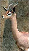
Male Gerenuk at San Diego Zoo. |
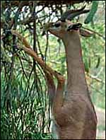 |
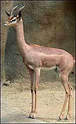
Typical feeding posture of Gerenuk at San Diego Zoo. |
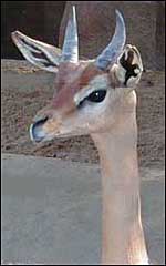 |
Male Gerenuk head at San Diego Zoo. |
Leuthold (1978) described the social organization and behavior of gerenuks. His estimate of the length of gestation was 6.5-7 months. Single young are born that weigh around 3 kg (Puschmann, 1989). Schomber (1963, 1966) reported a 203 day length of gestation from observations in zoological gardens.
3)
Implantation
Early stages of gerenuk placentation have not been described. I have had
available a single uterus of a dam from which the somewhat immature fetus
had been delivered by Cesarean section. The placenta had remained attached;
the1.24 kg male fetus was too immature to be viable. Histologic sections
of its kidneys show continued glomerulopoiesis that indicates immaturity
by at least one month. The uterus was bicornuate and the placenta occupied
both uterine horns. It had 80+ cotyledons that measured between 1 and
3 cm in diameters. Because of its attachment to the uterus, the placenta
was not weighed. Characteristically, the endocervix was filled with thick
mucus indicating that labor had not ensued. The umbilical cord was 5 cm
long.
This pregnancy came from a 65 kg dam that was 3 years old. She had fractured
her neck when frightened by a mandrill that had entered the enclosure.
Because of her late gestation, a Cesarean section was performed. The dam's
tissues were mildly autolyzed, thus the histologic sections are less well
preserved than are those of my usual placentas. The tips of the subchorionic
maternal endometrial tufts show quite extensive degeneration and focal
hemorrhages.
4) General Characterization of the Placenta
This is a typical polycotyledonary placenta, similar to that of most other
ungulates. It has an epitheliochorial relation to the maternal tissues.
There is no trophoblastic invasion of the uterus. The endometrium has
normally-configured glands beneath a layer of dense stroma upon which
the cotyledons are located. In between the cotyledons, the same appearance
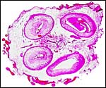
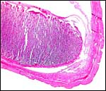
prevails, there being no discrete areolae. Another placenta was received in 2006 (S-6203) It measured 51x22x0.5 cm, had 48 cotyledons and a cord of 12.7 cmx0.3 cm. A representative placenta with its 48 cotyledons arranged in four rows is shown next. It comes from an animal that died with dystocia at term. Its umbilical cord was 15 cm long.
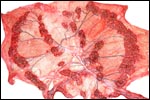 |
Near term gerenuk placenta with 48 cotyledons. |
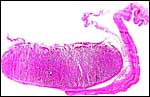 |
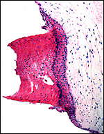
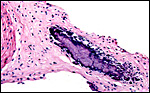
Implanted cotyledon of gerenuk. |
 |
Relation of central portion of cotyledon to maternal uterus below. |
 |
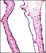
Enlarged fibrous tip of maternal septum with foal degenerative changes, lying between villi and beneath the pigmented subchorionic trophoblast. |
The villous epithelium consists of a single layer of trophoblast with numerous binucleate cells that are primarily located at the villous tips. Beneath the chorion, the trophoblast is more cylindrical and has a moderate amount of yellow pigmentation. The endometrial epithelium if flat and directly apposed to the trophoblast.
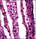 |
In the center is a slender villus with layer of trophoblast (one binucleate cell) in between maternal endometrial septa. |
The umbilical cord of this gestation was 5 cm long, had no spirals and had numerous squamous plaques on its surface. There were a central allantoic duct, four large blood vessels and numerous small vessels, especially concentrated around the duct. That of a term gestation was 15 cm long.
 |
Cross section of umbilical cord with central allantoic duct that is surrounded by numerous small blood vessels. The amnionic surface has numerous squamous plaques. |
 |
Keratinized squamous plaque on umbilical cord surface. |
This has not been described and, by the appearance of this uterus, it follows the general architecture of the ungulate uterine circulations.
8)
Extraplacental membranes
A minute focus of calcification was found in the surface chorionic membrane
of one cotyledon that, judging from analogy of other ungulate placentas
is likely to be the remains of a former yolk sac. The amnion is avascular,
while numerous blood vessels are present in the allantoic membrane. The
allantoic sac was large and lined by columnar epithelium.
 |
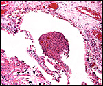
Amnion and vascularized allantois. |
 |
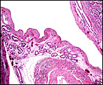
Margin of implanted cotyledon. This shows the bright red surface endometrium surrounding the entire cotyledon. |
 |
Focal area of calcification in chorion, possibly the remains of the yolk sac. |
There is no evidence of trophoblast infiltration of the uterus other than the surface of the fibrous-appearing superficial endometrium.
10)
Endometrium
There is no true decidua, the endometrial stroma, other than the glands,
has a very fibrous appearance during pregnancy. The fibrous superficial
portion of the endometrium surrounds the entire cotyledons, to reach the
underside of the chorion.
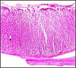
 |
Intercotyledonary endometrium with endometrial glands and myometrium in center. Cotyledons on both sides. |
There is no subplacenta.
12)
Endocrinology
Wasser et al. (2000) studied stress responses in gerenuk and other species
by measuring fecal glucocorticoid excretion with specific antibodies to
quantify cortisol metabolites. They found a typical stress response on
ACTH challenge. I no of no endocrine studies of pregnancy.
.
13) Genetics
The gerenuk has a very "primitive" karyotype, consisting of
2=60 acrocentric elements (Wurster & Benirschke, 1968). Superficially
the karyotype is identical to that of Bos taurus. No hybrids have
been described.
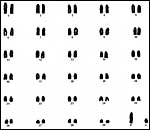 |
Representative karyotype of male gerenuk. |
I know of no studies of the gerenuk immune system other than the identification of Trypanosome brucei antibodies in gerenuks by Black et al. (1999). Backues et al. (1999) vaccinated many species, including the gerenuk, with a genetically engineered Mengo virus.
15)
Pathological features
Gerenuks are shy animals and easily frightened. Thus, Griner (1983) reported
myocardial infarcts as causes of death, and the animal discussed here
died from a fractured neck incurred by running into a wall because of
fright.
16)
Physiologic data
Clemens & Maloiy (1983, 1984) studied the digestive physiology of
a wide variety of African ruminants. They found a somewhat unusual gastrointestinal
composition in gerenuks, suggesting that they needed special attention;
these animals had the greatest fluid absorption in the colon. Wasser et
al. (2000) studied stress responses in gerenuk and other species by measuring
fecal glucocorticoid secretion with specific antibodies to measure cortisol
metabolites. They found a typical stress response on ACTH challenge.
17)
Other resources
Cell strains of fibroblasts are available by contacting Dr. Oliver Ryder
at the CRES
department of the San Diego Zoo (oryder@ucsd.edu),
18) Other remarks - What additional Information is needed?
Endocrine data are virtually absent and knowledge of early stages of implantation
would be useful.
Acknowledgement
The animal photographs in this chapter come from the Zoological Society
of San Diego. I appreciate also very much the help of the pathologists
at the San Diego Zoo.
References
Backues, K.A., Hill, M., Palmenberg, A.C., Miller, C., Soike, K.F. and
Aguilar, R.: Genetically engineered Mengo virus vaccination of multiple
captive wildlife species. J. Wildl. Dis. 35:384-387, 1999.
Black, S.J., Wang, Q., Makadzange, T., Li, Y.L., van Praagh, A., Loomis, M. and Seed, J.R.: Anti-Trypanosoma brucei activity of nonprimate zoo sera. J. Parasitol. 85:48-53, 1999.
Clemens, E.T. and Maloiy, G.M.: Digestive physiology of East African wild ruminants. Comp. Biochem. Physiol. A 76:319-333, 1983.
Clemens, E.T. and Moloiy, G.M.: Colonic absorption and secretion of fluids, electrolytes and organic acids in East African wild ruminants. Comp. Biochem. Physiol. A 77:51-56, 1984.
Crandall, L.S.: Record of African antelopes in the New York Zoological Park. Int. Zoo Yearb. 5:52-55, 1965.
Gotch, A.F.: Mammals - Their Latin Names Explained. Blandford Press, Poole, Dorset, 1979.
Griner, L.A.: Pathology of Zoo Animals. Zoological Society of San Diego, San Diego, California, 1983.
Jones, M.L.: Longevity of ungulates in captivity. Intern. Zoo Yearbk. 32:159-169, 1993.
Leuthold,
W.: On social organization and behaviour of the gerenuk Litocranius
walleri (Brooke 1878). Z. Tierpsychol. 47:194-216, 1978.
Puschmann, W.: Zootierhaltung. Vol. 2, Säugetiere. VEB Deutscher
Landwirtschaftsverlag Berlin, 1989.
Schomber, H.W.: Beiträge zur Kenntnis der Giraffengazelle (Litocranius walleri Brooke, 1978) Säugetier. Mitt. 11:1-44, 1963.
Schomber, H.W.: Giraffengazelle und Lamagazelle. Die Neue Brehm-Bücherei. A. Ziemsen, Wittenberg, 1966.
Wasser, S.K., Hunt, K.E., Brown, J.L., Cooper, K., Crockett, C.M., Bechert, U., Millspaugh, J.J., Larson, S. and Monfort, S.L.: A generalized fecal glucocorticoid assay for use in a diverse array of nondomestic mammalian and avian species. Gen. Comp. Endocrinol. 120:260-275, 2000.
Wurster, D.H. and Benirschke, K.: Chromosome studies in the superfamily Bovoide. Chromosoma 25:152-171, 1968.