 |
| (Clicking
on the thumbnail images will launch a new window and a larger version
of the thumbnail.) |
| Last updated: Sept 4, 2006. |
Oryx gazelle gazella
Order: Artiodactyla
Family: Bovidae
1) General Zoological Data
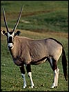
Of the three African Oryx species (Oryx dammah, Oryx gazella, and Oryx leucoryx), this Cape oryx' is perhaps still the most numerous, with an estimated 75,000 still in the wild (Nowak, 1999). Moreover, numerous zoos have breeding colonies. Gotch (1979) derived the common name from the German for Gemse =chamois and the Dutch bok =Buck. Another oryx subspecies (Oryx gazelle callotis), the fringe-eared oryx is considered in a separate chapter. It has many similarities to this species but has 58 chromosomes, vs . 56 in this animal. The longevity of the Gemsbok is listed as being 19 years (Weigl, 2006). Kumamoto et al. (1999) have discussed evolutionary relationships among the oryxes.
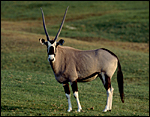 |
Adult Gemsbok at San Diego Zoo's Wild Animal Park. |
2) General Gestational Data
The gestation is about 8.5 months according to Nowak (1999). Birth weight of the singletons is ~12 kg.
3) Implantation
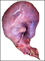
This specimen comes from a dam that died whilst giving birth. The head and feet of the fetus were extruding, but the fetus had been dead for some time before; and then the dam died. In this specimen, the corpus luteum and implantation were on the right side of the uterus; the left horn was completely unoccupied. This is the usual finding in pregnant Oryx species. It is a most unusual implantation because the major portion of the placenta was separated from the uterus by a large blood clot. Moreover, some portions of the cotyledons were much more degenerated than the rest, as will be seen. They were essentially bloodless and much compressed. No physical abnormality was found at autopsy, however.
This is a polycotyledonary placenta with approximately 90 cotyledons arranged in four rows. Implantation had occurred only in the right horn; the left horn was completely empty.
4) General Characterization of the Placenta
This placenta had largely separated by a huge retroplacental clot that had apparently caused the fetal demise. Because I did not separate the placenta from the uterus, its weight was not determined. Together the placenta and uterus weighed 3.05 kg. The female fetus had an 83 cm CR length and weighed ~9 kg. The cotyledons measured between 2 and 7 cm in width and 1 cm in thickness; the entire placenta was 95x28 cm in size. The pregnant dam weighed 198.5 kg.
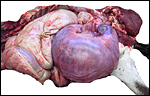 |
Pregnant right uterine horn in the abdomen. |
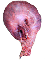 |
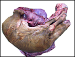
Unopened uterus from the right side with feet presenting from the cervix. |
 |
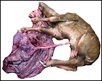
Unopened uterus from the left side with (pale) left uterine horn in the middle of the picture. |
 |
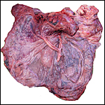
Uterus opened with cervix at to right. |
 |
After removal of the fetus the insertion of the umbilical cord is shown. |
 |
Placenta with central cord insertion whose four vessels are shown. |
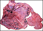 |
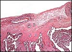
After removal of the membranes (right), the retroplacental hematoma (dark red) is seen behind the much flattened dead villi. |
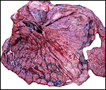 |
Higher magnification of the dead villi and retroplacental hematoma at left bottom. |
 |
The endometrium of the left uterine horn (right) is markedly edematous. The intramyometrial hemorrhage behind the detached placenta is at left. |
5) Details of fetal/maternal barrier
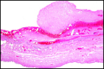
This is a polycotyledonary, epitheliochorial placenta with scattered binucleate trophoblast. The placenta intersects with endometrial caruncles that are laid out in roughly four rows. Invasion of the endometrium was not observed. Beneath some areas of the chorionic plate, pigmented cells of the so-called hemophagous organ' were found. The macrophages contained some rectangular refringent crystals, perhaps representing hematoidin.
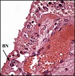 |
Villous surface with distended fetal vessel (right) and binucleate trophoblast at arrow. |
 |
The villi arise from the chorionic plate above in which the major fetal vessels course. |
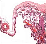 |
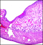
This section is from the edge of a cotyledon with membranes extending left to the next cotyledon. |
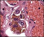 |
Collections of pigmented cells were found beneath the chorionic plate in several small regions, representing the hemophagous' organ. |
6) Umbilical cord
The umbilical cord measured 35 cm in length and had no caruncles. It had four vessels and a large allantoic duct. There were no spirals or other unusual features.
7) Uteroplacental circulation
This has not been studied.
8) Extraplacental membranes
The thin amnion was studded with numerous very small, white foci of squamous metaplasia. One such region is shown below. There was no decidua capsularis' in this specimen; most of the membranes were separated from the myometrium by blood clot. There was a large, vascularized allantoic sac that was lined by tall epithelium.
 |
The amnion attached to the chorion loosely, here shown over a large fetal vessel. |
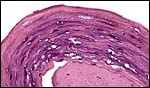 |
This is one small focus of squamous metaplasia of the amnion. |
9) Trophoblast external to barrier
There was none in this specimen of an implanted placenta.
10) Endometrium
The endometrium of the non-gravid horn was very edematous, but it had no decidual change. The gravid horn endometrium was largely replaced by relatively recent blood clot. Interestingly, some iron-positive staining hemosiderin macrophages were present near vessels, indicating older retroplacental bleeding to have occurred.
11) Various features
No unusual features other than the hemorrhage were identified.
12) Endocrinology
I am not aware of any specific studies.
13) Genetics
This Cape oryx' has 56 chromosomes with a fixed 2/17 fusion'. Kumamoto et al. (1999) have made detailed studies of oryx species and found that there is moderate Robertsonian fusion polymorphism among these species that, however, has not led to hybrid sterility. Gray (1972) also lists a number of hybrids occurring in zoos with Beisa oryx, O. gazella beisa , one of the subspecies, and they were also fertile. Kumamoto et al. (1999) also reviewed the possible speciation mechanism.
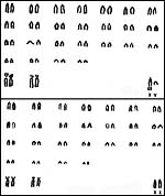 |
Karyotypes of male and female Gemsboks. (From Hsu & Benirschke, 1968). |
14) Immunology
I am not aware of any studies.
15) Pathological features
This fetus apparently died with/from dystocia and it is thus interesting to learn that Griner (1983) also reported a dystocia in this species, with uterine rupture and maternal peritonitis. In the present case, however, retroplacental hemorrhage must have occurred some time before labor commenced as many cotyledons were so compressed and degenerated, while only 2/3 of the placental mass was still viable. There was no evidence of any structural reason for the retroplacental hemorrhage, nor were there wounds from possible trauma. Nevertheless, some iron pigment was found, indicating some length of time of the separation.
Other pathology: Tuberculosis in O. gazella beisa has occurred in a zoo (Lomme et a., 1976). Flach et al. (2002) found serologic evidence of herpesvirus infection in captive oryx. Li et al. (2003) found serologic evidence of malignant catarrhal fever virus infection and identified a new virus. Goossens et al. (2005) surveyed various artiodactyls at Belgian zoos and identified various parasites that were detailed in their report. In New Mexico , Bender et al. (2003) found a high seroprevalence of malignant catarrhal fever virus, blue-tongue virus, respiratory syncytial virus, and parainfluenza virus. Kirkwood & Cunningham (1994) identified scrapie (spongiform encephalopathy) in a kudu, gemsbok, scimitar-horned oryx and in a nyala in Great Britain . Tapeworm infection has been reported from Namibia (Bohrmann, 1990). Boomker et al. (2000) have also reported on parasite infection in South African ungulates, including the gemsbok.
Kumamoto et al. (19999) reported on an aneuploid gemsbok with 2n=57,XXY.
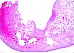
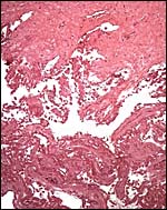 |
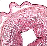
This is a section through one of the many completely infarcted - or compressed dead cotyledons. |
 |
The separation of cotyledon from the myometrium is shown here. The hematoma often also extended into the myometrial layers. |
 |
The non-gravid horn with edematous endometrium at right; the detached placenta at left. |
16) Physiologic data
The basic hematological values were published by Pospisil et al. (1984). Protocols for successful anesthesia were provided by Grobler et al. (2001); moreover, Ebedes (1975) has given detailed information on capture and translocation of gemsbok in Africa . Maloney et al. (2002) implanted data loggers' and measured brain and arterial blood temperatures in free-ranging gemsbok in Africa and concluded that the carotid rete does not protect the brain during hyperthermia.
17) Other resources
Cell lines of various oryx species are stored at CRES of the Zoological Society of San Diego and can be made available by contacting Dr. Oliver Ryder at oryder@ucsd.edu .
18) Other remarks What additional Information is needed?
Information on earlier stages and implanted placentas of living fetuses would be helpful to have.
Acknowledgement
The animal photograph in this chapter comes from the Zoological Society of San Diego.
References
Bender, L.C., Li, H., Thompson, B.C., Morrow, P.C. and Valdez , R.: Infectious disease survey of gemsbok in New Mexico . J. Wildl. Dis. 39:772-778, 2003.
Bohrmann, R.: Coenurus in the muscle of a gemsbok (Oryx gazella). Vet. Parasitol. 36:353-356, 1990.
Boomker, J., Horak, I.G., Watermeyer, R. and Booyse, D.G.: Parasites of South African wildlife. XVI. Helminths of some antelope species from the Eastern and Western Cape Provinces . Onderstepoort J. Vet Res. 67:31-41, 2000.
Ebedes, H.: The capture and translocation of gemsbok Oryx gazella gazelle in the Nami Desert with the aid of fentanyl, etorphine and tranquilizers. J. S. Afr. Vet. Assoc. 46:359-362, 1975.
Flach, E.J., Reid, H., Pow, I. and Klemt, A./: Gamma herpesvirus carrier status of captive artiodactyls. Res. Vet. Sci. 73:93-99, 2002.
Goossens, E., Dorny, P., Boomker, J., Vercammen, F. and Vercruysse, J.: A 12-month survey of the gastro-intestinal helminths of antelopes, gazelles and giraffes kept at two zoos in Belgium . Vet. Parasitol. 127:303-312, 2005.
Gotch, A.F.: Mammals Their Latin Names Explained. Blandford Press, Poole, Dorset , 1979.
Gray, A.P.: Mammalian Hybrids. A Check-list with Bibliography. 2 nd edition.
Commonwealth Agricultural Bureaux Farnham Royal, Slough , England , 1972.
Griner, L.A. : Pathology of Zoo Animals. Zoological Society of San Diego , San Diego , California , 1983.
Grobler, D., Bush, M., Jessup, D. and Lance, W.: Anaesthesia of gemsbok (Oryx gazella) with a combination of A3080, medetomidine and ketamine. J. S. Afr. Vet Assoc. 72:81-83, 2001.
Hsu, T.C. and Benirschke, K.: Atlas of Mammalian Chromosome. 2: Folio 94, 1968. Springer-Verlag , NY .
Kirkwood , J.K. and Cunningham, A.A.: Epidemiological observations on spongiform encephalopathies in captive wild animals in the British Isles . Vet. Rec. 24:296-303, 1994 (and p. 440).
Kumamoto , A.T., Charter, S.J., Kingswood , S.C. , Ryder, O.A. and Gallagher, D.S.Jr.: Centric fusion differences among Oryx dammah, O. gazella, and O. leucoryx (Artiodactyla, Bovidae). Cytogenet. Cell Genet. 86:74-80, 1999 (and p. 124).
Li, H., Gailbreath, K., Bender, L.C., West, K., Keller, J., Crawford, T.B.: Evidence of three new members of malignant catarrhal fever virus group in muskox (Ovibos moschatus), Nubian ibex (Capra nubiana), and gemsbok (Oryx gazella). J. Wildl. Dis. 39:875-880, 2003.
Lomme, J.R., Thoen, C.O., Himes, E.M., Vinson, J.W. and King, R.E.: Mycobacterium tuberculosis infection in two East African oryxes. J. Amer. Vet. Med. Assoc. 169:912-914, 1976.
Maloney, S.K., Fuller, A., Michell, G. and Mitchell, D.: Brain and arterial blood temperatures of free-ranging oryx (Oryx). Pflügers Arch. 443:437-445, 2002.
Nowak, R.M.: Walker 's Mammals of the World. 6 th ed. The Johns Hopkins Press, Baltimore, 1999.
Pospisil, J., Kase, F., Vahala, J. and Mouchova, I. : Basic haematological values in antelopes II. The Hippotraginae and the Tragelaphinae. Comp. Biochem. Physiol. A. 78:799-807, 1984.
Weigl, R.: Longevity of Mammals in Captivity; From the Living Collections of the World. [Kleine Senckenberg-Reihe 48]. E. Schwe izerbart'sche Verlagsbuchhandlung (Nägele und Obermiller), Stuttgart, 2005.