 |
| (Clicking
on the thumbnail images will launch a new window and a larger version
of the thumbnail.) |
| Last updated: June 16, 2004. |
Pygathrix nemaeus
Order:
Primates
Family: Cercopithecidae (or Colobidae)
1) General zoological data of species
Genus: Pygathrix. ("Kleideraffe", "Painted
Monkey"). These animals originate from Vietnam, Laos and there exist
the following species:
Red-shanked douc langur: Pygathrix nemaeus
Grey-shanked douc langur: Pygathrix cinerea
Black-shanked douc langur: Pygathrix nigripes
Breeding
colonies of the first species exist at the San Diego, USA, Zoo Köln,
Germany, and at Cuc Phuong National Park Endangered Primates Rescue Center,
Vietnam all species are maintained. The douc langur is one of several
species of leaf-eating monkeys with a partitioned stomach. These animals
are significantly endangered. Regarding their evolutionary relations,
see Starck (1995).
The placenta of one closely related species, the Delacours langur (Trachypithecus
delacouri) is also included in this discussion.
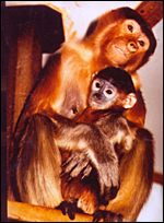
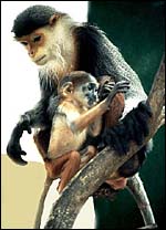 |
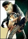
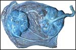
Douc langur female with young at San Diego Zoo. |
The length of gestation is 165-190 days, litter size is one, and the newborn weight is between 500-720 g. The maternal weight, not pregnant, is 6,785 kg. The placental weight at term gestation is 100-156 g, excluding membranes and cord. Some placentas, however, although with normal births, weigh as little as 72 g and are monodiscoid. In addition to these own data, I have received some additional data from Ulrike Streicher of animals born after 1998 at the Cuc Phuong Endangered Primate Research Center (EPRC) in Vietnam. Four male animals weighed 367, 475, 510, and 577 g respectively. An additional male of 200 g died on the second day of life because of maternal agalactia. Two females weighed 337 and 450 g, respectively. Two bilobed placentas were 130 and 133 g. In addition, a grey-shanked Douc langur placenta weighed 100g, had two disks (8 x 8 cm and 7 x 6 cm); it had a 24 cm umbilical cord and some marginal infarcts. An additional placenta received in May, 2002 from Dr. Streicher weighed 87.5 g, was bilobed (6.5 x 7.5 cm and 5 x 4 cm) with a newborn weighing 304 g. Its umbilical cord was 31 cm long, the mother weighed 5,100 g. This placenta, as others, had numerous infarcts.
Another placenta of a term neonatal demise from a male fetus weighed 68.4 g (34.7 + 33.7 g). The neonate weighed 475 g and the umbilical cord was 32 cm long. The main lobe contained several infarcts.
3) Implantation
This species has a two-lobed placenta with one lobe anterior, the other posterior in their simplex uterus. Indeed, the placentas of Cercopithecidae are all very similar and have been best described for the rhesus monkey and the baboon. The blastocyst attaches antimesometrially. There is no allantoic sac. Amnion development is by cavitation. Implantation is superficial, not interstitial. This is a hemochorial placenta, as that of all anthropoidea. The placentas of two related species have been described for: the proboscis monkey, Nasalis larvatus (Soma and Benirschke, 1977) and for Presbytis obscura (Burton, 1980).
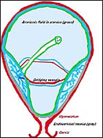 |
General situs of placenta in an uterus simplex. |
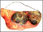 |
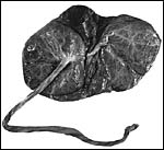
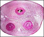
Mature placenta of Douc langur to show its two lobes and bridging vessels between the lobes. |
 |
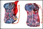
Another term placenta of Douc langur. |
 |
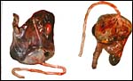
This placenta comes from a Delacours langur (Trachypithecus delacouri), a closely related species at Cuc Phuong, Vietnam. Kindly made available by Dr. T. Nadler, Cuc Phuong, Vietnam. |
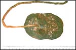
This usually is a bilobed placenta, as is present in many Cercopithecidae. The lobes are of approximately the same size. The umbilical cord inserts on the "primary lobe". From this insertion, the fetal blood vessels ramify over the primary lobe and then course over the membranes to the secondary lobe. The placenta is slightly cotyledonary and villous, the feto-maternal "barrier" is hemomonochorial. The basal plate has superficially invasive extravillous trophoblastic cells, much like human placentas, but less extensively so. There is slight vascular infiltration by trophoblast. The decidua basalis is separated from the villous tissue and frequent focal hemorrhages. There is frequently widespread, focal minute calcification in villi and fibrinoid. As will be seen from the following picture, not all placentas have two disks. This placenta comes from a normal delivery. It weighed only 72 g, with central cord insertion and a few marginal infarcts.
 |
Single-disked Douc langur placenta - normal fetus. |
The placenta s of a closely related species, the Delacours' langur (Trachypithecus delacouri), were kindly made available by Tilo Nadler of the Cuc Phuong National Park in Vietnam. They had the same morphology, two lobes and one is depicted above.
Likewise, a placenta of Trachypithecus laotum hatinhensis had identical morphology.
Measurements and weights of langur placentas |
Weight |
Lobes |
Cord |
|
| Pygathrix nemaeus | 72 g |
1 |
18 cm |
| Pygathrix nemaeus | 130 g |
2 |
23 cm |
| Pygathrix nemaeus | 133 g |
2 |
30 cm |
| Pygathrix nemaeus | 100 g |
2 |
24 cm |
| Pygathrix nemaeus | 87.5 g |
2 |
31 cm |
| Pygathrix nemaeus (stb.) | 68.4 g |
2 |
32 cm |
| Trachypithecus delacouri | 82 g |
2 |
21 cm |
| Trachypithecus laotum hatinhensis | 87 g |
2 |
? |
 |
Placenta of stillborn red-shanked douc langur. Provided by Dr. T. Nadler, Cuc Phuong, Vietnam. |
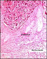 |
Placental floor with superficially invasive extravillous trophoblast (center) and underlying decidua basalis (right). |
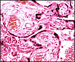 |
Low power view of villi. Fetal vessels, syncytial knots, intervillous space with maternal blood. |
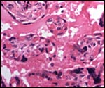 |
Villi with fetal capillaries that bulge into the intervillous space. |
 |
Term villi in intervillous space (M. ivs). F = fetal capillaries. Arrow points to a trophoblastic syncytial "knot". |
The villi are covered with syncytium that is markedly "knotted". Prominent vascular membranes are found with fetal vessels bulging under a thinned trophoblast.
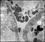 |
Electronmicrograph of villous structure of term Douc langur placenta. The borders of two villi are shown with the vacuolated curved syncytiotrophoblast. Maternal red blood cells (dark) in the intervillous space (center) and in fetal capillary (right). |
The length of the umbilical cord was 23, 24, and 30 cm respectively in three placentas measured by me, and they had few left-handed spirals. The umbilical cord of the single-disked organ shown above was 18 cm long and centrally inserted. The umbilical cord contains two arteries and one vein, which is usually located in the center between the arteries. There are no ducts. The cord inserts nearly centrally on the primary lobe. At term, each lobe weighs 50-55 g, and each measures approximately 8 x 7 x 1 cm. The "primary" lobe is usually slightly larger.
 |
Cross section of term umbilical cord. It is covered by amnionic epithelium; the vein is on top, arteries below. |
No descriptions exist but it appears to be similar to that of the rhesus monkey.
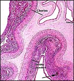
8) Extraplacental membranes
There is only a very thin layer of decidua capsularis that covers the chorion laeve; it contains no atrophic villi, as is the case in most simians. This contrasts with human placentas that always have atrophic villi in the free membranes, residua from the former interstitial implantation. The "bridging" fetal blood vessels (between the two lobes) are carried in the chorionic membrane. The amnion is loosely fused to the chorionic membrane. There is no allantoic or vitelline tissue. Some decidual necrosis is common here, but maternal vascular disease, as seen in human pre-eclampsia ("atherosis"), has not been found, despite the frequency of infarcts of the villous tissue.
 |
Rolled membranes between placental lobes. At arrow is a maternal, decidual spiral arteriole. |
No larger collections of extravillous trophoblast are present, as found in human placentas, but extravillous trophoblast infiltrates the superficial portion of the decidua basalis within the layer of fibrinoid. No giant cells are present in the decidua. There is focal and superficial vascular trophoblastic invasion of the decidua basalis. Often, much decidual necrosis is found in the decidua basalis, as are infarcts of the villous tissue. Also in contrast to ape and human placentation, the maternal arterioles are not significantly restructured by infiltrating extravillous trophoblast as is also the case in many cercopithecids and apes. Thus, the frequent infarcts must have a different basis than that which causes human placental infarcts in pre-eclampsia. These are due to thrombosis of the structurally altered vessels.
10) Endometrium
There is significant basal decidualization of the endometrium. Most of
the decidua basalis is separated from the villous tissue by a frequently
broad layer of fibrinoid and fibrin. Little trophoblastic infiltration
into the basal decidua occurs in any depth.
11) Various features
No metrial glands are present. There is, of course, no subplacenta.
12) Endocrinology
The estrous cycle is 28-30 days; the length of gestation is 165 -190 days.
Resnik et al.(1978) determined the total urinary estrogen levels in early
pregnancy and after fetal demise. A general review of steroids in pregnancy
was published by Pepe and Albrecht (1995). Czekala et al. (1981) measured
estrogens in urine and thus delineated the menstrual cycle. Estrogens
declined gradually during pregnancy although it was found that they were
of greater quantity than those of other cercopithecidae. In addition,
these authors reported that a large proportion of the total estrogen is
not the "usual" estrogenic hormone but is of an as yet unknown
nature This was also found to exist in Hanuman langurs, Presbytis
entellus. Gonadotropin measurements were positive in the first three
months of gestation but were difficult to interpret.
More recently, Ademmer et al. (2002) have determined the length of cycles
with steroid analysis of feces in three captive groups of douc langurs
at the Cologne zoo. They found an average cycle length of 28 days with
the follicular phase lasting 13 days, and the luteal phase was 11 days
long.
13) Genetics
The chromosome number is 2n=44 (40 metacentrics and 2 acrocentrics). The
X-chromosome is metacentric, the Y-chromosome is acrocentric. One small
metacentric pair has a significant secondary constriction (Hsu & Benirschke,
1975). Hybrids with other non-douc langur species have not been described,
although personal reports have been received that subspecific hybrids may
exist between the putative Vietnamese species of orphaned animals. Bogart
and Kumamoto (1978) have described an additional chromosomal band (?translocation)
in chromosome # 14 of a male animal that had sired abnormal offspring.
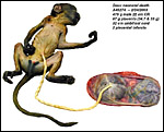
In letters I have since learned that a male Douc langur (2n=44) has produced a living offspring with a Proboscis monkey (2n=48) at Zoo Erfurt ( Germany). It was born on August 27, 1981 and died of a bezoar on July 1, 1986 (Personal communication Dr. Tilo Nadler, Cuc Phuong , Vietnam). A picture of this animal is shown next.
 |
Hybrid offspring of male Douc langur and this female Proboscis monkey. (Courtesy Dr. Tilo Nadler, Cuc Phuong , Vietnam. The animal was at Erfurt Zoo in Germany). |
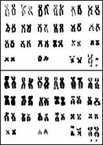 |
Chromosomes of male and female red-shanked Douc langurs. |
No immunological studies have been described except the depletion of B-cells in a calicivirus-infected young animal (Smith et al., 1985).
15)
Pathological features
Chorioamnionitis, an ascending infection of the intrauterine space, has
been observed in a few specimens. In addition, several spontaneous abortions
have been seen, one with hydrops fetalis (Resnik et al., 1978). Several
of our placentas have had typical and numerous infarcts, others have had
retroplacental hemorrhages and decidual necrosis. This is not apparently
related to the pre-eclampsia-like condition of humans and it is also not
restricted to primigravidae. Moreover, the maternal arterioles do not
show "atherosis" but, when infarcts are present, these vessels
are found to be thrombosed. In addition, hemosiderin is often found in
the fibrinoid floor. The San Diego colony has had chronic problems with
pulmonary acariasis, caused by the mite Pneumonyssus simicola.
The main problems in captivity, however, have been of a nutritional nature.
With the principal need for a folivorous diet and sensitivity to sugars,
lignin and fruit, these animals can be maintained only when appropriate
leafy food is available year-round. This topic has been addressed in an
important publication by Bauchop & Martucci (1968) [See chapter on
Hanuman langurs]. At the EPRC in Vietnam, a female with five successive
deliveries was found to completely lack milk for the neonates. The first
animal died, the others were hand-raised.
The
presence of a calicivirus was demonstrated in an orphaned 4-months-old
Douc langur that succumbed with bronchopneumonia, hepatic hemosiderosis
and B-cell depletion of nodes and spleen, among other pathologic findings
(Smith et al., 1985).
Several abortions occurring in the San Diego colony were associated with,
if not due to, chromosomal rearrangements (translocations). Meconium staining
(v.i.) is occasionally seen in newborns and their placentas.
Infection with Helicobacter is important and perhaps it contributes
to the frequent gastrointestinal problems suffered by these animals. In
surveys of animals at Cuc Phuong, however, it was found to be a regular
(?normal) component of fecal flora.
 |
Placenta of stillborn Douc langur with hydrops. |
 |
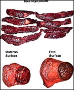
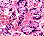
Cross sections of a term, delivered placenta from a Douc langur with a surviving infant. There are numerous infarcts (yellow) throughout this villous tissue, but apparently causing no harm to the fetal development. |
 |
Term gestation with neonatal death of Douc langur. |
 |
Same placenta under higher magnification. |
 |
Small placenta of douc langur with meconium staining and 8 infarcts. Kindly made available by Dr. U. Streicher at Cuc Phuong, Vietnam. |
Blood volume and pressures have not been published. There is an extensive consideration of the food intake and folivory in a recent publication by Ademmer et al (2002) that addresses the need for long periods of sleep in this species and the susceptibility to stress.
17)
Other resources
Frozen cell lines of tissue cultures from many different langurs are available
at the San Diego Zoo from Dr. Oliver Ryder at CRES
at oryder@ucsd.edu.
18) Other relevant needs
This is a significantly endangered animal whose reproductive history in
captivity has not been great. Several colonies have ended because of problems
with gestation and nutrition. The chromosomal problems alluded to here
need to be further studied. This is important especially as to their possible
relationship to neonatal and fetal deaths. The infarcts in the placenta
suggest possible maternal vascular disease in pregnancy. It must be different
from that of human atherosis, exhibiting only thrombosis. But the cause
of the thrombosis is uncertain. The future may bring embryo transfer into
related species that have a similar placentation. But, the overwhelming
problem is nutritional and needs to be addressed more fully.
19) Acknowledgment
I am grateful for important information received from Drs. T. Nadler and
U. Streicher at Cuc Phuong, and from the Cologne Zoo staff.
References
Ademmer, C., Klumpe, K., v.Maravic, I., Königshofen and Schwitzer, C.: Nahrungsaufnahme und Hormonstatus von Kleideraffen (Pygathrix n. nemaeus Linnaeus, 1771) im Zoo. Z. Kölner Zoo 45:129-135, 2002.
Bauchop, T. and Martucci, R.W.: Ruminant-like digestion of the langur monkey. Science 161:698-700, 1968.
Benirschke, K. and Bogart, M: Pathology of Douc langurs (Pygathrix nemaeus) at San Diego Zoo. XX. Internatl. Symp. Erkr. Zootiere, Dvur Kralove, CSSR, June 14-18. Akademie Verlag, Berlin, pp. 257-261, 1978.
Bogart M.H. and Kumamoto, A.T.: Karyotype abnormalities in two primate species, Pygathrix nemaeus and Lemur coronatus. Fol. Primatol. 30:152-160, 1978.
Burton, G.J.: Early placentation in the dusky leaf monkey (Presbytis obscura). Placenta 1:187-195, 1980.
Cuc Phuong National Park Endangered Primate Rescue Center, Vietnam. See: Traitel, D.A.: Building a safe haven for Douc langurs. Zoonooz (San Diego), October, pp. 10-15, 1996.
Czekala, N.M., Hodges, J.K. and Lasley, B.L.: Pregnancy monitoring in diverse primate species by estrogen and bioactive luteinizing hormone determinations in small volumes of urine. J. Med. Primatol. 10:1-15, 1981.
Griner, L..A.: Pathology of Zoo Animals. Zool. Soc. San Diego, pp. 366-367, 1983.
Herrmann, H.-W. and Pagel, T.: Phong Nha-Ke Bang - das Regenwaldschutzprojekt des Kölner Zoos in Vietnam. Z. Kölner Zoo 43(2):79-88, 2000. {Includes reference to Vietnam breeding/rescue station.]
Hick, U.: Breeding and maintenance of douc langurs at Cologne zoo. In: Martin, R.D. ed.: Breeding endangered species in captivity. Academic Press, London, pp. 223-233, 1975.
Hösli, P.: Chromosomes of clothes monkey, (Pygathrix nemaeus). Schweiz. Arch. Tierh. 112:338, 1970.
Hsu, T.C. and Benirschke: An Atlas of Mammalian Chromosomes. 9:Folio 450, 1975.
Lippold, L. K.: The douc langur: a time for conservation. In: Rainier III and Bourne, G.H.: Primate Conservation. Academic Press, NY, pp. 513-538, 1977.
Lippold, L.K.: International Douc Langur Studbook Keeper. Department of Anthropology, San Diego State University, San Diego, CA 92182-0377. Douc Press, P.O. 232344, San Diego, CA 92193.
Lippold, L., Czekala, N.M., Lasley, B.L. and Benirschke, K.: Steroid monitoring of langur pregnancy. Amer. Soc. Primatol. (abstr.) Seattle, April, 1977.
Mossman, H.W.: Vertebrate Fetal Membranes. MacMillan, Hound mills, UK, 1987.
Pepe, G.J. and Albrecht, E.D.: Actions of placental and fetal adrenal steroid hormones in primate pregnancy. Endocr. Reviews 16:608-648, 1995.
Puschmann, W.: Zootierhaltung. Säugetiere. Vol. 2. VEB Landwirtschaftsverlag Berlin, Germany, 1989.
Ramsey, E.M.: The Placenta. Human and Animal. Praeger, NY, 1982.
Resnik, R., Robinson, P.T., Lasley, B.L. and Benirschke, K.: Intrauterine fetal demise associated with consumption coagulopathy in a Douc langur monkey (Pygathrix nemaeus). J. Med. Primatol. 7(4):249-253, 1978.
Ruempler, U.: Haltung und Zucht von Kleideraffen (Pygathrix nemaeus nemaeus Linnaeus, 1771) im Kölner Zoo. Z. des Kölner Zoo 34(2):47-65, 1991.
Ruempler, U.: Husbandry and breeding of Douc langurs Pygathrix nemaeus nemaeus at Cologne Zoo. Int. Zoo Yb. 36:73-81, 1998.
Smith, A.W., Skilling, D.E., Anderson, M.P. and Benirschke, K.: Isolation of primate calicivirus Pan paniscus type 1 from a Douc langur (Pygathrix nemaeus L.). J. Wildl. Dis. 21(4):426-428, 1985.
Soma, H. and Benirschke, K.: Observations on the fetus and placenta of a proboscis monkey (Nasalis larvatus). Primates 18:277-284, 1977.
Starck, D.: In: Lehrbuch der Speziellen Zoologie, Vol. II, Teil 5/1:Säugetiere, pp.574-576. Gustav Fischer, Jena, 1995.
Wurster, D. and Benirschke, K.: Chromosomes of some primates. Mammal. Chromos. Newsl. 10:3, 1969.
Zneimer, S., Kumamoto, A.T. and Benirschke, K.: Banding patterns of the chromosomes of two langur species. Pygathrix nemaeus and Presbytis entellus: A comparative study. Chromosome Inform. Serv. 26:19-22, 1979.