(Clicking on the thumbnail images below will launch a new window and a larger version of the thumbnail.)

Atlantic bottlenose dolphin
Tursiops truncatus

Order: Cetacea
Family: Delphinidae

1) General zoological data


This is the common dolphin, shown often in aquaria. It is widely distributed, but lives primarily in coastal waters. Adult animals weigh around 200 kg. They are gray, with a pale/white underside. The bottlenose(d) dolphin is not an endangered species. Large schools are found offshore and, occasionally, further out in the ocean and up rivers. They are well-known animals in aquaria and are easily trained. Dolphins reproduce well in captivity and, following the two workshops referenced below; most aquaria now maintain successful breeding colonies.

2) General gestational Data

Births take place usually in the summer and singletons are the rule. They are generally born every other year. The length of gestation is about 11-12 months. Newborns weigh around 10 kg and measure 98-126 cm in length. Lactation lasts 7 to 18 months. Sexual maturity is reached around 5 years in females; longevity is about 25 to 30 years. Ovulation is most likely a spontaneous event and is also mostly left-sided; it is probably not induced by copulation. The placenta weighs, on average, 2,200-2,800 g including membranes and umbilical cord, at term, and with surviving newborns.

   
  Mature female bottlenose dolphins in San Diego.
     
 
3) Implantation

The cetacean placenta is an epitheliochorial, diffuse organ that implants all over both uterine horns. It fills the entire uterus, with the umbilical cord inserting near the endocervical os. The time of implantation following fertilization is unknown. The main body of the fetus occupies one horn, while the tail occasionally extends into the other horn. The intrauterine position of the cetacean fetus has been illustrated by Slijper (1956, 1966). The position of the fetus is generally such that the tail delivers first. Wislocki & Enders (1941) were the first to describe the placenta of bottlenose dolphins.

4) General Characterization of the Placenta

The cetacean placenta is a diffuse, epitheliochorial organ without free membranes. It is extremely thin and covered diffusely with villi, possibly slightly reduced over the tubal ends and thinned also over the compressing rump of the fetus. There are no cotyledons. The villous surface inderdigitates with the endometrial glands without invading the endometrium. One may see ridges, or rugae on the villous surface in the very freshly delivered placenta. They correspond presumably to endometrial counterparts and were already illustrated and discussed by Turner (1872) in his description of an Orca placenta. Turner provided a long detailed analysis of that species' placenta, which differs little from the placenta of delphinidae. Mossman (1987) stated that implantation is mesometrial. No very young placentas or blastocysts have been described. Nevertheless, since the placentas of cetacea are generally very similar, the photographs by Stump et al. (1960) in humpback whales and ours (Benirschke et al., 1980) in spinner dolphins (Stenella longirostris) may give an indication of those in bottlenose dolphins. A tiny embryo is enclosed in hugely expansive delicate membranes. There is a large allantoic sac that surrounds the amnionic cavity. A large allantoic duct carries urine from the urachus to this allantoic sac. The allantois has no vasculature. The duct, as well as the allantoic sac, often contains brown debris made of birefringent crystals. These are known as "hippomanes". They dissolve in acid and are chemically composed of calcium oxalate hydrate (CaC2O4.2.25 H2O). The allantoic sac is lined with a flat epithelium that is focally squamoid. The amnion is covered with a single-layered epithelium, with occasional foci of keratinized squamous metaplasia that are occasionally pigmented, especially over the umbilical cord.

   
  Dolphin situs in utero.
     
 
5) Details of fetal/maternal barrier


The entire villous apparatus is covered with trophoblast; there are no denuded areas. The endometrium is covered with epithelium that is not eroded. Thus, the barrier is a complete epitheliochorial one. The trophoblast is cuboidal and has frequent vacuoles.

6) Umbilical cord

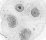
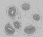
The umbilical cord is short, between 30 and 40 cm. It maximally has one spiral turn. The cord inserts near the endocervical canal and ruptures during delivery spontaneously near its fetal insertion. The normal umbilical cord contains two large arteries and two large veins, without major anastomoses between any of the vessels (unlike Hyrtl's anastomosis, present in the umbilical cord of humans, that connects the two arteries). The umbilical vessels branch at the placental insertion dichotomously towards the periphery of the placenta. One exceptional dolphin that I studied had only three umbilical vessels. It came from a growth-restricted fetus that died within a year. Although associated anomalies are common in humans lacking a cord vessel, this animal had no anomalies. In addition to the major umbilical vessels, there is a profusion of small and medium-sized blood vessels in the umbilical cord, as many as up to 200 in cross-sections. Many have no musculature and are then scattered irregularly through the cord. They have no particular predilection to locate around the allantoic duct. Injection of the large veins and arteries discloses that the small vessels originate from the large ones and that they communicate among the large vessels. The exits from large vessels are very sparse. The function of these small vessels is unknown. The central portion of the umbilical cord carries an irregularly shaped allantoic duct with sparse, and usually very thin epithelium. Well-preserved cords may show some squamous allantoic epithelium and adjacent smooth muscle bundles. There is very little vascularization of this duct. The Wharton's jelly is sparse and unusually firm. On the umbilical surface, foci of squamous metaplasia ("caruncles") are common. They are often pigmented.

   
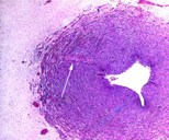
  Umbilical artery with arrow pointing at the rare branching vessel.
     
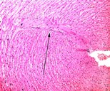
  Higher magnification of the branching vessel in the umbilical cord.
     
  Cross-section of an umbilical cords with four large and numerous tiny (black) blood vessels.
     
  Umbilical cord with three vessels and large allantoic duct (right)
     
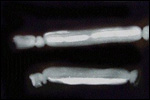
  Barium sulfate injected cord vessels with tiny branches communicating among large vessels.
     
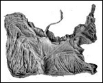
  Spontaneously delivered dolphin placenta, seen from the fetal side to show the insertion of the umbilical cord and amnionic/allantoic membranes.
     
  Delivered dolphin placenta with cord insertion above, near the middle.
     
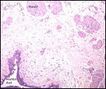
  Allantoic duct (left bottom) lined by squamous epithelium and with adjacent bundles of smooth muscle.
     
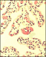
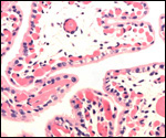
  Terminal villi of normal dolphin placenta.
     
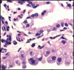
  Trophoblastic surface of dolphin placenta.
Note the vacuolation of the single-layered trophoblast
     
  Immature villi of bottlenose dolphin. Note the vacuolated
trophoblast and the extensive subtrophoblastic capillaries.
     
 
7) Uteroplacental circulation

There are no specific publications dealing with either the uterine circulation or its physiology, but the arrangement of the pelvic vasculature was discussed by Rommel and colleagues (1993). They described that the uterus is surrounded by a complex countercurrent system of blood vessels, which, they suggested, is being used for cooling purposes. There is also an extensive consideration of various other circulatory aspects in their description.

8) Extraplacental membranes

There are no free membranes, but amnion and allantoic sacs are contained within the chorionic sac. The allantoic duct within the umbilical cord is occasionally lined by squamous epithelium and accompanied by bundles of smooth muscle.

9) Trophoblast external to barrier

Since this is so superficial an implantation - really only an appositional one - there are no trophoblastic cellular elements external to the endometrium.

10) Endometrium

No decidua is present in cetacea. There is an "enormous development of superficial capillaries as well as large numbers of racemose glands" in the endometrium(Slijper, 1966). The microscopic anatomy of the female genital tract, including the endometrium, has been described for marine mammals in general by Simpson and Gardner (1972). It includes a description of the arterial supply, as well as of the cervix and the ovaries. We have found the non-pregnant uterus (of formerly pregnant dolphins) to weigh 190 g, with 17-18 cm uterine horns.

11) Various features

None.

12) Endocrinology

The corpus luteum of pregnancy persists to term and then becomes a corpus albicans. Corpora lutea of non- pregnant animals involute to become corpora atretica. When ovaries of cetacea are sectioned, the scars of the corpora lutea of pregnancy (corpora albicantia) can be enumerated; they give an accurate indication of the number of past pregnancies. Slijper (1966) recorded occasional cetacean twin pregnancies, both monozygotic and dizygotic.

Seasonal testosterone levels and testicular sizes have been reported by Harrison and Ridgway (1971), Schroeder and Keller (1989), and by Wells (2000). Progesterone and estrogen determinations have been reported in pregnant and nonpregnant dolphins (Sawyer-Steffan et al. 1980, 1983). A complete review of the current status of hormonal measurements and the reproductive anatomy of male and female dolphins is available from Robeck (2000). Some of these studies indicate the much greater frequency of ovulation in the left-sided ovary. The reason for this unilaterality of ovulation is not known.

Orphaned neonates may induce lactation (Ridgway et al., 1995); the same authors also described the composition of dolphin milk.

13) Genetics

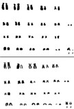
The diploid chromosome number (2n) is 44, with 30 metacentric and 12 acrocentric elements (Hsu & Benirschke, 1973). The X-chromosome is submetacentric, the Y-chromosome is minute.
Recent studies by Nikaido et al. (1999) have shown the close genetic relationship between hippopotamus and cetacea.


14) Immunology

No information is available.

15) Pathological features

Occasional congenital anomalies have been observed in dolphin fetuses. An anencephalic fetus was diagnosed sonographically (Brook, 1994). Cardiac anomalies (transposition of great vessels) in one of dizygotic twin was reported by Gray and Conklin (1974). Other neonatal diseases (umbilical infections) were summarized by Miller and Bossart (2000). In the placenta, other than the 3-vessel cord described above, abnormal features described so far are restricted to the infection with Brucella delphini, as described by Miller et al. (1999). This infection has led to abortion, fetal and neonatal deaths. The sparse data on late abortions and infectious diseases have been summarized by van Bonn (2000). A few ovarian and uterine tumors have been reported in cetacea (Cowan, 2000). In Stenella spp. a variety of interesting findings were reported by Perrin et al. (1989), such as caudal regression, megalencephaly, omphalocele, maxillary hypoplasia with mandibular left, and monozygotic twinning as well.

   
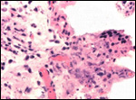
  Villi of immature Brucella-infected placenta. There is inflammation and degeneration of villi.
     
 
16) Physiologic data

Various physiologic data, endoscopy, semen cryopreservation, environmental contaminants and their effects, and contraceptive methods were discussed in a variety of contributions of the workshop edited by Duffield and Robeck (2000). Reddy et al. (1998) described the content of chemical contaminants in dolphins.


17) Other resources

A substantial number of formalin-fixed specimens of pregnant uteri, placentas and fetuses of various cetacea is available for scientific study through W.F. Perrin, Southwest Fisheries Center, La Jolla Laboratory, National Marine Fisheries Service, NOAA, La Jolla, California, 92037. Reviews of dolphin holdings in the world are given in the two workshops referred to earlier: Ridgway and Benirschke (1977), and Duffield and Robeck (2000).

18) Other remarks - What additional Information is needed?

Few data exist on placentas of other cetacea. The next picture is of a Beluga, other cetacean species should be studied as well. They would be of interest. There are also surely more pathologic features to be identified. The function of the large number of cord blood vessels, and how they are being circulated might be studied.

   
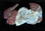
  Delivered term placenta of Beluga (Delphinapterus leucas).


Acknowledgement

Specimens were largely obtained through the courtesy of Dr. S.H. Ridgway, San Diego.

References

Benirschke, K., Johnson, M.L. and Benirschke, R.J.: Is ovulation in dolphins, Stenella longirostris and Stenella attenuata, always copulation-induced? Fishery Bull. 78:507-528, 1980.

Benirschke, K.: Past and future investigations to enhanced understanding of cetaceans. In, Bottlenose Dolphin Reproduction Workshop. San Diego, California June 3-6, 1999. D. Duffield and T. Robeck, eds. AZA Marine Mammal Taxon Advisory Group, Silver Spring, MD, 2000.

Bonn, W. van: Infectious diseases and late abortions. pp. 279-287, In, Bottlenose Dolphin Reproduction Workshop. San Diego, California June 3-6, 1999. D. Duffield and T. Robeck, eds. AZA Marine Mammal Taxon Advisory Group, Silver Spring, MD, 2000.

Brook, F.: Ultrasound diagnosis of anencephaly in the fetus of a bottlenose dolphin (Tursiops aduncas). J. Zoo Wildlife Med. 25:569-574, 1994.

Cowan, D.F.: Pathological evidence of reproductive disease and/or dysfunction in wild cetaceans, pp. 175-182, In, Bottlenose Dolphin Reproduction Workshop. San Diego, California June 3-6, 1999. D. Duffield and T. Robeck, eds. AZA Marine Mammal Taxon Advisory Group, Silver Spring, MD, 2000.

Duffield, D.A. , Ridgway, S.H. and Sparkes, R.S.: Cytogenetic studies of two species of porpoise. Nature 213:189, 1967.

Duffield, D. and Robeck, T. eds.: Bottlenose Dolphin Reproduction Workshop. San Diego, California June 3-6, 1999. Marine Mammal Taxon Advisory Group, Silver Spring, MD, 2000

Gray, K.N. and Conklin, R.H.: Multiple births and cardiac anomalies in the bottle-nosed dolphin. J. Wildl. Dis. 10:155-157, 1974.

Harrison, R.J. and Ridgway, S.H.: Gonadal activity in some bottlenose dolphin (Tursiops truncatus). J. Zool. London 165:355-366, 1971.

Hsu, T.C. and Benirschke, K.: An Atlas of Mammalian Chromosomes. Vol. 7, Folio 331, 1973.

Miller, W.G., Adams, L.G., Ficht, T.A., Cheville, N.F., Payeur, J.P., Harley, D.R., House, C. and Ridgway, S.H.: Brucella-induced abortion and infection in bottlenose
dolphins (Tursiops truncatus). J. Zoo and Wildlife Med. 30:100-110, 1999.

Miller, D.L. and Bossart, G.D.: Reproductive and fatal neonatal diseases in cetaceans from Florida Oceanaria 1979-1999. pp. 183-186, In, Bottlenose Dolphin Reproduction Workshop. San Diego, California June 3-6, 1999. D. Duffield and T. Robeck, eds. AZA Marine Mammal Taxon Advisory Group, Silver Spring, MD, 2000.

Mossman, H.W.: Vertebrate Fetal Membranes. Comparative Ontogeny and Morphology; Evolution; Phylogenetic Significance; Basic Functions; Research Opportunities. The MacMillan Press, Ltd. Houndmills, 1987.

Nikaido, M., Rooney, A.P. and Okada, N.: Phylogenetic relationships among certartiodactyls based on insertions of short and long interspersed elements: Hippopotamuses are the closest extant relatives of whales. Proc. Natl. Acad. Sci. USA 96:10261-10266, 1999.

Nowak, R.M. and Paradiso, J.L.: Walker's Mammals of the World. 4th ed. Vol. II. The Johns Hopkins University Press, Baltimore and London, 1983.

Pabst, D.A., Rommel, S.A., McLellan, W.A., Williams, T.M. and Rowles, T.K.: Thermoregulation of the intra-abdominal testes of the bottlenose dolphin (Tursiops truncatus) during exercise. J. Exp. Biol. 198:221-226, 1995.

Perrin, E.V.D., Benirschke, K. and Perrin W.F.: Monstrous dolphins - Malformations in marine mammals. Teratology 39:p.51, abstr. # 472, 1989.

Prasad, N., Mumford, D.M., Barsales, P.B., Whitman, T. and Wilbur, J.R.: Cytogenetic studies of dolphin (Tursiops truncatus) by an extended tissue culture technique. Experientia 26:1167, 1968.

Reddy, M., Echols, S., Finklea, B., Busbee, D., Reif, J. and Ridgway, S.H.: PCBs and chlorinated pesticides in clinically healthy Tursiops truncatus: Relationships between levels in blubber and blood. Marine Pollution Bull. 36:892-903, 1998.

Ridgway, S.H. and Benirschke, K., eds.: Breeding Dolphins; Present Status, Suggestions for the Future. Report MNC 76/07, Marine Mammal Commission, Washington, DC, November, 1977.

Ridgway, S., Kamolnick, T., Reddy, M. and Curry, C.: Orphan-induced lactation in Tursiops and analysis of collected milk. Marine Mamm. Sci. 11:172-182, 1995.

Robeck, T.R.: Advances in the understanding and manipulation of bottlenose dolphin reproduction. pp. 109-131, In, Bottlenose Dolphin Reproduction Workshop. San Diego, California June 3-6, 1999. D. Duffield and T. Robeck, eds. AZA Marine Mammal Taxon Advisory Group, Silver Spring, MD, 2000.

Rommel, S.A., Pabst, D.A. and McLellan, W.A.: Functional morphology of the vascular plexuses associated with the cetacean uterus. Anat. Rec. 237:538-546, 1993.

Sawyer-Steffan, J.E. and Kirby, V.L.: A study of serum steroid hormone levels in captive female bottlenose dolphins, their correlation with reproductive status, and their application to ovulation induction in captivity. Natl. Techn. Inf. Serv. PB 80-177199, 1980.

Sawyer-Steffan, J.E., Kirby, V.L., and Gilmartin, W.C.: Progesterone and estrogens in the pregnant and non-pregnant dolphin, Tursiops truncatus, and the effects of induced ovulation. Biol. Reprod. 28:897-901, 1983.

Schroeder, J.P. and Keller, K.V.: Seasonality of serum testosterone levels and sperm density in Tursiops truncatus. J. Exp. Zool. 249:316-321, 1989.

Simpson,J.G. and Gardner, M.B.: Comparative microscopic anatomy of selected marine mammals. Chapter 5 in, Mammals of the Sea. Biology and Medicine. S.H. Ridgway, ed. C.C. Thomas, Springfield, Illinois, pp. 298-418, 1972.

Slijper, E.J.: Some remarks on gestation and birth in cetacea and other aquatic mammals. Hvalradets Skrifter 41:1-62, 1956.

Slijper, E.J.: Functional morphology of the reproductive system in Cetacea. Chapter 15 in, Whales, Dolphins, and Porpoises. K.S. Norris, ed. Univ. Calif. Press, Berkeley, 1966.

Stump, C.W., Robins, J.P. and Garde, M.L.: The development of the embryo and membranes of the humpback whale, Megaptera nodosa (Bonaterre). Austral. J. Marine and Freshw. Res. 11:365-386, 1960.

Turner, W.: On the gravid uterus and on the arrangement of the foetal membranes in the Cetacea (Orca gladiator). Trans. Roy. Soc. Edinburgh 26:467-504, 1872.

Walen, K.H. and Madin, S.H.: Comparative chromosome analysis of the bottle-nosed dolphin (Tursiops truncatus) and the pilot whale (Globicephala scammonii). Am. Nat. 99:349, 1965.

Wells, R.S.: Reproduction in wild bottlenose dolphins: Overview of patterns observed during a long-term study. pp. 57-74, In, Bottlenose Dolphin Reproduction Workshop. San Diego, California June 3-6, 1999. D. Duffield and T. Robeck, eds. AZA Marine Mammal Taxon Advisory Group, Silver Spring, MD, 2000.

Wislocki , G.B. and Enders, R.K.: The placentation of the bottle-nosed porpoise (Tursiops truncatus). Amer. J. Anat. 68:97-114, 1941.


back to top

© 2002. All rights reserved.
welcome | home | intro | placentation | glossary | author contact me: kbenirsc@ucsd.edu