(Clicking on the thumbnail images below will launch a new window and a larger version of the thumbnail.)
 
Last updated:
Oct 18, 2005.


Deer Species
Cervus, Odocoileus, etc.

Order: Artiodactyla
Family: Cervidae

1) General zoological data of species


There are at least 38 species in 17 recent genera that constitute the group of Cervidae, according to Nowak & Paradiso (1983). This group reaches back to about 40 MYA when speciation commenced, details of which may be read in Vrba & Schaller, (2000).

The cervids can be grouped into five major families: Moschinae, Hydropotinae, Muntiacinae, Cervinae, and Odocoilinae. A review by Groves & Grubb (1987), however, now excludes the Moschinae. Most, but not all cervid species, have antlers. Deer are widely scattered over the world and differ much in size and phenotype. Likewise, their gestation varies tremendously, from 160 days in Moschus, to 10 months in Capreolus, the latter is perhaps so long because of its exceptional delayed implantation. Plotka (1998) summarized this information in a well-referenced table. In contrast to other polycotyledonary artiodactyls (e.g. sheep, cow), the deer species have relatively few cotyledons. For instance, the white-tailed deer (Odocoileus virginianus) possesses only three caruncles per uterine horn (6 altogether). Consequently, the placenta has only three cotyledons since twins are common. Most cervids have 1 to 2 young, very rarely more. Each develops in its own uterine horn. The placental cotyledons and, especially the chorionic membranes, are closely approximated in most twins. When he described the exceptional uterus with triplets, Mossman (1987) drew attention to our need to know more about this junctional zone of the two blastocysts.

Deer species have a worldwide distribution and there are numerous breeding colonies, of many different species, in zoological gardens and on ranches. Several species have also been commercially ranched in recent years, for instance the red deer in New Zealand, and other species in other countries. Also, Chinese muntjacs have escaped in England and compose now a feral population in that country.

The taxonomy of cervinae, indeed of all cervidae, has been confusing and is now being studied with molecular probes (Miyamoto et al., 1990; Randi et al., 2001). These have revealed some surprising findings. For instance, the Canadian and European wapiti/elk are much more divergent than heretofore believed. For that reason, they may now have to be given species sanctity. Elaphurus should probably not be classified in a separate genus, and so on. These studies are not complete as yet and have been accomplished mostly by the use of mtDNA; other studies will follow, using cDNA and other molecular techniques. They promise to bring some surprises. I would be gratified if they correlated with the varied chromosomal numbers found in these species, as there is some early indication for this.

2) General gestational data

Since there is much difference in maternal size, the fetal and placental weights also vary widely. For instance, a wapiti newborn (usually one) weighs 13-18 kg, while a mule deer fawn at birth is around 1.5-3 kg, depending on whether it is a singleton or a twin; other weights will be referred to subsequently. Of all deer species that have been studied in some detail, the roe deer is the exception in its gestational properties - it has a strikingly delayed implantation.

In captivity, the life span in most cervids is up to around 15-20 years; in the wild it is less.

   
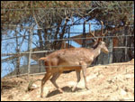
  Red deer (Cervus elaphus barbarus).
     
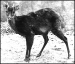
  Brocket deer (Mazama sp.).
     
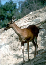
  White-tailed deer (Odocoileus virginianus).
   
 
3) Implantation


The deer placenta is "oligocotyledonary", with maximally 10 cotyledons. Most of their placentas possess only 4-6 cotyledons. This contrasts with cow, sheep, and other ruminants, which have up to ten times that many cotyledons. Moreover, the shape of cotyledons varies somewhat in ruminants. Most deer species have convex cotyledonary shapes (see Mossman, 1987). This contrasts with the goat, for instance that have a concave cotyledonary structure (Please see the chapter on Cretan goat). The early attachment of deer blastocysts is mesometrial and it is confined to the uterine caruncles. These are usually in mesometrial rows. In between the cotyledons is the allantochorionic membrane that is covered by villus-free trophoblast. This area is the site of the areolae, modifications of endometrium with focal endometrial secretion and absorption of nutrients (uterine milk) by the trophoblast. Most cervid placentas have their umbilical cords attached to the chorion at the mesometrial aspect of the uterus. All have a large allantoic sac, but the yolk sac is transitory. Chapman and Dansie (1969) studied implantation in feral Chinese muntjac that had escaped in England. They found that the majority of their fetus are in the right uterine horn. That is not the case in all cervids.

   
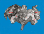
  General situs of deer placentas.


4) General characteristics of placenta


Several detailed accounts of cervid placentas are available. These are comprehensive studies but they are only a few common cervid species. More superficial descriptions are available for some other species, but many ruminant species' placentas have never been described and may show surprising differences when they might become available. The more detailed accounts come from Sinha et al. (1969) who reported on the placenta of white-tailed deer, while Aitken et al. (1973) gave a fine account of the roe deer implantation. The placenta of fallow deer (Dama dama) had an exhaustive description by Hamilton et al. (1960), which also lists all theretofore-examined species. They give specific details for a few other deer species, recognizing that there is great structural similarity of all cervid placentas. We cannot confirm the suggestion attributed to Weldon (1884) that the musk deer (Moschus moschiferus) has a diffuse placenta; ours had seven typical cotyledons. Mossman (1987) who quoted this note must have been misled, as Weldon's paper is actually about the four-horned antelope (Tetracerus quadricornis) rather than the musk deer.

For the white-tailed deer, Odocoileus virginianus, the placentation study by Sinha et al. (1969) gives the best description of cervid placentas. It is based on the study of 42 pregnant uteri. Moreover, their article provides reference to all prior placental studies, carried out on a variety of other deer species. Most of these are quite superficial, when compared with their report. Of these 42 uteri, 46% contained twins. Singleton placentas overlapped the two horns with 6 cotyledons, while in twins, each has only three cotyledons, as there are only 3 caruncles (rarely four) per uterine horn. The chorions of these twins were always fused in later gestation (as is shown in the specimen below - from Wurster & Benirschke, 1967). Remarkably, in two sets of twins and one set of triplets, their placental vasculature is said to have been fused. This is contrary to my observations made by injection of dyes on similar material. Moreover, neither freemartinism nor chimerism has been documented in this species. When we searched for chimerism in "antlered does", a relatively common occurrence in this species, we found none. These exceptional animals are assumed to have antler development because of sporadic adrenal tumors with androgen production. Sinha's report also assumed that one of the triplets was a set of monozygotic twins (MZ), without adequate support, however. More detailed study of twinning in a deer species comes from Kurnosov and was done in the elk (1962). He found close approximation of sacs, and identified occasional monochorionic twins. But only minute anastomoses were found and these were confined to MZ twins (same sex, single corpus luteum). Although Kurnosov was looking for freemartins, none were found. Additional support for the lack of anastomoses in cervid twin placentas comes from the study of Hamilton et al. (1960) who also used colored dye injections.

The cotyledons are typical (see photo below). There is intensive interdigitation of villi with the caruncular epithelial folds. Thus, an intimate contact between trophoblast and endometrial epithelium is established. The caruncles are composed of stroma and blood vessels, covered by endometrial epithelium. There are no glands. The remainder of the endometrium is glandular and has PAS+ secretions. Much "uterine milk" was observed between these epithelia in the fine-structural study. These authors differentiated four trophoblastic cell types: Cuboidal cytotrophoblast; columnar cells at the pits of villi; tall columnar storage cells at the arcades; and binucleated giant cells (diplokaryocytes) that have been the topic of much further study. These 20-50?m-large cells derive by chromosome duplication that is combined with a lack of cell division. They come from uninuclear trophoblastic precursors that are located mostly at the tips and sides of the villi. These features have been reported for all other ruminants (see Lawn et al., 1969) studied. These authors consistently saw the binucleated cells to be separate from the maternal epithelium but at that time they did not know what their function might be.

This study of binucleate cells contrasts with Wooding's more recent studies of placentas in cow, goat, sheep, roe deer and Chinese water deer (1982, 1983). He showed that 15-20% of trophoblast is composed of binucleate cells and that they are present throughout pregnancy. He also reported the fusion of these cells with endometrium. When this occurs, the cells deliver their characteristic granules of placental lactogen into or near to the maternal circulation. Wooding also confirmed the suspected migration of mature diplokaryocytes. Lee et al. (1983) had been convinced that there was no fusion. They raised antibodies against the granular content, found it to cross-react with deer as well as cow antigen, and found migration of the cells. In contrast to other studies, theirs showed some antibody-positive cells also to reside in between caruncles, in the free chorioallantoic membrane. The apparent fusion of the binucleate cells with endometrium is one of the reasons why, today, the deer placenta is often referred to as being synepitheliochorial. In later studies, Wooding et al. (1997) reaffirmed the migration and fusion (to trinucleate cells), location, percentage distribution, and lactogen production for all polycotyledonary artiodactyls studied. They are an apparently characteristic feature of these species, and the lactogen is presumably needed for pregnancy maintenance.

Sinha et al. (1969) then showed numerous electronmicroscopic features of the trophoblast with its microvillous surfaces. They observed PAS+ material and lipid in several cell types and interpreted some phagocytic activity in degenerating foci of the arcades. In the intercotyledonary membranes, "areolae" were found to correspond to the openings of endometrial glands, which contained "uterine milk" and a few red blood cells, apparently for absorption by the opposing tall trophoblastic cells.

   
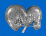
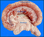
  Bicornuate uterus pregnant with twins of white-tailed deer (Odocoileus virginianus).
     
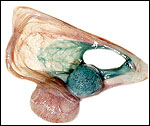
  Bicornuate uterus pregnant with twins of white-tailed deer (Odocoileus virginianus).
     
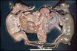
  Twin gestation in white-tailed deer, in December (New Hampshire). Note three cotyledons per animal of approximately equal size in different uterine horns.
     
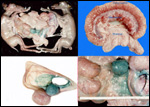
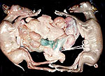
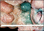
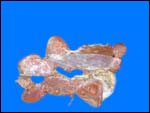
  Composite of the same white-tailed deer specimen, injected with green dye. Note the intimate fusion of the cotyledons and chorions, but an absence of anastomoses between the twin circulations. At top right is a complete "placentome".
     
  For a larger image of the composite above, click this thumbnail.
     
  For a larger image of the composite above, click this thumbnail.
     
  For a larger image of the composite above, click this thumbnail.
     
  For a larger image of the composite above, click this thumbnail.
   
  The placental development of the red deer, Cervus elaphus, was described by McMahon et al. (1997). Twelve uteri were studied between 27 and 55 days of gestation. Gastrulation was complete by 27 days and the trophoblast had covered both uterine horns. Trophoblastic "plaques" were seen by day 34 and, by day 41, the placentomes had formed. Placentation was otherwise deemed to be similar to that of other ruminants. Hamilton et al. (1960) had 15 such uteri and found four cotyledons in each uterine horn.
   
  Placenta of Barbary red deer, Cervus elaphus barbarus.
     
  Placenta of Barbary red deer, Cervus elaphus barbarus.
     
  Placenta of singleton wapiti, Cervus elaphus, with 8 cotyledons, 4 in each horn.
     
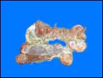
  Placenta of singleton Père David's deer neonate, Elaphurus davidianus. As discussed next, there are five large cotyledons with flat, ovoid shape and a relatively long cord.
     
  Placenta of singleton Père David's deer neonate, Elaphurus davidianus. As discussed next, there are five large cotyledons with flat, ovoid shape and a relatively long cord.
   
 
The placenta of the Père David's deer, Elaphurus davidianus, has been studied by Harrison & Hamilton (1952). They had four specimens of pregnant uteri at their disposal. They found three elongated caruncles in the right uterine horn and two in the left horn of two non-pregnant animals. The placentomes of this deer species are the largest observed to date. They are more flat than those of other cervids with their more convex placentomes. Harrison & Hamilton found that the fetuses were present in the right horn, but the left horn was equally enlarged. There were five placentomes (cotyledon + caruncle), mostly situated on the posterior wall of the uterus, and on the septum. They also observed large allantoic sacs and the usual excrescences of the amnion, representing squamous metaplasia. The umbilical cords measured 10-12 x 2-3 cm in length and width, and contained two arteries, two veins and the allantoic duct. A number of minute vessels were also present in the cords, interpreted to supply the mesenchyme of the cord. The general structure of the cotyledon was essentially similar to that found in the white-tailed deer described above, except that they were larger. These authors estimated the number of villi per placentome to be 10,000 - 12,000 stem villi, and 46,000 - 47,000 secondary villi. They also referred to the ease with which cotyledons can be peeled away from the caruncles. In contrast to the usual findings on the surface of human placentas where arteries pass over the veins, in cervid specimens both modes occur with equal frequency. The investigators observed the numerous giant cells with two nuclei and their "discrete large and small granules", lack of brush border and the presence of intraepithelial fetal capillaries. The endometrial surface epithelium lacked such capillaries. There is a detailed consideration also of the complexity of the fetal and maternal vasculatures. Finally, they observed that the free chorionic membrane is always covered by a single layer of trophoblast, is never villous, and is in contact with the endometrium; areolae were also described, similar to those of the white-tailed deer.

I have had two full term placentas of an Elaphurus davidianus for study. The stillborn fetus of the last specimen weighed 15 kg. The placenta weighed 450 g, had 5 cotyledons and measured 16-17 x 14.5-6.5 x 1-0.8 cm. The umbilical cord was 20 cm long, 2 cm in diameter, and it had no twists. It contained 4 vessels and the allantoic duct. Additional information is shown in the table below.

The placentation of the roe deer, Capreolus capreolus, has not been studied recently. Hamilton et al. (1960) described the placentas of 22 pregnant uterine specimens. There were twins in 84%, of which they believed that at least 7.7% were monozygotic. Interestingly, although all twins had the same sex, no circulatory anastomoses were found. The usual number of placentomes was eight, four in each horn. Aitken et al. (1973) studied the blastocyst and endometrium in the diapause of delayed implantation. Aitken (1975) later expanded these electronmicroscopic studies on blastocysts. This is of interest, of course, because roe deer have a delayed implantation of about 5 months. The blastocysts lose their zona pellucida and remain in the uterus for 5 months before implanting on the caruncular ridges to form the basis for their cotyledonary placenta. Prior to implantation, there is much embryonic growth. The trophoblastic surface of the blastocysts was covered with microvilli but the cytoplasm of the trophectoderm was strikingly devoid of organelles, other than containing some mitochondria. Later, the blastocysts became elongated and, when there was more embryonic development, many more organelles became evident in the trophoblast, including granules and lipid. The elongation of the blastocyst was interpreted as being due to endometrial secretions. The endometrium had a glandular structure except for four aglandular "caruncular ridges". Surprisingly, no major changes took place in the structure of the corpora lutea during the diapause. Because much increased estrogen secretion occurred during this time, one might have expected morphologic differences to account for this endocrine change. So far, the presumed endocrine signal for implantation has remained elusive.

Hamilton et al. (1960) made an exhaustive effort to describe the anatomy of the fallow deer, Dama dama, having over 200 pregnant uteri at their disposal. In addition, they dissected a variety of other pregnant deer, some results of which are summarized at the end of the table below. Furthermore, they did histology, electronmicroscopy of all structures and they displayed their findings of color-injected cotyledons in superior photographs.

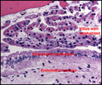
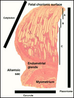
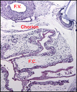
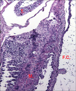
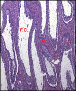
I encountered no additional new findings of placentation in cervids. It is perhaps important to mention though that these authors divided the cotyledons into three zones: The subchorionic comprising 1/5 of villi, was referred to as the "storage zone"; next comes an even narrower "zone of attrition"; the bulk (towards the uterine wall) was made up of the "zone for physiologic exchange". These areas have been depicted in a photograph below. It is true, however, that Mossman (1987) took exception to these terms. He indicated that in the "storage zone" (A in the photo) there accumulates glycogen with advancing gestation. This region, as he pointed out, is the area of atrophy of maternal crypts with columnar cytotrophoblast, an area more like a syndesmochorial region. Hamilton et al. (1960) also drew attention to intraepithelial capillaries (in the trophoblastic surface -see photo below).


Because of the very unusual features identified in the placentation of the Chinese Tufted Deer (Elaphodus cephalophus) a separate chapter is available on that species (see Tufted deer).
The placenta of an Eld's deer (Cervus eldii) weighed 550 g and had 5 large, flat cotyledons. Its umbilical cord measured approximately 15 cm in length.
   
  Tip of villus of delivered placenta from wapiti to show the intraepithelial capillaries in the trophoblast layer.
     
  The placenta of a Javan rusa deer (Cervus timorensis) became available during the extraction of a female neonate with dystocia. The placenta had 3 very large cotyledons (15 x 13 cm) and weighed 750 g with all appendages. Its cord was 17 cm long and 2 cm in width. Its surface and that of the amnion had a remarkable degree of keratin formation (Squamous metaplasia). The stillborn female fetus (7,500 g) had large cystic ovaries and a bicornuate uterus which contained one small and 2 large caruncles in each horn.

Although Hamilton et al. (1960) did not find hematomas, as other authors had, I can confirm that hemosiderin, representing the remains of hematomas, is frequent at the margin of cotyledons. There is early necrosis when the centers of the caruncles develop.
   
 
Weights, Number of Cotyledons, and Cord Lengths
 
Species
Placental weight
Neon Weight
# Cotyledons
Cord
Calamian deer (Axis calamianensis)
150 g
3
6 cm
Calamian deer (Axis calamianensis)
300 g
4
9 cm
Calamian deer (Axis calamianensis)
3
Calamian deer (Axis calamianensis)
75.5 g
2 (?complete)
Musk deer (Moschus moschiferus)
35 g
7
14 cm
Barbary red deer (Cervus elaphus barbarus)
880 g
4,475 g
6
Bactrian wapiti (Cervus elaphus bactrianus)
1,559 g
8
13 cm
Père David's deer (Elaphurus davidianus)
650 g
5
20 cm
Père David's deer (Elaphurus davidianus)
450
15kg
5
20 cm
Siberian reindeer (Rangifer tarandus)
400 g
6
13 cm
Siberian reindeer
200 g
6
Siberian reindeer
550 g
6
12 cm
White-tailed deer (Odocoileus virginianus)
6
Musk deer (Moschus moschiferus)
35 g
7
14 cm
Eld's deer (Cervus eldii)
550 g
5
15cm
Javan rusa deer (Cervus timorensis)
750g
7.5 kg
3
17 cm

(Personal observations from delivered placentas. (above)

Species
Placental weight
Neon Weight
# Cotyledons
Cord
Fallow deer (Dama dama)
8
Père David's deer (Elaphurus davidianus)
5
Roe deer (Capreolus capreolus)
8
Red deer (Cervus elaphus)
8
Reindeer (Rangifer tarandus)
6
Sika deer (Cervus nippon)
6
Indian spotted deer (Cervus axis)
6

(Preceding data are from Hamilton et al., 1960)


  Structure of one-half placentome (Eld's deer). Fetus.


Structure of one-half placentome (Eld's deer). Uterus.
Zone of storage.

(F.V.= Fetal vessel; F.C.= Fetal connective tissue of villi;
M.= Maternal tissue; T.= Trophoblast)


Structure of one-half placentome (Eld's deer). Uterus.
Zone of attrition.

(F.V.= Fetal vessel; F.C.= Fetal connective tissue of villi;
M.= Maternal tissue; T.= Trophoblast)


Structure of one-half placentome (Eld's deer). Uterus.
Zone of exchange.

(F.V.= Fetal vessel; F.C.= Fetal connective tissue of villi;
M.= Maternal tissue; T.= Trophoblast)


5) Details of barrier structure


All cervidae have an epitheliochorial, cotyledonary placenta whose size varies with the size of the animal and offspring. In general, the number of cotyledons is very small in cervids when compared to other ruminants. Because of the apparent fusion of the binucleate trophoblastic cells with some endometrial epithelium, the barrier is now often referred to as being "synepitheliochorial". The trophoblast is cuboidal to columnar and differs in fine structural morphology between the cotyledonary regions and that of the intercotyledonary membranes (see above, Sinha et al., 1969). The fusion of diplokaryocytes with endometrium has also been referred to above.

   
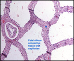
  This is a cross section of the villous tissue of a cotyledon in an Eld's deer (Cervus eldi). Note the widely scattered capillaries in the very loose connective tissue of villi.

T=trophoblast; M=maternal tissue (caruncle).
     
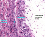
  Fetal/maternal apposition at the bottom of the caruncle crypt in an Eld's deer (Cervus eldi). These show the tip of a villus (left and bottom right), interdigitating with the caruncle. The diplokaryocytes/giant cells are readily identified. Note the very loosely constructed villous cores in which fetal capillaries are widely dispersed.
     
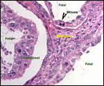
  Tips of villi at implantation site, interdigitating with caruncular clefts. Note the large number of giant cells and mitoses.

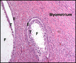
E=endometrium; F=fetal villous connective tissue; T=trophoblast. (Cervus eldi).
     
  Tips of villi at implantation site, interdigitating with caruncular clefts. Note the large number of giant cells and mitoses.

E=endometrium; F=fetal villous connective tissue; T=trophoblast. (Cervus eldi).
     
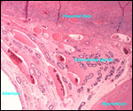
  Margin of cotyledon with endometrial glands next to cotyledon. There is much hemosiderin in the superficial endometrium. (Eld's deer).
     
 
6) Umbilical cord


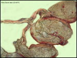
The white-tailed deer has two arteries, two veins and an allantoic duct (Sinha et al., 1969). This is true of the other deer species that I have examined and for what studies have been detailed in publications. Arteries and veins branch, in parallel, to supply the cotyledons. Harrison & Hamilton (1952) found the same to be true in Père David's deer. There are no good descriptions of the lengths of umbilical cords, other than for what is compiled in the table above from a few delivered placentas. They may be longer in utero, but that apparently has not been measured. Spiral turns are not observed, and there are relatively fewer small blood vessels than what has been observed in some other taxa, dolphins for instance. Some are clearly derived from the major cord vessels.
   
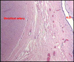
  Portion of umbilical cord of reindeer (Rangifer tarandus). A.D.=allantoic duct. Note the large number of scattered small blood vessels, mostly around allantois.


7) Uteroplacental circulation


There are no functional studies, but Harrison & Hamilton (1952) have given a detailed description of the vasculature in Père David's deer both, for the caruncular supply and for that of cotyledons. In their later paper on fallow deer, Hamilton et al., (1960) provided details on the villous circulation of that species' placenta. Basically, an artery enters the stem villus and divides into a plexiform net of capillaries that spreads out over the secondary villi. The blood then returns by a single vein that exits the stem villus.

8) Extraplacental membranes

The amnion is a delicate, translucent membrane that fuses with the allantois in the first half of pregnancy. It is composed of a connective tissue layer upon which there is a flat, squamous epithelium. Its structure has been thoroughly investigated by Sinha et al. (1970) who reported findings that were similar to those of Harrison and Hamilton (1952) on Père David's deer. The remarkable aspect of many ungulates is the presence of "pustules" or "plaques" on the amnion (Mossman calls them "verrucoids"), areas of epithelial proliferation and layering. Keratohyaline granules can be identified in the cells. This "squamous metaplasia" is also frequent in human placentas; is has no consequence. These authors are mistaken, however, when they liken these normal epithelial outgrowths to human amnion nodosum, which is very differently composed. Human amnion nodosum occurs with prolonged oligohydramnios (usually because of deficient fetal urination). It results from amnion epithelial degeneration and impaction of vernix caseosa. Because of their observation of micropinocytotic vesicles, the authors considered the capacity of the amnionic epithelium to absorb water. There are no blood vessels in the connective tissue, however. The epithelial cells contain glycogen.

The large allantoic membrane is fused with the amnion in those places where the two layers appose each other. It is highly vascularized. The allantoic epithelium is cuboidal to columnar; there are no villi between the cotyledons. The allantoic membrane often degenerates towards the tubal end of the sac. Allantoic fluid - mostly fetal urine - contains much fructose.

In the intercaruncular "free membranes", the trophoblast is cuboidal to columnar, has few diplokaryocytes, and forms regions that are referred to as "areolae". These are opposite endometrial gland openings and serve as sites of absorption of uterine milk.

9) Trophoblast external to barrier

There is no invasion of trophoblast into the endometrium or myometrium, other than the putative fusion of diplokaryocytes with endometrial epithelium to form trinucleated cells described earlier.

10) Endometrium

There is no true decidualization of the endometrium.

11) Various features

There is no subplacenta, nor are there metrial glands.

12) Endocrinology

The parameters on reproductive endocrinology of some cervids have been summarized by Plotka (1998). They are also considered by Puschmann (1989). They appear to be reasonably similar for most cervid species. Most cervids are polyestrous and very seasonal. The pineal gland, via melatonin secretion, is thought to regulate the seasonality and its hormonal cycles. In males, the growth of antlers in these "pecora" is regulated by testosterone and is very well studied. In females, an LH surge follows a period of estrogen production. Pregnancy often occurs in the first year of life in cervidae, and a relatively constant progesterone blood level accompanies it. Importantly, the levels in uterine vein blood are not quantitatively different, ruling out a placental contribution. Also, in those species in which oophorectomy has been performed during gestation, this has led to abortion. This indicates the dependency upon ovarian luteal secretion of progesterone. Estrogen levels are low during pregnancy and rise towards parturition. Other than that a few reports of failure to abort following oophorectomy occur, there is no good evidence that the placenta produces progesterone. In looking for estrogen production by blastocysts, Gadsby et al. (1980) found aromatase activity primarily in pigs, and very little estrogen production by the allantochorion shortly after implantation of roe deer placentas.

I have had the opportunity of examining the fetal ovaries of a Javan Rusa deer (Cervus timorensis) at term that expired during extraction for dystocia. They had numerous follicle cysts and a few atretic follicles, suggesting that fetuses in utero are exposed to gonadotropic stimuli.

13) Genetics

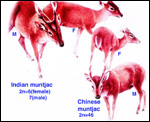
Numerous hybrids have been described among many quite different cervidae. Some are fertile; other are sterile. The reader is referred to the compilation by Gray (1972). The most remarkable hybrid perhaps has occurred (in captivity, of course) between Muntiacus muntjac and Muntiacus reevesi. That is so unusual because of their marked chromosomal difference (2n=6/7 to 46). This hybrid was produced in China (Shi et al., 1980) and was viable but sterile (Shi & Pathak, 1981).

   
  The two muntjac species ("barking deer") with their marked difference in chromosome number but only slight phenotypic variation. (After Grzimek).
     
 
The chromosome number of cervidae is variable. The usually high chromosomal number in many deer species (around 2n=70) presumably reflects their closer relationships, while a few "outliers" are also somewhat removed from the mainstream of cervidae (see Yang et al, 1997). We have established the following chromosome numbers: Moose (70); Axis deer - chital (66); Hog deer (68); Roe deer (70); Barasingha deer (56); Red deer (68); Sambar deer 60; 64-65); Fallow deer - Dama (68); Père David's deer (68); Chinese water deer (Hydropotes inernis) (70); Red brocket deer (Mazama) (50); Reindeer (70); Pudu (70); White-tailed deer (70); Mule deer (70); Chinese muntjac (46); Indian muntjac (6/7) [see Hsu & Benirschke]. Since then, numerous chromosomal banding studies have been done on some of these species and additional species have also been studied: Tufted deer (Elaphodus cephalophus) 2n=46-48 (Shi et al., 1991); white-lipped deer (Cervus albirostris Przewalski) 2n=66 (Wang et al., 1982), and several other muntjacs. Eld's deer (Cervus eldi) has 2n=58 (Neitzel, 1979, she also lists another group of deer species). Sika deer (Cervus nippon hortulorum Swinhoe) has 2n=64-68 (Gustavsson & Sundt, 1969). Another complete listing may be found in Groves & Grubb (1987).

Evolutionary relationships have been discussed at the beginning of this chapter. Modern studies are beginning to provide further insight into disputed areas. Thus, the mtDNA of muntjacs was explored by Lan & Shi (1993). Repetitive DNA of cervids was used for evolutionary questions by Bogenberger et al. (1987). Chromosome "painting" led to a better understanding of muntjac chromosomal fusions (Yang et al., 1995). mtDNA was used in studying hybrids between mule and white-tailed deer (Carr et al, 1986), and so forth.

Genetic heterozygosity positively influenced fetal size and growth in white-tailed deer, while number of fetuses correlated negatively (Cothran et al., 1983).

14) Immunology

Lymphocytic infiltration of the uterine mucosa/epithelium is a frequent, if not regular, feature of ruminant uteri. Lee et al. (1995) studied this in six species of deer. They found a significant cellular increase to occur from early implantation to mid-gestation. They observed also that the size of granules of these cells varies greatly and that they increase with gestation. The investigators observed that these lymphocytes tend to be close to regions where the trophoblastic binucleated cells fuse with the endometrial epithelium. Here they produce the trinucleate cells and give up their granules. The precise reason for these changes remains unknown but "immune recognition" is suspected..

15) Pathological features

Fetal demise of one of twins has been described in an elk uterus (Saunders, 1955), and Mansell & Cringan (1968) found two dead female fetuses with a live female triplet in the uterus of a white-tailed deer. The dead fetuses were enclosed in a single chorion (? monozygotic) and their cotyledons had detached. They referred to other cases of this fetal absorption and attributed it to poor nutrition. We have seen this in axis deer and found no good explanation for the demise.

Major die-offs can occur in island deer due to overcrowding. This was described for sika deer by Christian et al. (1960). In Texas, abnormal antler growth of white-tailed deer was the result of hypogonadism, presumed to result from some poison (Taylor et al, 1964).

Numerous infections have been described in a variety of deer: Toxoplasmosis in mule deer (Dubey, 1982); Theileria cervi infection in white-tailed deer (Schaeffler, 1963); Pneumostrongylus tenuis infection of white-tailed deer (Anderson, 1965), and others. Most important at present is the transmission of the spirochetal organism Borrelia burgdorferi that causes Lyme disease and is transmitted from wild mice to man by the deer tick Amblyomma americanum (e.g. Ebel et al., 2000).

Ratcliffe (1968) reported abnormal antler growth and "wasting" in captive white-tailed deer and related it to abnormal pituitary/adrenal activity. Trophoblastic tumors have not been described, nor are ascending intrauterine infections features of cervid gestations.

The San Diego Zoo has traditionally held many cervid species and, consequently, Griner (1983) gathered much pathological material. Most deaths he recorded were due to trauma, anesthesia, fracture, fights and old age. Dental disease was not uncommon in captivity and outbreaks of malignant catarrhal fever and of blue tongue virus infection were recorded. Only two congenital heart diseases, one cleft palate and one congenital goiter were observed as anomalies, and very rare neoplasms (one lymphosarcoma; a biliary carcinoma in an axis deer, even though deer have no gall bladder) were found. Some parasites posed a continued problem, but reproductive pathology occurred rarely.

16) Physiological data

The placenta of some deer species produces a pregnancy-specific protein B PSPB) which Huang and colleagues (1999) found to be similar, in preparations from moose and elk, to that extracted from cow and sheep placentas. The growth of mule deer fetuses and embryos was methodically recorded by Hudson & Browman (1959). Serum protein profiles of white-tailed and mule deer were studied by Cowan & Johnston (1962). They found great similarity in quantities, but different electrophoretic mobility in these sympatric Odocoileus species.

I am not aware of any studies of blood flow, blood volume, or blood pressures records. It is well known, however, that many deer species have a physiologic tendency to develop sickled erythrocytes. In contrast to human sickle cell anemia patients whose sickling occurs in oxygen-poor environments, that of deer species is initiated by increased oxygenation. The sickle cells are very similar to human sickled red blood cells, with tactoids and similar shapes, but there is no disease associated with deer sickling. It is due to hemoglobin polymorphism. (Undritz et al., 1960; Pritchard et al., 1963; Naik et al., 1964; Kitchen et al., 1964).

17) Other resources

For a wide variety of cervid species the "Frozen Zoo" of CRES at San Diego Zoo possesses fibroblast cell lines and most have had chromosomal analysis.

18) Other relevant features and information needed in future

Several species of deer have become farm animals, especially so with the advent of a complete understanding of their reproductive physiology. Thus, Fletcher (2001) gives examples of farming of Cervus elaphus, C. e. canadensis, Dama dama and D. d. mesopotamica, with successful embryo transfer and other artificial breeding techniques. We know too little of the endocrinology and cord lengths. Also, do placental abnormalities occur?


References


Aitken, R.J.: Ultrastructure of the blastocyst and endometrium of the roe deer (Capreolus capreolus) during delayed implantation. J. Anat. 119:369-384, 1975.

Aitken, R.J., Burton, J., Hawkins, J., Kerr-Wilson, R., Short, R.V. and Steven, D.H.: Histological and ultrastructural changes in the blastocyst and reproductive tract of the roe deer, Capreolus capreolus, during delayed implantation. J. Reprod. Fertile. 34:481-493, 1973.

Anderson, R.C.: The development of Pneumostrongylus tenuis in the central nervous system of white-tailed deer. Path. Vet. 2:360-379, 1965.

Bogenberger, J.M., Neitzel, H. and Fittler, F.: A highly repetitive DNA component common to all Cervidae: its organization and chromosomal distribution during evolution. Chromosoma 95:154-161, 1987.

Carr, S.M., Ballinger, S.W., Derr, J.N., Blankenship, L.H. and Bickham, J.W.: Mitochondrial DNA analysis of hybridization between sympatric white-tailed deer and mule deer in west Texas. Proc. Natl. Acad. Sci. USA 83:9576-9580, 1986.

Cell lines are available from CRES at the San Diego Zoo under: www.sandiegozoo.org

Chapman, D.I. and Dansie, O.: Unilateral implantation in the muntjac deer. J. Zool. Lond. 159:534-536, 1969.

Christian, J.J., Flyger, V. and Davis, D.E.: Factors in the mass mortality of a herd of sika deer, Cervus nippon. Chesapeake Sci. 1:79-95, 1960.

Cothran, E.G., Chesser, R.K., Smith, M.H. and Johns, P.E.: Influences of genetic variability and maternal factors on fetal growth in white-tailed deer. Evolution 37:282-291, 1983.

Cowan, I.M. and Johnston, P.A.: Blood serum protein variations at the species and subspecies level in deer of the genus Odocoileus. Systematic Zool. 11:131-138, 1962.

Dubey, J.P.: Isolation of encysted Toxoplasma gondii from muscles of mule deer in Montana. JAMA 181:1535, 1982.

Ebel, G.D., Campbell, E.N., Goethert, H.K., Spielman, A. and Telford, S.R.: Enzootic transmission of deer tick virus in New England and Wisconsin sites. Amer. J. Trop. Med. Hyg. 63:36-42, 2000.

Fletcher, T.J.: Deer: New domestic farm animals defined by controlled breeding. In press, 2001.

Gadsby, J.E., Heap, R.B. and Burton, R.D.: Oestrogen production by blastocyst and early embryonic tissue of various species. J. Reprod. Fertil. 60:409-417, 1980.

Gray, A.P.: Mammalian Hybrids. Second edition. A Check-List with Bibliography. Commonwealth Agricultural Bureaux, Farnham Royal, Slough, UK, 1972.

Griner, L.A.: Pathology of Zoo Animals. Zoological Society of San Diego, 1983.

Groves, C.P. and Grubb, P.: Relationships of living deer. In, Biology and Management of the Cervidae. C.M. Wemmer, ed. Smithsonian Inst. Press, Washington, 1987, pp. 21-59.
Gustavsson, I. And Sundt, C.O.: Three polymorphic chromosome systems of centric fusion type in a population of Manchurian Sika deer (Cervus nippon hortulorum Swinhoe). Chromosoma 28:245-254, 1969.

Hamilton, W.J., Harrison, R.J. and Young, B.A.: Aspects of placentation in certain Cervidae. J. Anatomy 94:1-33, 1960.

Harrison, R.J. and Hamilton, W.J.: The reproductive tract and the placenta and membranes of Père David's deer (Elaphurus davidianus Milne Edwards). J. Anat. 86:203-224, 1952.

Hsu, T.C. and Benirschke, K.: An Atlas of Mammalian Chromosomes. Springer-Verlag, New York, 1975.

Huang, F., Cockrell, D.C., Stephenson, T.R., Noyes, J.H. and Sasser, R.G.: Isolation, purification, and characterization of pregnancy-specific protein B from elk and moose placenta. Biol. Reprod. 61:1056-1061, 1999.

Hudson, P. and Browman, L.G.: Embryonic and fetal development of the mule deer. J. Wildlife Manag. 23:295-304, 1959.

Kitchen, H., Putnam, F.W. and Taylor, W.J.: Hemoglobin polymorphism: Its relation to sickling of erythrocytes in white-tailed deer. Science 144:1237-1239, 1964.

Kurnosov, K.M.: Interfetal placental connections of the elk in embryonic parabiosis. Proc. Akad. Nauk. SSR Doklady, Biol. Sect. 142:92-94, 1962 (in English).

Lan, H. and Shi, L.: Restricted endonuclease analysis of mitochondrial DNA of muntjac and related deer. In, Deer of China, N. Ohtaishi and H.-I. Sheng, eds. Elsevier Science Publ., 1993.

Lawn, A.M., Chiquoine, A.D. and Amoroso, E.C.: The development of the placenta in the sheep and goat: an electronmicroscope study. J. Anat. 105:557-578, 1969.

Lee, C.S., Gogolin-Ewens, K. and Brandon, M.R.: Comparative studies on the distribution of binucleate cells in the placentae of the deer and cow using monoclonal antibody, SBU-3. J. Anat. 147:163-179, 1986.

Lee, C.S., Wooding, F.B. and Morgan, G.: Quantitative analysis of intraepithelial large granular lymphocyte distribution and maternofetal cellular interactions in the synepithelichorial placenta of the deer. J. Anat. 187:445-460, 1995.

McMahon, C.D., Fisher, M.W., Mockett, B.G. and Littlejohn, R.P.: Embryo development and placentome formation during early pregnancy in red deer. Reprod. Fertil. Dev. 9:723-730, 1997.

Mansell, W.D. and Cringan, A.T.: A further instance of fetal atrophy in white-tailed deer. Canad. J. Zool. 46:33-34, 1968.

Miyamoto, M.M., Kraus, F. and Ryder, O.A.: Phylogeny and evolution of antlered deer determined from mitochondrial DNA sequences. Proc. Natl. Acad. Sci. USA 87:6127-6131, 1990.

Mossman, H.W.: Vertebrate Fetal Membranes. MacMillan, Houndmills, 1987.

Naik, S.N., Bhatia, H.M., Baxi, A.J. and Naik, P.V.: Hematological study of Indian spotted deer (Axis deer). J. Exper. Zool. 155:231-236, 1964.

Neitzel, H.: Chromosomenevolution in der Familie der Hirsche (Cervidae). Bongo 3:27-38, 1979.

Nowak, R.M. and Paradiso, J.L.: Walker's Mammals of the World, Vol. II. 4th edition. The Johns Hopkins University Press, Baltimore, 1983.

Plotka, E.D.: Deer. In, Encyclopedia of Reproduction. E. Knobil and J.D. Neill, eds. Academic Press, San Diego 1998, Vol. I, pp.842-857.

Pritchard, W.R., Malewitz, T.D. and Kitchen, H.: Studies on the mechanism of sickling of deer erythrocytes. Exp. Molec. Pathol. 2:173-182, 1963.

Puschmann, W.: Zootierhaltung. Vol. 2, Säugetiere. VEB Deutscher Landwirtschaftsverlag, Berlin, 1989.

Randi, E., Mucci, N., Claro-Hergueta, F., Bonnet, A. and Douzery, E.J.P.: A mitochondrial DNA control region phylogeny of the Cervinae: speciation in Cervus and implications for conservation. Animal Conserv. 4:1-11, 2001.

Ratcliffe: Environment, behavior and disease: Observations and experiments at the Philadelphia Zoological Garden. Trans. & Studies Coll. Physic. Philadelphia 36:7-21, 1968.

Saunders, J.K.: Fetus in yearling cow elk, Cervus canadensis. J. Mammal. 36:145, 1955.

Schaeffler, W.F.: Serologic tests for Theileria cervi in white-tailed deer and for other species of Theileria in cattle and sheep. Amer. J. Vet. Res. 24:784-791, 1963.

Shi, L., Ye, Y., Duax, X.: Comparative cytogenetic studies on the red muntjac, Chinese muntjac, and their F1 hybrids. Cytogenet. Cell Genet. 26:22-27, 1980.

Shi, L. and Pathak, S.: Gametogenesis in a male Indian muntjac x Chinese muntjac hybrid. Cytogenet. Cell Genet. 30:152-156, 1981.

Shi, L., Yang, F. and Kumamoto, A.: The chromosomes of tufted deer (Elaphodus cephalophus) Cytogenet. Cell. Genet. 56:189-192, 1991.

Sinha, A.A., Seal, U.S., Erickson, A.W. and Mossman, H.W.: Morphogenesis of the fetal membranes of the white-tailed deer. Amer. J. Anat. 126:201-242, 1969.

Sinha, A.A., Seal, U.S., Erickson, A.W.: Ultrastructure of the amnion and amniotic plaques of the white-tailed deer. Amer. J. Anat. 127:369-396, 1970.

Taylor, D.O.N., Thomas, J.W. and Marburger, R.G.: Abnormal antler growth associated with hypogonadism in white-tailed deer in Texas. Amer. J. Vet. Res. 25:179-185, 1964.

Undritz, E., Betke, K. and Lehmann, H.: Sickling phenomenon in deer. Nature 187:333-334, 1960.

Vrba, E.S. and Schaller, G.B., eds.: Antelopes, Deer, and Relatives. Fossil Record, Behavioral Ecology, Systematics, and Conservation. Yale University Press, New Haven, 2000.

Wang, Z., Du, D.R., Xu, J. and Che, Q.: Karyotype, C-banding and G-banding patterns of white-lipped deer (Cervus albirostris Przewalski). Acta Zool. Sin. 28:250-255, 1982.(in Chinese).

Weldon, W.F.R.: Note on placentation of Tetraceros quadricornis. Proc. Zool. Soc. London pp.2-4, 1884. (Cited by Mossman, 1987).

Wooding, F.B.: The role of the binucleate cell in ruminant placental structure. J. Reprod. Fertil. Suppl. 31:31-39, 1982.

Wooding, F.B.: Frequency and localization of binucleate cells in the placentomes of ruminants. Placenta 4:527-539, 1983.

Wooding, F.B., Morgan, G. and Adam, C.L.: Structure and function in the ruminant synepitheliochorial placenta: central role of the trophoblast binucleate cell in deer. Microsc. Res. Tech. 38:88-99, 1997.

Wurster, D.H. and Benirschke, K.: Chromosome studies in some deer, the springbok, and the pronghorn, with notes on placentation in deer. Cytologia 32:273-285, 1967.
Yang, F., Carter, N.P., Shi, L. and Ferguson-Smith, M.A.: A comparative study of karyotypes of muntjacs by chromosome painting. Chromosoma 103:642-652, 1995.

Yang, E., O'Brien, P.C.M., Wienberg, J., Neitzel, H., Kin, C.C. and Ferguson-Smith, M.A.: Chromosomal evolution of the Chinese muntjac (Muntjacus reevesi). Chromosoma 106:37-43, 1997.


back to top

© 2005.
welcome | home | intro | placentation | glossary | author | contact