| |
9)
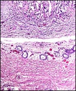
Trophoblast external to barrier
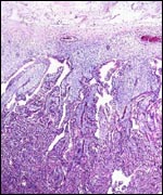
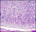
There is no extravillous trophoblast or infiltration of the endometrium
by fetal cells.
10)
Endometrium
No true decidua is being formed.
11)
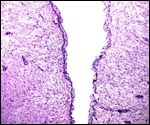
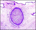
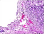
Various features
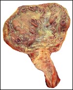
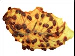
The endometrium beneath the implanted cotyledon is not infiltrated by
trophoblast and appears histologically fibrous.
12)
Endocrinology
No studies have been performed. The fetal ovaries were very small and
unstimulated.
13)
Genetics
The Cuvier's gazelle has 32 chromosomes in females, and 33 chromosomes
in males. This is due to the Robertsonian translocation of the X-chromosome
to an autosome (Kumamoto & Bogart, 1984). These authors also found
minor banding variations among several animals. Vassart et al. (1995)
further delineated the cytogenetic arrangements of gazelle chromosomes,
using the caprine banded karyotypes as the presumed ancestral form. These
two groups of authors both found the karyotypes to be similar to that
of Gazella leptoceros, despite their significantly different phenotypes.
Hybrids are unknown.
Inbreeding ("depression"), however, may be a problem in this
species. Roldan et al. (1998) and Gomendio et al. (2000) studied the ejaculatory
semen quality of this species and that of the less endangered G. dama.
Their findings suggested that, while the quality of Cuvier's gazelle semen
was inferior, it had not yet reached devastating proportions.
14)
Immunology
No studies are known to me.
15)
Pathological features
This animal died from peritonitis due to intestinal perforation. It also
had gastric ulcers and massive whipworm infestation. Toxoplasmosis has
been reported by Stiglmair-Herb (1987), and a variety of helminthes were
found in animals captive in Spain (Ortiz et al., 2001a). These authors
worked with a large number of animals (58) and experimented with various
anthelminthic regimes (Ortiz et al., 2001b).
16)
Physiologic data
No studies are known to me other than the collection of hematologic and
biochemical data by Casado et al. (1991).
17)
Other resources
Cell lines of numerous animals are available from CRES
at San Diego Zoo by contacting Dr. Oliver Ryder at: oryder@ucsd.edu.
18)
Other remarks - What additional Information is needed?
Term placentas, their weights, and dimensions are still unknown. It would
also be of interest to know if hybridization with slender-horned gazelles
occurs and whether they would be fertile.
Acknowledgement
The animal photographs in this chapter come from the Zoological Society
of San Diego. I appreciate also very much the help of the pathologists
at the San Diego Zoo.
References
Casado, A., de la Torre, R., Lopez-Fernandez, E. and Ruiz del Castillo,
B.: Comp. Biochem. Physiol. B.: 99:637-640, 1991.
Furley,
C.W.: Reproductive parameters of African gazelles: Gestation, first fertile
matings, first parturition and twinning. Afr. J. Ecol. 24:121-128, 1986.
Gomendio,
M., Cassinello, J. and Roldan, E.R.: A comparative study of ejaculate
traits in three endangered ungulates with different levels of inbreeding:
fluctuating asymmetry as an indicator of reproductive and genetic stress.
Proc. Roy. Soc. Lond. B Biol. Sci. 267:875-883, 2000.
Gotch,
A.F.: Mammals - Their Latin Names Explained. Blandford Press, Poole, Dorset,
1979.
Jones,
M.L.: Longevity of ungulates in captivity. Int. ZooYbk. 32:159-169, 1993.
Krölling,
O.: Uber den Bau der Antilopenplazentome. Z. mikrosk. anat. Forschg. 27:216-232,
1931.
Kumamoto,
A.T. and Bogart, M.H.: The chromosomes of Cuvier's gazelle. Chapter 10,
pp. 100-108, in: One Medicine, Springer-Verlag, NY , 1984.
Nowak,
R.M.: Walker's Mammals of the World. 6th ed. The Johns Hopkins Press,
Baltimore, 1999.
Olmedo,
J.E., Escos, J. and Gomedio, M.: Reproduction de Gazella cuvieri en captivité. Mammalia 49:501-507, 1985.
Ortiz,
J., Ruiz de Ybanez, M.R., Garijo, M.M., Goyena, M., Espeso, G., Abaigar,
T. and Cano, M.: Abomasal and small intestinal nematodes from captive
gazelles in Spain. J. Helminthol. 75:363-365, 2001a.
Ortiz,
J., Ruiz de Ybanez, M.R., Abaigar, T., Garijo, M.M., Espeso, G., and Cano,
M.: Oral administration of mebendazole failed to reduce nematode egg shedding
in captive African gazelles. Onderstepoort J. Vet. Res. 68:79-82, 2001b.
Roldan,
E.R., Cassinello, J., Abaigar, T. and Gomendio, M.: Inbreeding, fluctuating
asymmetry, and ejaculate quality in an endangered ungulate. Proc. R. Soc.
Lond. B Biol. Sci. 265:243-248, 1998.
Stiglmair-Herb,
M.T.: Microparasitosis (toxoplasmosis) in mountain gazelles (Gazella
g. cuvieri). Berl. Munch. Tierärztl. Wochenschr. 100:273-277,
1987 (in German).
Vassart,
M., Séguéla, A. and Hayes, H.: Chromosomal evolution in
gazelles. J. Hered. 86:216-227, 1995.
|
