 |
| (Clicking
on the thumbnail images will launch a new window and a larger version
of the thumbnail.) |
| Last updated: April 25, 2008 |
Coquerel’s Sifaka
Propithecus (verreauxi) coquereli
Order: Primates
Family: Indridae (formerly Indriidae)
1) General Zoological Data
This is a severely endangered Madagascan primate from the Northwestern region of Madagascar whose taxonomic status is described by Wilson & Reeder (1993) in the way as is indicated above. Groves (2001) referred to this ‘sub-species’ as belonging to ‘the Propithecus verreauxi group’. According to Gotch (1979) ‘indri’ originates from the Malagasy word meaning ‘look’, or ‘there it is’; ‘sifac’ is also a Malagasy word and appears to simulate the sound the animals produce (Nowak, 1999). C. Coquerel was a French zoologist (1822-1867). Few breeding colonies exist; most notable is the one at the Duke Lemur Center, Durham, NC whence this placenta comes. Sifakas are herbivorous and frugivorous, and they prefer high trees in which to roam and sleep during the night. The maximal lifespan for Coquerel’s sifaka in captivity is 30 years (Weigl, 2005). Puschmann (1989) provided information for their captive care and breeding.
Thenius & Hofer (1960) discussed the evolution of primates extensively and made much reference to the prosimians in general. Prosimian ancestors arrived in Madagascar in the Old Tertiary (~50MYA) and speciated into a variety of genera; now most of these animals are severely endangered (see also Starck, 1995).
 |
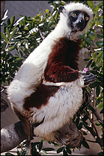
Coquerel’s sifaka from the web. |
 |
Female Coquerel’s sifaka at San Diego Zoo. |
2) General Gestational Data
Sifakas weigh between 3 and 7 kg (Nowak, 1999) and deliver single young after a gestation period of 130-141 days. They are reported to weigh only ~ 40 g. In that case, this stillborn fetus is extremely heavy. Perhaps this relates to the somewhat edematous-appearing placenta. Unfortunately, the fetus was frozen for future study and is not yet dissected.
3) Implantation
No early development has been described.
4) General Characterization of the Placenta
This placenta was made available from a Cesarean section of a stillborn sifaka by the Duke Lemur Center. The placenta weighed 48 g (after fixation) and came to me formalin-fixed with a very short central umbilical cord. The neonate of this primigravid female weighed 120 g (normal for this Center is reported to be 100 g, but the literature indicates that neonates weigh only ~ 40 g), and this was full-term gestation. The fetus was delivered by Cesarean section because, when the mother felt ‘uncomfortable’, sonography showed the absence of fetal cardiac activity. The amniotic fluid was somewhat discolored and there was acute inflammation of the membranes.
This is a diffuse, villous, epithelio-chorial placenta with areas that suggest the formation of small cotyledons. It has only very rarely been studied, earliest and very briefly, by Anthony (1908). He had access to a near-term pregnant uterus of Propithecus verreauxi typicus, but his description is of a typical Coquerel sifaka. He asserted that the placenta extended into both uterine horns. At the ‘cephalic pole’, Anthony found the placental villi to be sparse and described the placenta as being bell-shaped. Mossman (1987) only reviewed this species minimally but confirmed the findings from the sparse literature. Unlike some features of other Strepsirrhini, I found no typical or complete areolae in this placenta and there was also no pigment; nevertheless, three areas were present in numerous slides prepared that could have been areolae, at least structurally. They contained necrotic debris which contained also birefringent crystals; they are depicted below. These areas were enclosed by a thin layer of muscular tissue. The villous capillaries are often located directly beneath the single-layered trophoblastic epithelium and lie in a somewhat edematous stroma that contains numerous cells that are interpreted as being macrophages. Nevertheless, they are CD 68-, CD1a-, and S100- negative and may thus not be true macrophages. Frequently, the trophoblast is indented by the capillaries. A thin, vascularized allantoic sac was present that connected to the fetal bladder by a small umbilical allantoic duct.
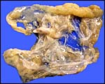 |
Gross appearance of the formalin-fixed placenta with centrally located umbilical cord. |
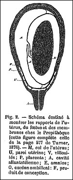 |
The original diagram from the description by Anthony (1908). |
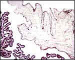
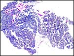 |
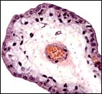
Low-power view of the somewhat lobulated placenta. |
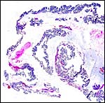 |
The ‘thinner’ portions of the placenta (?cephalic) are still covered with trophoblast. |
5) Details of fetal/maternal barrier
The villi have a single layer of trophoblast which contained occasional mitoses. The fetal capillaries are close to the trophoblast and often bulge into it. Many trophoblastic cells appeared to be edematous and a few had distinct eosinophilic, granular cytoplasmic inclusions. They are also shown below.
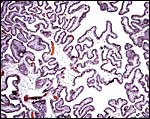 |
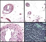
Somewhat edematous villous structures. |
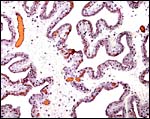 |
Note the proximity of capillaries to the trophoblastic surface. |
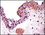 |
The indentation of trophoblast by capillaries is variable. |
 |
Single villus to show the single layer of trophoblast with mitosis at 2 o’clock. |
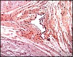
 |
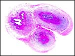
These are the two areas of possible ‘areolae’, with polarizing light illumination in panel “D” to show the rectangular crystals. |
 |
The trophoblast has focally eosinophilic granular inclusions in the cytoplasm. |
6) Umbilical cord
The cord was short and measured here approximately 5 cm in length. It had three vessels, a small central allantoic duct and there was no squamous metaplasia on its surface. There were no ‘smaller’ blood vessels as are seen in many other animal placentas (e.g. the dolphin).
 |
Cross section of the umbilical cord; vein at left. |
 |
Allantoic duct in the center of the umbilical cord. Other than the three large umbilical vessels, there are no smaller blood vessels associated with the duct. |
 |
A small mural thrombus was found in a surface fetal blood vessel. |
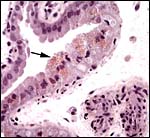
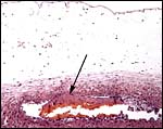
7) Uteroplacental circulation
This unusual placenta with acute inflammatory reaction in the membranes and fetal demise had a mural thrombus in one of the surface vessels (arrow). This may be the result of prenatal infection. No other data are available to my knowledge.
8) Extraplacental membranes
The allantoic sac is large and irregularly formed. As Anthony (1908) already reported, it remains until term gestation and has some ‘diverticula’. The amnion was focally markedly necrotic; most areas were very edematous and contained numerous apparent macrophages; they did not stain with CD 68, however, as would be expected. In addition, foci of acute chorioamnionitis were present.
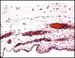
 |
Membranes and a small portion of villous tissue. |
 |
Edematous ‘membranes’ with vascularized allantoic membrane below and the degenerating amnion above. |
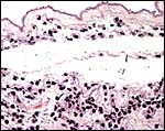 |
Marginal membranes with acute chorioamnionitis. |
 |
Low-power magnification of the marginal membranes with acute chorioamnionitis. |
9) Trophoblast external to barrier
There is no infiltration of the myometrium in this epitheliochorial placentation.
10) Endometrium
The females have a bicornuate uterus that is described as being only ‘mildly’ bicornuate, and there is no development of decidua, as was also stated by Anthony (1908).
11) Various features
Males have a baculum.
12) Endocrinology
I have been unable to find any data.
13) Genetics
This sifaka has 48 chromosomes (Poorman, 1983). A banded karyotype from CRES is shown here that was already depicted by O’Brien et al. (2006). Hybrids have not been reported.
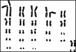 |
Female sifaka chromosome set from CRES, as published by O’Brien et al. (2006). |
14) Immunology
I have not been able to find any information.
15) Pathological features
Griner (1983) examined 6 female sifakas that died soon after arrival at San Diego Zoo from inadequate diet and ‘tympanitis’; early acute gastritis was described in a female sifaka of this group by Scott (1992). The present animal was stillborn and must have been edematous; it had chorioamnionitis in the placental membranes that suggested the reason for the cloudy amniotic fluid and possibly also the demise.
16) Physiologic data
There are no data.
17) Other resources
Cell strains are present at CRES (San Diego Zoo) and can be obtained by contacting Dr. Oliver Ryder (oryder@ucsd.edu).
18) Other remarks – What additional Information is needed?
Early stages of placental development are badly needed, as well as an implanted placenta. There are no endocrine or physiological data.
Acknowledgement
One animal photograph in this chapter comes from a web site on Sifakas, the other is from the San Diego Zoo. The placenta was kindly made available by Dr. Sarah Zehr of the Duke Lemur Center, 3705 Erwin Road, Durham, NC 27705-5000.
References
Anthony, R.: Note sur un foetus de Propithèque et ses membranes. Ann. Sc. Nat. (Ser.9) 7:243-248, 1908. (in French).
Gotch, A.F.: Mammals – Their Latin Names Explained. Blandford Press, Poole, Dorset, 1979.
Griner, L.A.: Pathology of Zoo Animals. Zoological Society of San Diego, San Diego, California, 1983.
Groves, C.: Primate Taxonomy. The Smithsonian Institution, Washington, DC. 2001.
Mossman, H.W. and Duke, K.L.: Comparative Morphology of the Mammalian Ovary. University of Wisconsin Press, Madison, Wisconsin, 1973.
Nowak, R.M.: Walker’s Mammals of the World. 6th ed. The Johns Hopkins Press, Baltimore, 1999.
O’Brien, S.J., Menninger, J.C. and Nash, W.G., eds.: Atlas of Mammalian Chromosomes. Wiley-Liss; A. John Wiley & Sons, Inc. Hoboken, N.J., 2006.
Poorman, A.P.: The banded chromosomes of Coquerel’s sifaka, Propithecus verreauxi coquereli (Primates, Indriidae). Intern. J. Primatol. 4-419-425, 1983.
Puschmann, W.: Zootierhaltung. Vol. 2, Säugetiere. VEB Deutscher Landwirtschaftsverlag Berlin, 1989.
Scott, G.B.D.: Comparative Primate Pathology. Oxford University Press, 1992.
Starck, D.: Säugetiere. In, Lehrbuch der Speziellen Zoologie (A. Kaestner, Founder). Vol. II Wirbeltiere. Teil 5/2. Gustav Fischer Verlag, Jena, 1995.
Thenius, E. and Hofer, H.: Stammesgeschichte der Säugetiere. Springer-Verlag, Berlin, 1960.
Weigl, R.: Longevity of Mammals in Captivity; From the Living Collections of the World. [Kleine Senckenberg-Reihe 48]. E. Schweizerbart’sche Verlagsbuchhandlung (Nägele und Obermiller), Stuttgart, 2005.
Wilson, D.E. and Reeder, D.A.M.: Mammal Species of the World. A Taxonomic and Geographic Reference. 2nd ed. Smithsonian Institution Press, Washington, DC, 1993.