Angolan Colobus Monkey
Colobus angolensis

Order: Primates
Family: Cercopithecidae

1) General Zoological Data


There are five members of the black and white colobus monkeys (Nowak, 1999), of which the Guereza (Colobus guereza) is perhaps the most widely studied species. The name "colobus" derives from the Greek 'kolobos', meaning docked or mutilated, because of the very short or absent thumb (Gotch, 1979). The variability of short digits in various primates is further discussed by Tague (2002).

The Angolan colobus monkey is widely distributed in several countries of East Africa. They are large, leaf-eating primates whose complex, subdivided stomach is specialized to produce fatty acids from the exclusively gastric leaf fermentation. These large monkeys have a beautiful black and white plumage, while newborns are a pure white. The plumage has led to an extensive fur trade with marked reduction of all populations. The animals live in small family groups although larger aggregations have been observed. There is good evidence of chimpanzee hunting of these animals.

The phylogeny of the colobinae has been described in detail by Groves (1970). In his discussion of the "forgotten leaf-eaters", Groves links the Asiatic and Indian forms (Pygathrix, Rhinopithecus, Nasalis, etc.) together with the African forms in part because of their gastric peculiarities, their limb structure, nasal and skull features. Moreover, most have 2n=44, with very similar chromosomes (a few have 2=48). They would all appear to be closely related forms. DNA studies conducted by Page et al. (1999) also suggest a division of the Asian from the African colobinae.

Many zoos now keep these leaf-eating primates that were once thought to be problematic for zoos. The dietary constraints are perhaps the most important problems encountered, as Hollihn (1973) pointed out.


Adult and young Colobus guereza monkeys at San Diego Zoo's Wild Animal Park.

     
  Subadult Angolan colobus monkey at San Diego Zoo.
     
  Newborn male colobus monkey. Note the congested face of the, normally white, newborn due to his face presentation in labor.
     
  The longevity of C. angolensis is around 17 years, according to Jones (1977).

2) General Gestational Data

Colobus monkeys normally have a single offspring after a gestation of around 170-180 days. Newborns weigh around 180-200 g and the placental weight of this birth (129 g) is apparently within normal range. Estrus lasts 1-3 days. Adults weight between 5.4 and 14.5 kg (Nowak, 1999).

3) Implantation

No information is published of early implantation but since the placenta has two disks, it is assumed that one disk attached anterior, the other posterior in the uterus. General remarks on comparative placentation in the cercopithecidae can be found in an earlier contribution (Benirschke & Miller, 1982). Baur (1970) calculated that the villous surface of a colobus placenta is a remarkable 383cm2/cm3 while that of humans was calculated to be only 248 cm2/cm3. The total surface was 1.7-5.7 m2 (human12 m2).

4) General Characterization of the Placenta

I have had an opportunity to study four placentas of various colobus monkeys. They had the following dimensions:

   
Number
1 Disk
2 Disk
Combined
1 Disk
2 Disk
Cord
3971
55 g
65 g
120 g
10x8cm
8x9cm
21cm
4038
90 g
45 g
135 g
12 cm
8 cm
NA
11660
105 g
10 cm
44270
67 g
62 g
129 g
10x9 cm
9x8 cm
3.5 cm
6805
103.9 g
(single disk)
9.6x8.6
47.6 cm
   
  The placenta of a term male neonate was last available. The mother had labored all night, was exhausted and for that reason a Cesarean section was performed; a face presentation was found. The male neonate died shortly after birth. It weighed 750 g, and measured 23 cm in CR length. The placenta had two disks that were connected by four blood vessels running in the membranes on both sides of the uterus. It weighed 129 g (one disk 67 g, the other 62 g). They measured 10 x 9 x 1 cm and 9 x 8x 1 cm respectively. There were several infarcts as is a common finding in cercopithecids (see chapters on Douc langur and Guenons). The first two placentas of this table, however, were free of infarcts. The short umbilical cord was inserted centrally on the smaller disk.The last placenta was monodiscoid and had a relatively long cord.

This is a hemochorial placenta with identical histological features as seen in Douc langurs, macacs and other cercopithecidae. It is superficially invasive.
   
  Monodiscoid placenta of Angolan Colobus monkey.
     
  The fetal surface of the delivered placenta of this stillborn animal with two lobes.
     
  Maternal surface of the two lobes. The yellow-white infracts are scattered in both lobes.
     
  5) Details of fetal/maternal barrier

Cercopithecidae, as the colobus monkey, have a villous hemochorial placenta with the tertiary villi constructed very much like those of humans. They are covered by syncytium, beneath which resides a layer of cytotrophoblast. Mitoses are only seen in the cytotrophoblast; they are absent in the syncytium. The "anchoring villi" at the maternal base of the placenta are connected by "extravillous trophoblast" (also referred to as "X-cells") that produces major basic protein. In the connective tissue of the villi are a few macrophages ("Hofbauer cells"). The extravillous trophoblast infiltrates superficially into the decidua basalis.
   
  Fetal surface of the placenta with loosely attached amnion and fetal vessels in the chorion.
     
  Marginal portion of term colobus placenta with infarct and intervillous thrombus.
     
  This is the maternal surface with the placental implantation. The large number of invasive extravillous trophoblast ("X-cells") attaches to the basal endometrium.
     
  Low-power microscopic appearance of a section of this delivered placenta.
     
  Margin of the placenta with focal old retroplacental hematoma and marginal infarct.
     
  "Anchoring" villi with extravillous trophoblast that attach villi to the decidua below.
     
  Major stem villus at left. The darkly-stained cells are the syncytiotrophoblast cells.
     
  6) Umbilical cord  
     
  Cross section of umbilical cord with two arteries and one vein.
     
  This fetus' umbilical cord was only 3.5 cm long and it was not spiraled. Surely, however, much more cord was present originally and was not submitted for study. The cord contained two arteries and one vein. There were no remnants of ducts. The longest cord measured was 47.6 cm long.

7) Uteroplacental circulation

No implanted placenta has yet been described. One may assume, however, that the vascular relations are similar to the widely studied rhesus monkey placenta. See the chapters on macacs, guenons, Douc langur.

8) Extraplacental membranes

   
  Fetal placental surface showing the loose attachment of the amnion to the chorion and the decidua capsularis and some extravillous trophoblast. As in other cercopithecidae, there are no atrophied villi as seen in human membranes.
     
  Mural thrombosis in connecting vessels between the disks.
     
  Mural thrombi in membranous connecting vessels.
     
  The "free" membranes of colobus monkey placentas lack the atrophic villi seen in human gestations. There is, however, an extensive decidua capsularis. Four large fetal blood vessels spanned from one lobe to the other in the membranous chorion. The amnion is avascular and loosely attached to the chorionic membrane. It is possible that the mural thrombi observed in these connecting membranous vessels of this placenta are the result of pressure by the fetus upon these blood vessels.

9) Trophoblast external to barrier

No implanted placenta has been described. More likely than not, there is no invasion of the myometrium or even major vessels by trophoblast except for the spiral arterioles. This has been beautifully depicted by Houston (1969) for the baboon.

10) Endometrium

The decidual formation is typical of all primates and the basal endometrium has a typical Nitabuch fibrinoid layer.

11) Various features

No subplacenta or metrial glands exist.

12) Endocrinology

I know of no relevant endocrine studies of this species.

13) Genetics

Colobus monkeys have 2n= 44 chromosomes composed of only metacentric or submetacentric elements. There are no acrocentrics with the exception of the Y chromosome (Bender & Chu, 1963; Chiarelli, 1963). In our comparisons of the Y chromosome between different Colobus species, the Y-chromosome of C. angolensis is larger and has more distal heterochromatin than other species. One hybrid between two subspecies is listed by Gray (1972). Lang (1973) described a surviving hybrid between C. polykomos vellerosus and C. abyssinicus occidentalis. "Swarms of hybrids between C. polykomos and C. vellerosus were mentioned by Nowak (1999). Lee et al. (1999) found different chromosomal localizations of human gamma-X sequences among golden monkeys and colobus.

14) Immunology

A new immunodeficiency virus was discovered in guerezas by Courgnaud et al. (2001).

15) Pathological features

A variety of pathologic features have been described, aside from the chorioamnionitis present in this animal's placenta. Most likely, this inflammation is related to the prolonged labor. Most cercopithecidae, including the colobus monkey, have infarcts in the placenta without showing clinical evidence of preeclampsia, which is the commonest cause of infarcts in human gestations. Also in contrast to human placentas in cases of preeclampsia, the maternal spiral arterioles in these primates do not show the changes of "atherosis" (see chapters on Douc langur and guenons).

An outbreak of measles was described in guerezas by Scott & Keymer (1975). Gastric dilatation of their complex stomachs has been described (Soave, 1978; Farah et al., 1993). An infection with bovine tubercle bacilli caused death of two colobus monkeys and 4 white rhinoceroses in a zoo (Stetter, et al., 1995). Aerosolic spread of the organisms at cleaning was blamed. A free-living ameba (Balamuthia mandrillaris) caused fatal infection of several primates, including a guereza, in our zoo (Rideout et al., 1997). Sialolithiasis, masquerading as a dental disease, was described by Ensley et al. (1981). Viral papillomas of hand and feet were described in guerezas by Boever & Kern (1976), and subsequently in several animals by Rangan et al. (1980). DNA from a penile papilloma in colobus monkeys was then characterized by Reszka et al. (1991) and shown to be similar to human papilloma viruses. Fatal infection with herpesvirus was reported by Loomis et al. (1981) in a colobus monkey housed near rhesus monkeys, the usual viral carriers. Shigellosis became a serious disease of primates at the National Zoo, including colobus monkeys (Banish et al., 1993). Eberhard et al. (2001) identified cyclospora from a variety of African primates, including colobus monkeys (Cyclospora colobi) and showed lack of seasonality of infection, lack of cross-infection between different species, and they characterized the DNA.
Schultz (1956) referred to the frequency of dental disease in colobinae. Griner (1983) described occasional lung mites (acariasis), and arteriosclerosis with chronic nephritis in a 23 year old colobus monkey. Colitis and pulmonary edema were mentioned to occur in colobinae by Scott (1992) who also discussed the gastric morphology.

   
  This shows a typical placental infarct of villous tissue.
     
  Intervillous thrombi are common and contain fetal and maternal blood.
     
  At left is normal placental tissue, at right is one of the infarcts.
     
  Fetal surface with mild inflammatory infiltration of the chorion.
     
  Marked leukocytic infiltration of the decidua capsularis on the membranes.
     
  16) Physiologic data

I know of no relevant data

17) Other resources

Cell lines are available of several colobus species from CRES at the San Diego Zoo by contacting Dr. Oliver Ryder at oryder@ucsd.edu.

18) Other remarks - What additional Information is needed?

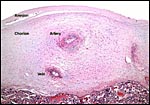
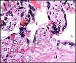
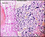
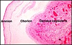
Early implantations, a pregnant uterus to delineate depth of trophoblast invasion, and endocrine data would be desirable.

Acknowledgement

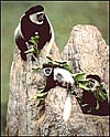
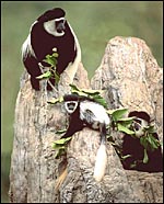
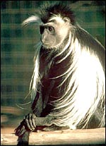
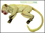
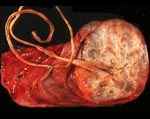
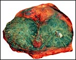
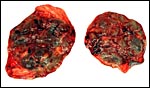
The animal photographs in this chapter come from the Zoological Society of San Diego. I appreciate also very much the help of the pathologists at the San Diego Zoo.

References

Banish, L.D., Sims, R., Sack, D., Montali, R.J., Phillips, L. Jr. and Bush, M.: Prevalence of shigellosis and other enteric pathogens in a zoological collection of primates. JAVMA 203:126-132, 1993.

Baur, R.: Uber die Relation zwischen Zottenoberfläche der Geburtsplacenta und Gewicht des Neugeborenen bei verschiedenen Säugetieren. Z. Anat. Entwickl.-Gesch. 13131-38, 1970.

Bender, M.A. and Chu, E.H.Y.: The chromosomes of primates. Chapter 7 (pp.261-310) in, Evolutionary and Genetic Biology of Primates, Vol. 1. J. Buettner-Janusch, ed. Academic Press, NY 1963.

Benirschke, K. and Miller, C.J.: Anatomical and functional differences in the placenta of primates. Biol. Reprod. 26:29-53 1982.

Boever, W.J. and Kern, T.: Papillomas in black and white colobus monkeys (Colobus polykomus). J. Wildl. Dis. 12:180-181, 1976.

Chiarelli, B.: Comparative morphometric analysis of the primate chromosomes. III. The chromosomes of the genera Hylobates, Colobus, and Presbytis. Caryologia 16:637-648, 1963.

Courgnaud, V., Pourrut, X., Bibollet-Ruche, F., Mpoudi-Ngole, E., Bourgeois, A., Delaporte, E. and Peeters, M.: Characterization of a novel simian immunodeficiency virus from guereza colobus monkeys (Colobus guereza) in Cameroon: a new lineage in the nonhuman primate lentivirus family. J. Virol. 75:857-866 200.

Eberhard, M.L., Njenga, M.N., DaSilva, A.J., Owino, D., Nace, E.K., Won, K.Y. and Mwenda, J.M.: A survey for Cyclospora spp. in Kenyan primates, with some notes on its biology. J. Parasitol. 87:1394-1397, 2001.

Ensley, P.K., Fagan, D. and Reichard, T.: Sialolithiasis in a Colobus monkey. JAVMA 179:1297-1299, 1981.

Farah, I.O., Chege, G.K. and Riday, A.M.: Acute gastric dilatation in two black and white colobus monkeys. J. Med. Primatol. 22:278-279, 1993.

Gotch, A.F.: Mammals - Their Latin Names Explained. Blandford Press, Poole, Dorset, 1979.

Gray, A.P.: Mammalian Hybrids. A Check-list with Bibliography. 2nd edition. Commonwealth Agricultural Bureaux Farnham Royal, Slough, England, 1972.

Griner, L.A.: Pathology of Zoo Animals. Zoological Society of San Diego, San Diego, California, 1983.

Groves, C.P.: The forgotten leaf-eaters, and the phylogeny of the colobinae. Pp. 555-586, in: Old World Monkeys. Evolution, Systematics and Behavior. Napier & Napier, eds. Academic Press, NY, 1970.

Hollihn, U: Remarks on the breeding and maintenance of Colobus monkeys Colobus guereza, Proboscis monkeys Nasalis larvatus and Douc langurs Pygathrix nemaeus in zoos. Intern. Zoo Yearb. 13:185-188, 1973.

Houston, M.L.: The development of the baboon (Papio sp.) placenta during the fetal period of gestation. Amer. J. Anat. 126:17-30, 1969.

Jones, M.: Personal communication, 1977.

Lang E.M.: Ein Colobus-Bastard. Zool. Garten 43:161-1, 1973

Lee, C., Stanyon, R., Lin, C.C. and Ferguson-Smith, M.A.: Conservation of human gamma-X centromeric satellite DNA among primates with an autosomal localization in certain Old World monkeys. Chromosome Res.7:43-47 1999.

Loomis, M.R., O'Neill, T., Bush, M. and Montali, R.J.: Fatal herpesvirus infection in patas monkeys and a black and white colobus monkey. JAVMA 179:1236-1239, 1981.

Nowak, R.M.: Walker's Mammals of the World. 6th ed. The Johns Hopkins Press, Baltimore, 1999.

Page, S.L., Chiu, Ch. and Goodman, M.: Molecular phylogeny of Old World monkeys (Cercopithecidae) as inferred from gamma-globin D sequences. Mol. Phylogenet. Evol. 13:348-359, 1999.

Rangan, S.R.S., Gutter, A., Baskin, G.B. and Anderson, D.: Virus associated papillomas in Colobus monkeys (Colobus guereza). Lab. Anim. Sci. 30:885-889 1980

Reszka, A.A., Sundberg, J.P. and Reichman, M.E.: In vitro transformation and molecular characterization of Colobus monkey venereal papillomavirus DNA. Virology 181:787-72, 1991.

Rideout, B.A., Gardiner, C.H., Stalis, I.H., Zuba, J.R., Hadfield, T. and Visvesvara, G.S.: Fatal infections with Balamuthia mandrillaris (a free-living amoeba) in gorillas and other Old World primates. Vet. Pathol. 34:15-22, 1997.

Schultz, A.H.: The occurrence and frequency of pathological and teratological conditions and of twinning among non-human primates. Pp. 966-1014, in: Primatologia. Vol. I. H. Hofer, A.H. Schultz, and D. Starck, eds. Karger, Basel, 1956.

Scott, G.B.D.: Comparative Primate Pathology. Oxford University Press, 1992.

Scott, G.B. and Keymer, I.F.: The pathology of measles in Abyssinian Colobus monkeys (Colobus guereza): a description of an outbreak. J. Pathol. 117:229-233, 1975.

Soave, O.A.: Observations on acute gastric dilatation in nonhuman primates. Lab. Anim. Sci. 28:331-334, 1978.

Stetter, M.D., Mikota, S.K., Gutter, A.F., Monterroso, E.R., Dalovisio, J.R., Degraw, C. and Farley, T.: Epizootic of Mycobacterium bovis in a zoological park. J. Amer. Vet. Med. Assoc. 207:1618-1621, 1995).

Tague, R.G.: Variability of metapodials in primates with rudimentary digits: Ateles geoffroyi, Colobus guereza, and Perodicticus potto. Amer. J. Phys. Anthropol. 117:195-20, 2002.


back to top

© 2004. All rights reserved.
welcome | home | index | intro | placentation | glossary | author | contact