(Clicking on the thumbnail images below will launch a new window and a larger version of the thumbnail.)


Two-toed sloths
Choloepus didactylus & C. hoffmanni

Order: Xenarthra
Family: Melanonychidae

1) General Zoological Data


This family contains the species: Choloepus didactylus and C. hoffmanni. They differ from the three-toed sloths by having only two toes on their forearms, but having three on their legs. The designation "Choloepus" indicates this. It refers to being "maimed", because of the lack of a third front digit, when compared with three-toed sloths, according to Gotch (1979). They are South American animals, with a distribution of Hoffmann's sloth over the central Americas, and the two-toed sloth being confined to northern Brazil and Colombia (Wetzel, 1982). This author, and others (Nowak, 1999) now assign this new and perhaps unusual family name to these two animals; they were formerly listed as Choloepidae. The reasons are their relationships to extinct families of sloths. These relations and their putative evolution from larger ancestors have been reviewed in the molecular studies of Greenwood et al. (2001), and also by Delsuc et al. (2001). Several subspecies have been nominated for both species. They vary mostly in pelage and distribution but have not necessarily been fully defined by the various authors. The animals are herbivorous and, while Hoffmann's sloth has a mostly nocturnal activity, this is in contrast to his mostly diurnal three-toed cousins. These sloths are also somewhat larger, weighing up to 8.5 kg. Many of the references that I have cited in the chapter on the three-toed sloth, Bradypus, also apply to this chapter. They should be consulted in that chapter. There are, however, also a number of significant differences, especially chromosomal differences that also set the two toed sloths apart from Bradypus.

The longevity of two-toed sloths is 29 years, according to Moeller (1975, quoted by Wetzel, 1982) but it may even be longer in Hoffmann's sloth (up to 32 years, according to Nowak's sources). Pregnancy in C. hoffmanni lasts 11.5 months (309 days) according to Eisenberg & Maliniak (1978; 1985) who used special techniques to verify the gestations. It produces normally one offspring (Meritt, 1975). There is virtually no external sexual dimorphism in the two-toed sloths. Therefore, gender assignment has been difficult and is now aided by the DNA study of hair samples (Murata & Masuda, 1996). Main predators of sloths are the large carnivores and eagles. The animals are not currently endangered. While they have been relatively easily adapted to zoo maintenance, breeding in captivity has occurred only in a few zoos (McCrane, 1966). The Hoffmann's sloth placenta that is discussed in this chapter was kindly supplied by Dr. D.A. Meritt who, at Lincoln Park Zoo, Chicago, has bred the animals successfully. He has also written a detailed review of the animal's physiology, diet, immobilization and provided access to other physiologic studies (Meritt, 1985). This placenta was also depicted by Soma (1976), and it was described in a previous contribution (Benirschke & Powell, 1985).
   
  Choloepus didactylus at San Diego Zoo. The two forefoot digits and three leg digits are well visible.
     
  Another photograph to show two and three digits.
     
  Choloepus didactylus.
     
  2) General Gestational Data

One young is produced after a pregnancy that lasts up to 11.5 months in Hoffmann's sloth (Eisenberg & Maliniak, 1978). These authors were forceful in their interpretation of the gestational length and cited all previous records. Some of those pregnancies cited were also very long. But, they dismissed suggestions of sperm storage and delayed implantation to account for the long gestation. Neonates weigh 350-454 g. Other observers have suggested that pregnancy is shorter for C. didactylus. Veselovsky (1966), who recorded birth weights of 376, 408 and 309 g, observed a birth 5 months and 20 days after mating in C. didactylus. Thus, there may be marked differences in reproduction of these two species that need further attention.

3) Implantation

The exact time of implantation has not been studied. The only really useful data come from Heuser & Wislocki (1935) on three-toed sloths, of which they had several early specimens available for histologic study. But, the timing of implantation after mating is still unknown. Implantation occurs in a manner that is not dissimilar to that of apes, deep into the endometrium. Subsequently, there is much modification of the blastocyst shell and of its relation to the endometrium. There is no evidence for delayed implantation, as occurs in other Xenarthra (Eisenberg & Maliniak, 1985).

4) General Characterization of the Placenta

The placenta of these two species is essentially the same. It is also very similar to that of Bradypus. The placenta implants at the fundus in the unicornuate uterus and it has the same bulging cotyledons on its surface. I have had two specimens of C. didactylus and one of C. hoffmanni available for study (Benirschke & Powell, 1985). Several others are known from the literature. The specimens of C. didactylus were collected at the San Diego Zoo. The first placenta, from which the electron micrographs stem, came from a recently acquired animal that died from diarrhea due to salmonellosis. It was in generally poor health and nutrition on arrival. The female weighed 4.04 kg and had a 6 x 4 cm gastric ulcer. The fetus weighed 110 g (shown in the B&W photograph below) and had a placental weight of 109 g. The placenta measured 9 cm in width and had a 12 cm long umbilical cord. The fetus was 20 cm in length and possessed remarkably large adrenal glands (0.311 g each). The second maternal specimen weighed 5 kg and also died from salmonellosis shortly after arrival.

The specimen of Hoffmann's sloth was given to me by Dr. D.A. Meritt of Chicago. It comes from a term delivery, measured 20 x 16 x 1 cm and had a 9.5 cm long umbilical cord.

   
  Placenta of Choloepus hoffmanni with the typically bulging cotyledons. (Courtesy Dr. Dennis Meritt, Chicago). Some atrophy of lobules is seen at right.
     
  Closer detail of the term placenta from a Hoffmann's sloth (courtesy Dr. D.A. Meritt, Chicago).
     
  Choloepus didactylus gestation from the San Diego Zoo. It is within the uterus (left).
     
  Choloepus didactylus gestation from the San Diego Zoo.
     
  Becher (1921) considered the earlier publication on the placenta of Hoffmann's sloth by Turner. The findings were essentially identical to those that he found in a specimen of Bradypus. For that reason, and in order not to be too repetitive, the reader is referred to the chapter on the three-toed sloth. In my observations, no significant differences were ascertained when I compared the gross or microscopic features. This is a multicotyledonary, but fused placenta, in which the cotyledons (lobes) are separated by thin maternal (decidual) septa that contain large maternal blood vessels. It is more lamellar than villous, and the placenta is essentially endothelio-chorial in nature. There is no vitelline tissue, and the amnionic sac directly apposes the chorion. The chorionic membrane has many small blood vessels. The placental location is mostly fundal but extends on the posterior uterine surface distally.

5) Details of fetal/maternal barrier

We were able to prepare electron micrographs from one placenta that was freshly obtained at the San Diego Zoo (Benirschke & Powell, 1985). A representative illustration follows. While it is true that there are small remnants of a vascular (maternal) basement membrane, and occasional other remnants of connective tissue persist, the major relationship between mother and trophoblast is an apposition of syncytium to the hypertrophied maternal endothelium. The trophoblast has a microvillous surface and is largely, if not exclusively, composed of syncytiotrophoblast. As has often been remarked by Wislocki (see three-toed sloth chapter), the maternal endothelium is very prominent. Moreover, within the villous connective tissue, there are numerous pigmented "epithelioid" cells. These appear to be macrophages, filled with an electron-dense granular material. They should probably be equated to the primates' Hofbauer cells. The nature of the granular content is unknown.

   
  Site of implantation with septum between two cotyledons.
     
  During gestation, many lobules undergo degenerative changes, such as this cotyledon.
     
  Cross section through placental labyrinth. E=endothelium; M= maternal vessel.
     
  Electron micrograph of "barrier" in C. didactylus. BM=basement membrane, C.T.= connective tissue. The latter is the reason why this placenta has been considered to be syndesmochorial.
     
  At arrows are the so-called epithelioid cells of the villous stroma = Hofbauer cells = macrophages. They are pigmented in H&E preparations.
     
  6) Umbilical cord

The umbilical cord has two arteries and one vein. It is about 10 cm long at term and is not twisted. No remnants of ducts were identified. The surface is smooth and clad with thin amnionic epithelium. Few spirals were detected in our specimens.

7) Uteroplacental circulation

This has not been studied.

8) Extraplacental membranes

There is a thick decidua basalis within which the placenta separates at delivery. The membranes have a definite decidua capsularis on the outside of the chorion laeve. The amnion is also very similar to that of primates, being very thin and avascular. No allantoic sac exists at term and, in the young specimens of Bradypus examined by Wislocki (see chapter on three-toed sloths), the allantoic diverticulum was rudimentary and it disappeared soon.

   
  Membranes apposed to the uterine wall. Note the numerous small blood vessels in the chorion.
     
  9) Trophoblast external to barrier

There is no extravillous trophoblast, and the invasion of maternal spaces is limited to the decidua. Here, the trophoblast destroys decidual cells, which accounts for much of the debris that is found at the base. It then remodels the decidua and maternal blood vessel walls so as to form the trabeculae, or lamellae, which are apposed by the fetal components. Maternal blood vessel invasion does not occur.

10) Endometrium

The sloths have an unicornuate, pear-shaped uterus, as is seen in the next photograph. Its endometrium changes in pregnancy to become classical decidua, in which the placenta separates at delivery.

   
  Pregnant uterus of C. didactylus before opening.
     
  11) Various features

No subplacenta exists, but decidua basalis is left after delivery, whence the endometrium regenerates.

12) Endocrinology

No endocrine studies are known to me. The histology of fetal adrenal and testes, however, is identical to that of the three-toed sloth and this aspect should be looked up in that chapter. The fact that the adrenal glands are so large and apparently hyperactive in fetal life suggests that some kind of fetal endocrine stimulus exists in utero. This is also the case in nine-banded armadillos where this has been studied to some extent (Moser & Benirschke, 1962).

   
  Adrenal glands (0.311 g each), kidneys (0.55 & 0.85 g), and testes (0.065 & 0.058 g) of a fetal C. tridactylus that weighed 110 g.
     
  13) Genetics

The karyotype of these two species has been confusing and it is very different from, and more complex than that of three-toed sloths. Especially the sex-determining chromosomes have been a problem in their clear delineation. Because it is difficult to determine the gender of two-toed sloths externally, Murata & Masuda (1996) developed a technique that employs DNA from hair for sex assignment. They amplified the SRY region and were able to determine maleness in newborns.

Several authors have studied the chromosomes of Hoffmann's sloths and found initially 2n=49, with the Y-chromosome translocated to a small autosome. Females, however, also have only 49 chromosomes, with a single X-chromosome. Two animals (perhaps representing subspecific hybrids) were supplied to us for study by Meritt (1977). Both, one male and one female, had 50 chromosomes. Jorge et al. (1985) found further variations, and identified animals with chromosome numbers between 2n=49 and 51 for Hoffmann's sloths. The animals that they studied were from specified regions in South America.

The situation is different in C. didactylus. Sonta et al. (1980) found the two sexes to have 53 chromosome and a somewhat similar XO female sex determination. They found a translocated Y/A as well. The most comprehensive study undertaken on this species is that of Jorge et al. (1985). They found six different karyotypes in animals from specified, different regions in South America. Their chromosome numbers varied from 53 to 64 elements. It is thus certain that there may well be subspecies/species differences in different regions of South America that require much more intensive study.

No interspecific hybrids have been produced, but Meritt (1977) referred to probable intersubspecific crosses in his study. Furthermore, the very unusual chromosomal distribution found in these sloths suggests that the nominated subspecies may actually be good species. Much further work is needed before the situation is resolved.

14) Immunology

I know of no studies.

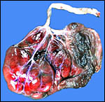
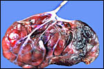
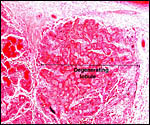
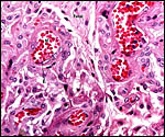
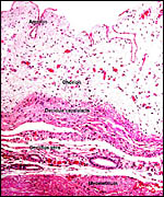
15) Pathological features

Seymour et al. (1983) accounted for the viruses isolated from sloths. Griner (1983) found infection and malnutrition to be problems in captivity.

16) Physiologic data

Meritt (1973) provided dietary details for feeding sloths in captivity, and Bush & Gilroy (1979) gave data on how to bleed sloths, and provided hematologic data at the same time. Gilmore et al. (2000) reviewed the general physiology of both types of sloths. In a later contribution (2001) they updated this information and gave data on infectious organism harbored by sloths. Clinical problems encountered in captivity were described by Diniz & Oliveira (1999). Effective means of immobilization of Hoffmann's sloths were described by Meritt (1972, 1974, 1985). The same observer also made observations that allow identification of recently parturient females in a group of animals (Meritt, 1976). Since parturition had not been witnessed and mothers were difficult to identify, he used vaginal orifice swabs and Hemastix to identify the mother of newborns.

17) Other resources

Cell strains are kept of some animals at CRES in the San Diego Zoo and can be obtained by contacting Dr. Oliver Ryder at: oryder@ucsd.edu

18) Other remarks - What additional Information is needed?

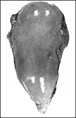
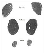
Endocrine studies are badly needed. The nature of the xenarthran fetal adrenal glands is of particular interest. Accurate data on the length of gestation of both species are also essential, as they differ so much in the few reports available. Are the chromosome differences indicative of "good" species? Are "hybrids" among them infertile?

Acknowledgement

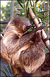
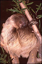
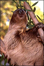
The animal photographs in this chapter come from the Zoological Society of San Diego. I appreciate also very much the help of the pathologists at the San Diego Zoo.

References

Becher, H.: Zur Kenntnis der Placenta vom Bradypus tridactylus. Z. Anat. Entwicklungsgesch. 61:114-136, 1921.

Benirschke, K. and Powell, H.C.: On the placentation of sloths. Pp. 237-241, In, Montgomery, G.G., ed.: The Evolution and Ecology of Armadillos, Sloths, and Vermilinguas. Smithsonian Institution, Washington, 1985.

Bush, M. and Gilroy, B.A.: A bleeding technique from nonpalpable vessels in anesthetized two-toed sloths (Choloepus didactylus) - plus hematologic data. J. Zoo Anim. Med. 10:26-27, 1979.

Delsuc, F., Catzeflis, F.M., Stanhope, M.J. and Douzery, E.J.: The evolution of armadillos, anteaters and sloths depicted by nuclear and mitochondrial phylogenies: implications for the status of the enigmatic fossil Eurotamandua. Proc. Roy. Soc. Lond. B Biol. 268:1605-1615, 2001.

Diniz, L.S. and Oliveira, P.M.: Clinical problems of sloths (Bradypus sp. and Choloepus sp.) in captivity. J. Zoo Wildl. Med. 30:76-80, 1999.

Eisenberg, J.F. and Maliniak, E.: Reproduction of the two-toed sloth Choloepus hoffmanni in captivity. Amer. Soc. Mamm. Abstr. Techn. Pap. 58th Ann. Meet. 41-42, 1978 (cited by Nowak, 1999).

Eisenberg, J.F. and Maliniak, E.: Maintenance and reproduction of the two-toed sloth Choloepus didactylus in captivity. Pp. 327-331, in, The Evolution and Ecology of Armadillos, Sloths, and Vermilinguas. G.G. Montgomery, ed. Smithsonian Institution, Washington, 1985.

Gilmore, D.P., da-Costa, C.P. and Duarte, D.P.: An update on the physiology of two- and three-toed sloths. Brazil. J. Med. Biol. Res. 33:129-146, 2000.

Gilmore, D.P., da Costa, C.P. and Duarte, D.P.: Sloth biology: an update on their physiological ecology, behavior and role as vectors of arthropods and arboviruses. Braz. J. Med. Biol. Res. 34:9-25, 2001.

Gotch, A.F.: Mammals - Their Latin Names Explained. Blandford Press, Poole, Dorset, 1979.

Greenwood, A.D., Castresana, J., Feldmaier-Fuchs, G. and Paabö, S.: A molecular phylogeny of two extinct sloths. Mol. Phylogenet. Evol. 18:94-103, 2001.

Griner, L.A.: Pathology of Zoo Animals. Zoological Society of San Diego, San Diego, California, 1983.

Heuser, C.H. and Wislocki, G.B.: Early development of the sloth (Bradypus griseus) and its similarity to that of man. Contrib. Embryol. Carnegie Inst. Washington 25:1-13, 1935.

Jorge, W., Meritt, D.A. and Benirschke, K.: Chromosome studies in edentates. Cytobios 18:157-172, 1978.

Jorge, W., Orsi-Souza, A.T. and Best, R.: The somatic chromosomes of Xenarthra. Pp.121 in, The Evolution and Ecology of Armadillos, Sloths, and Vermilinguas. G.G. Montgomery, ed. Smithsonian Institution, Washington, 1985.

McCrane, M.P.: Birth, behavior and development of a hand-reared two-toed sloth. Int. Zoo Ybk. 6:153-163, 1966.

Meritt, D.A.: Edentate immobilisation at Lincoln Park Zoo, Chicago. Intern. Zoo Ybk. 12:218-220, 1972.

Meritt, D.A.: Edentate diets. II. Two-toed sloths. Lab. An. Sci. 23:543-543, 1973.

Meritt, D.A.: A further note on the immobilisation of sloths. Intern. Zoo Ybk. 14:160-61, 1974.

Meritt, D.A.: Edentates. The Ark (Lincoln Park Chicago) 2:2-14, 1975.

Meritt, D.A.: A simplified technique for diagnosing parturition in Hoffmann's sloth. Intern. Zoo Ybk. 16:152-153, 1976.

Meritt, D.A.: The natural history, behavior, nutrition, physiology, reproduction, development and management of Hoffmann's sloth, Choloepus hoffmanni (Peters). M.A. Thesis, Northeastern Illinois University, 124 pp., 1977. (Kindly supplied by author).

Meritt, D.A.: The two-toed Hoffmann's sloth, Choloepus hoffmanni Peters. In, The Evolution and Ecology of Armadillos, Sloths, and Vermilinguas. G.G. Montgomery, ed. Smithsonian Institute Press, Washington, D.C., pp. 333-341, 1985.

Moser, H.G. and Benirschke, K.: The fetal zone of the adrenal gland in the nine?banded armadillo, Dasypus novemcinctus. Anat. Rec. 143:47?60, 1962.

Murata, K. and Masuda, R.: Gender determination of the Linné's two-toed sloth (Choloepus didactylus) using SRY amplified from hair. J. Vet. Med. Sci. 58:1157-1159, 1996.

Nowak, R.M.: Walker's Mammals of the World. 6th ed. The Johns Hopkins Press, Baltimore, 1999.

Seymour, C., Peralta, P.H. and Montgomery, G.G.: Viruses isolated from Panamanian sloths. Amer. J. Trop. Med. Hyg. 32:1435-1444, 1983.

Soma, H.: In chase of trophoblast: A comparison between human and other mammalian placentas. J. Tokyo Med. Coll. 34:873-883, 1976 (in Japanese).

Sonta S.-i., Hayata, I., Sasaki, M. and Kondo, N.: The karyotypes and sex-determining mechanism in the two-toed sloth, Choloepus didactylus. Chromos. Inf. Serv. 28:15-17, 1980.

Veselovsky, Z.: A contribution to the knowledge of the reproduction and growth of the two-toed sloth Choloepus didactylus at Prague Zoo. Int. Zoo Ybk. 6:147-153, 1966.

Wetzel, R.M.: Systematics, distribution, ecology, and conservation of South American edentates. Pp. 345-375, in "Mammalian Biology in South America. Special Publication Series of the Pymatuning Laboratory of Ecology, University of Pittsburg, Vol. 6, 1982.


back to top

© 2002. All rights reserved.
welcome | home | index | intro | placentation | glossary | author | contact