(Clicking on the thumbnail images below will launch a new window and a larger version of the thumbnail.)
Domestic Cat
Felis catus


Order: Carnivora
Family: Felidae

1) General zoological data of species


Domestic cats are abundant and many genetic varieties ("breeds") exist, with color, hair and other differences. There are numerous breeding colonies as well as cat fancier clubs and Societies. The ultimate origin of cats has been studied extensively and is summarized by Nowak & Paradiso (1983). The domestic cat is believed to have derived from Felis (silvestris) lybica (Starck, 1995).

Detailed phylogenetic considerations have been published by Thenius (1967), and Collier & O'Brien considered the phylogeny of felidae from investigating their immunological distances. Robinson (1977) has reviewed the general genetics of domestic cats and their great variation in the standard text on cat genetics.

2) General gestational data

The domestic cat has a 63-65 day gestation and produces litters of variable size, usually 4, but ranging from 1-8. Its placenta has often been investigated and a large literature exists on cat pregnancies, diseases, and genetics. The newborn weight is from 60-110 g, maternal weight (not pregnant and at term) is around 6,000 g, depending on the breed. Sexual maturity of females is reached between 7 and 12 months of age. Fox et al. (1984) have discussed the reproductive physiology. Domestic cats may live to be over 30 years.

The placenta of a term fetus weighs 15-20 g without cord and membranes.

   
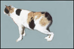
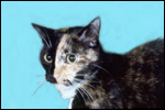
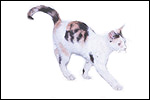
  Average female domestic "calico" or tortoiseshell cat.
     
 
3) Implantation

In the domestic cat, implantation occurs on days 12-14 post coitum and the trophoblast is superficially invasive (Leiser, 1979). Numerous publications have described early implantation. They were summarized by Denker et al. (1978). These authors suggested that, as in other species, there is dissolution (not rupture) of the zona pellucida and that proteinases are responsible for implantation. The blastocyst is much expanded before implantation and is oval. The embryonic disk attaches antimesometrially. There is an early yolk sac placentation that is soon replaced by the large allantoic placenta. The allantoic sac fuses with amnion and thus, this thin allantoic membrane possesses blood vessels. Vessels are classically lacking from the amnionic membrane. A girdle-like trophoblastic invasion occurs to produce the final placental shape. There is early invasion of the endometrial glandular mouths by trophoblast. This is particularly well illustrated by Kehrer (1973). He also illustrated the growth of the "lamellar" tongues of placental tissue in relation to the uterine axis. Later, the endometrial glands distend with secretion and contain degenerating epithelium. Adjacent to the placental girdle small hematomas of maternal blood develop that are, however, much smaller in cats than found in dog placentas. The portions of placenta lateral to the girdle are referred to as "paraplacenta". At this site, the trophoblast adheres to the uterine epithelium, thus making this area to have a truly epithelio-chorial relation. Rosettes, or chorionic vesicles, found here are numerous. Malassiné (1977) studied the erythrocytophagocytosis of maternal red blood cells by trophoblast at this site and suggested that this is an important site of iron absorption. This is, of course, the green/brown border of the zonary placental tissue. Whether much other transport occurs across this region is uncertain.
   
  General situs of the cat placenta: Maternal tissue - red; chorion - blue; amnion - purple; allantois - yellow; vitelline tissue - green; white triangle is exocoelom (adopted from Tiedemann, 1979).
     
 
4) General characteristics of placenta

The cat has a zonary placenta without cotyledons and it has a relatively small marginal hematoma, more so than the tiger but less than the dog. The materno-fetal barrier is endothelial-chorial, and it is superficially invasive into the endometrium but not into the myometrium.
   
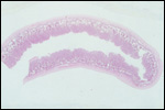
  Complete cross section of uterus with implanted cat placenta.
     
 
5) Details of barrier structure

The cat has an endothelial-chorial placental barrier. This was confirmed by electronmicroscopy. A very detailed description comes from Malassiné (1974) that shows the diminution of maternal endothelium towards the end of gestation. The labyrinth has very straight trophoblastic lamellae that cover the fetal mesenchyme, which contains the fetal blood vessels and some connective tissue cells. The trophoblastic surface is syncytial, beneath which are some cytotrophoblastic cells. They rest on a basement membrane above the fetal connective tissue. The syncytium directly abuts the maternal capillary endothelium, making this an endothelial-chorial barrier. Only Zhemkova (1962) has considered this to be a hemochorial placenta by virtue of her cytologic investigations. It employed Barr body identification of cells at the border zone in male conceptuses. Wislocki (1920) injected colloidal dyes and found them to be taken up by the maternal endothelium; the dyes did not to cross the placenta. He also affirmed the endothelio-chorial relation. Moreover, electronmicroscopy by Wynn and Björkman, 1968, Malassiné, 1974, and others have shown there to be a maternal endothelial layer. The transport of nutrients and other substances through this seemingly simple barrier is controversial. It is especially unclear what the origin is of the basement membrane material is, and what the nature of the debris is that is found here. These unresolved aspects have been studied by Björkman (1973), and demonstrated in his electronmicrographs, but they have not found a final resolution.

The feline placental labryrinth has characteristic giant cells. These are not found in most other endotheliochorial placentas. Dempsy & Wislocki (1956) considered these cells to have derived from endometrial connective tissue cells, and Malassiné (1974) showed them to decrease towards term. They have a pale cytoplasm and large, single nuclei. Wynn & Björkman (1968) made the point that the labyrinth of the cat has also a substantial amount of acellular material, perhaps an "intermediate layer" resembling a basement membrane. They drew the analogy of this material to the "fibrinoid" in human placentas. The cells were investigated in some detail by Malassiné (174) and considered again to represent decidual giant cells. He did not ascribe great functional importance to these cells.

Mossman (1987) asserted that the cytotrophoblastic cells are laden with debris, which originates from maternal erythrocytes, but Malassiné (1977) who studied the erythrocytophagocytosis in greater detail, found this to occur principally at the periphery of the paraplacenta.
   
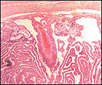
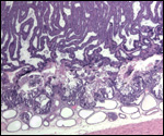
  Floor of placenta attached to uterus showing giant cell border (top left), large maternal blood vessel and trabecular villous structure.
     
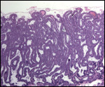
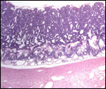
  Higher power photograph of fetal surface of cat placenta showing the trabecular nature with dark trophoblastic cells lining maternal blood channels and delicate fetal connective tissue.
     
  Lower power of the same cat's placental disk with uterus below. The endometrium has some cystic glands, next to the trophoblastic giant cells. Note the absence of trophoblast invasion in the endo/myometrium.
     
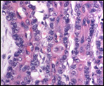
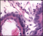
  The dark nuclei are trophoblast; in between these two layers flows the maternal blood. The connective tissue (top left and in between the rows of trophoblast) carries fetal capillaries.
     
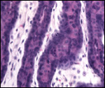
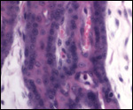
  High power view of placental trabeculae. The delicate connective tissue is the fetal portion. The dark cells are the trophoblast with the light purple being the cytoplasm of trophoblast; in between flows the maternal blood. A mitosis is seen in the cytotrophoblast near the top middle of the picture.
     
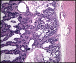
  "Floor" of the cat placenta with myometrium at right and very little endometrium. The border between villous tissue (left half) and uterus is a layer of giant cells, many with degenerative changes, which I believe to be trophoblast.
     
  Another border between myometrium (bottom) and placenta (top). Distended endometrial glands are next to the "invading" trophoblast.
     
  The dark streak in the center is where maternal blood is next to the trophoblastic surfaces. Fetal capillary and connective tissue above, with nucleated fetal red cell.
     
  Another high power view of trophoblastic columns. The purple centers are maternal blood spaces. Fetal connective tissue with capillaries and a nucleated red cell are obvious.
     
 
6) Umbilical cord

There are two pairs of vessels in the umbilical cord, two arteries and two veins. In addition there is an allantoic duct. The average length of the umbilical cord is 2-3 cm and 0.3-0.5 cm in diameter. It inserts at the margin of the zonary organ. There are no spirals, no vitelline duct, and no additional vessels or structures such as caruncles.

7) Uteroplacental circulation

No data are known to us.

8) Extraplacental membranes

The amnion has a very thin epithelium and no vasculature. It is attached to the chorion and, in some areas, to the large allantoic sac, which has an extensive vasculature, and a relatively thick epithelium that becomes thinner with advancing gestation. Tiedemann (1979) made the most detailed study of the epithelia of the three extraplacental saccular structures. He described, using fine-structural tools, that the amnionic epithelium degenerates completely by day 54 of gestation and thus the amnionic cavity becomes lined by connective tissue, an exceptional circumstance. The exocoelomic epithelium, he believed, to be responsible for the many macrophages in the membranes. The yolk sac epithelium was considered to be the most active epithelium of the membranes. In the same contribution he described the composition of the various fetal fluids. This author also showed that the various structures have often been mistaken one for another and discussed the difficulty one has with their individual identification. The schematic presentation above was drawn following the diagram he presented.

9) Trophoblast external to barrier

Since nidation is quite superficial, there are no invasive trophoblastic cells in the myometrium.

10) Endometrium

There is no typical decidualization of the endometrium as that seen in primate gestations. There are, however, transformations of endometrial connective tissue cells to fairly typical "giant cells" that abut the trophoblast. Wynn & Björkman (1968) and Malassiné (1974) have studied these cells. Ramsey (1982), on the other hand, was not so certain that they are of decidual nature and was unwilling to rule out trophoblastic derivation. My impression is that they are trophoblastic in origin.

11) Various features

Adjacent to the girdle-like placenta, the sac is covered with trophoblast that abuts the uterine epithelium. These projecting ends of the oval-shaped organ are referred to as "paraplacenta" which contains chorionic "rosettes".

12) Endocrinology

The cat, as the rabbit and some other species, is a "reflex ovulator"; i.e. she ovulates upon copulation. This allows good timing of early gestations. Oophorectomy or hypophysectomy leads to abortion (Tienhoven, 1983), Apparently no definitive studies on the production of gonadotropins or estrogens have been undertaken in cat placentas or gestational blood. Courrier (1945) stated that there are no gonadotropins found in the urine of pregnant cats.

In their very detailed endocrine study of Pallas' cat (Otocolobus mamul), Brown et al. (2002) discussed the endocrine findings of other feline species. For specific findings of the seasonality and the entrainment of cycles in this endangered species, with their effect on estrogen and progesterone section, the extensive description by these authors need to be read.

13) Genetics

Domestic cats, as well as most other felids, have 38 chromosomes that have been well delineated. Only some South American species of cats have 36 chromosomes. This is presumably due to autosomal chromosome fusions. Hybrids of domestic cats have been described to have occurred with Leopard cats, Jungle cats, Steppe cats and with bobcats (Gray, 1972).

We have seen 39 chromosomes with XXY sex chromosomes ("Klinefelter Syndrome" - in humans) in an infertile but otherwise normal tiger (Please see also the chapter on tiger). This genotype, 39,XXY, is a well-recognized error in domestic cats. Triploidy and chimerism are more common. They are easily recognized in male tortoiseshell cats. The reason is that the coat colors orange and black are located on the X chromosome in allelic positions. Thus, in order to exhibit black and orange, a cat needs to have two X chromosomes (Norby & Thuline, 1965). When this occurs in males, the presence of an extra X (e.g. 39,XXY) or a chimeric genetic content (e.g. 38,XX/XY or triploidy) are assured. Triploidy is apparently not uncommon in cats (Chu et al., 1964; Centerwall & Benirschke, 1975), nor is chimerism (Malouf et al., 1967). There is controversy as to the nomenclature of the gene conferring "black" upon cats (Miller & Hollander, 1986).

We have also seen an autosomal trisomy in a fetus of a domestic cat (Benirschke et al., 1974), and Johnstone et al. (1982) described X-monosomy in a Burmese cat with features similar to the human condition.

Genetic studies of cat chromosomal errors have been reported many times. Murphy et al. (1999a) reported virtual complete conservation of the X-chromosome, including complete synteny between cat and man. The Y chromosome was also found to exceedingly similar. Other genetic studies compare chromosomes 12 and 22 (Murphy et al., 1999b). O'Brien et al. (1997) reviewed the evolution and comparative genomics of cats. Other karyological studies come from Hsu & Rearden (1965), Hare et al. (1966), Wurster-Hill & Gray (1973) and Roubin et al. (1973).

Newman et al. (1985) described protein polymorphisms in a variety of felids. Numerous specific loci have been assigned (O'Brien et al., 1986; Cho et al., 1997a-d), and O'Brien (1980) recorded the biochemical genetic variations of cats.

More recent information on genes and linkage maps are contained in a report of a symposium on "Advances in Canine and Feline Genomics: Comparative Genome Anatomy and Genetic Disease" in J. Hered. 94, Issue 1 (January), 2003. It contains a paper on 78XX/77X mosaics.

14) Immunology

We have no knowledge of publications on MHC molecules, NK cells, or other immune cell populations.

15) Pathological features

An extensive review of diseases and pathological processes is to be found by Fox et al. (1984), and Benirschke et al. (1978). There are numerous infectious diseases and genetic errors reported. Abortions, metritis, retained placenta, etc. all occur. Huxtable et al. (1979) described the placenta in a habitually aborting cat; no infectious agents were found, only degenerative features that were not diagnostic. Feline infectious peritonitis, a viral disease, is a prominent problem (Jones, 1975; Evermann et al., 1981). Similarly, infectious anemia, a disease caused by the blood parasite Haemobartinella felis, occurs quite frequently (Hayes & Priester, 1973). Among neoplasms, mast cell tumors are prominent (Garner & Lingeman, 1970).

A number of "storage disorders" (inborn errors) have also been described (e.g. Sandström et al., 1969; Baker et al., 1971). While most litters are probably "fraternal" (multizygotic) offspring, monozygotic twin offspring probably also occur, as is evidenced by occasional two-headed conceptuses.

   
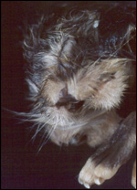
  Two-headed neonatal cat.
     
 
In addition, numerous disorders of the reproductive system, such as hydrometra, endometrial hyperplasia, alterations due to hormones, etc. have been described in this well-studied species. They are ably summarized by McEntee (1990).

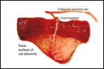
A litter of American Shorthairs that I observed delivered 4 days prematurely, with three pups, one of which was stillborn and meconium-stained. It was still enclosed in its sac when I received the specimen. The placenta of the stillborn was smaller (12 g) than that of the two survivors (18 and 21 g). It also had a zone that was much narrower than the usually quite uniform width of the ring-shaped placenta. Histologically, some areas of this placenta had massive distension of the maternal blood channels; making these areas appear to be hemorrhagic. Bordering these areas of "hemorrhage" was decidual necrosis with focal thrombosis of maternal blood vessels and infiltration with polymorphonuclear leukocytes. Organisms were absent and the fetal surface was not inflamed. The fetal ovaries were replete with oocytes, thus a chromosomal error appears to be an unlikely cause of fetal death. This is inferred, as most human neonates with trisomies or other major rearrangements have no mature oocytes. The dissection of the fetus also showed no abnormalities, other than growth retardation. At the peripheral end of this placenta the sac tapered off, suggesting that this was the peripheral-most implantation. Those placentas are shown next.
   
  This is the maternal aspect of the placenta from one of the normal fetuses.
     
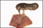
  Stillborn fetus "en caul", head is on the right.
     
  A short cord attached the dead fetus to the placenta. The allantoic sac is visible beneath the amnion.
     
  Fetal surface of stillborn's placenta with amnionic sac at left.
     
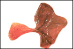
  Maternal aspect of the stillborn fetus' placenta. Note the hemorrhagic-appearing villous tissue and the irregular width. The membrane that extends at the top is amnion, not the umbilical cord.
     
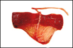
  Zonary placenta of stillborn with former head region at right.
     
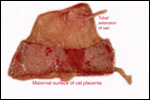
  This is the maternal aspect of a normal cat placenta.
     
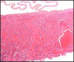
  Histologic appearance of the stillborn fetus' placenta with massive distension of the maternal blood channels.


16) Physiological data

I do not know any relevant data.

17) Other resources

Cell lines are available from the "Frozen Zoo" of CRES at the Zoological Society of San Diego. Reinert & Smith (1966) described the establishment of an experimental cat breeding colony.

References

Baker, H.J., Lindsey, J.R., McKhann, G.M. and Farrell, D.F.: Neuronal GM1 gangliosidosis in a Siamese cat with beta-galactosidase deficiency. Science 174:838-839, 1971.

Benirschke, K., Garner, F.M. and Jones, T.C.: Pathology of Laboratory Animals. 2 Vol. Springer-Verlag, New York, 1978.

Benirschke, K., Edwards, R. and Low, R.J.: Trisomy in a feline fetus. Amer. J. Vet. Res. 35:257-259, 1974.

Björkman, N.: Fine structure of the fetal-maternal area of exchange in the epitheliochorial and endotheliochorial types of placentation. Acta anat. 86 (Suppl. 1):1-22, 1973.

Brown, J.L., Graham, L.H., Wu, J.M., Collins, D. and Swanson, W.F.: Reproductive endocrine responses to photoperiod and exogenous gonadotropins in the Pallas' cat (Otocolobus manul). Zoo Biol. 21:347-364, 2002.

Centerwall, W.R. and Benirschke, K.: An animal model for the XXY Klinefelter syndrome in man: Tortoiseshell and calico male cats. Amer. J. Vet. Res. 36:1275-1280, 1975.

Cho, K.-W., Satoh, H., Youn, H.-Y., Watari, T., Tsujimoto, H., O'Brien, S.J. and Hasegawa, A.: Assignment1 of the cat immunoglobin heavy chain genes IGHM and IGHG to chromosomes B3q26 and T cell receptor chain gene TCRG to A2q12-q13 by fluorescence in situ hybridization. Cytogenet. Cell Genet. 79:118-120, 1997.

Cho, K.-W., Satoh, H., Youn, H.-Y., Watari, T., Tsujimoto, H. and Hasegawa, A.: Assignment of the feline c-mycgene (MYC) to cat chromosome F2q21.2 by fluorescence in situ hybridization. Cytogenet. Cell Genet. 78:135-136, 1997.

Cho, K.-W., Okuda, M., Endo, Y., Satoh, H., Kang, C.-B., Watari, T., Tsujimoto, H. and Hasegawa, A.: Assignment1 of the cat p53 tumor suppressor gene (TP53) to cat chromosome E1p14-p13 by fluorescence in situ hybridization. Cytogenet. Cell Genet. 79:145-146, 1997.

Cho, K.-W., Tsujimoto, H., Hasegawa, A. and Satoh, H.: The cat immunoglobulin lambda light chain gene maps to chromosome D3p12-p11. Mammal. Genome 9:178-179, 1997.

Chu, E.H.Y., Thuline, H.C. and Norby, D.E.: Triploid-diploid chimerism in a male tortoiseshell cat. Cytogenet. 3:1-18, 1964.

Collier, G.E. and O'Brien, S.J.: A molecular phylogeny of the felidae: Immunological distance. Evolution 39:473-487, 1987.

Dempsey, E.W. and Wislocki, G.B.: Electronmicroscopic observations on the placenta of the cat. J. Biophys. and Biochem. Cytol. 2:734-754, 1956.

Denker, H.-W., Eng, L.A. and Hammer, C.E.: Studies on the early development and implantation in the cat. II. Implantation: Proteinases. Anat. Embryol. 154:39-54, 1978.

Evermann, J.F., Baumgartner, L., Ott, R.L., Davis, E.V. and McKeirnan, A.J.: Characterization of a feline infectious peritonitis virus isolate. Vet. Pathol. 18:256-265, 1981.

Fox, J.G., Cohen, B.J. and Loew, F.M.: Laboratory Animal Medicine. Academic Press, Orlando, Florida, 1984.

Huxtable, C.R., Duff, B.C., Bennett, A.M., Love, D.N. and Butcher, D.R.: Placental lesions in habitually aborting cats. Vet. Pathol. 16:283-291, 1979.

Jones, B.R.: Feline infectious peritonitis: a review. New Zealand Vet. J.: 23:221-225, 1975.

Garner, F.M. and Lingeman, C.H.: Mast-cell neoplasms of the domestic cat. Path. Vet. 7:517-530, 1970.

Gray, A. P.: Mammalian Hybrids. A Check-list with Bibliography. 2nd ed. Commonwealth Agricultural Bureaux, Farnham Royal, 1972.

Hare, W.C.D., Weber, W.T., McFeely, R.A. and Yang, T.-J.: Cytogenetics in the dog and cat. J. Small Anim. Pract. 7:575-592, 1966.

Hayes, H.M. and Priester, W.A.: Feline infectious anaemia. Risk by age, sex and breed; prior disease; seasonal occurrence; mortality. J. small Anim. Pract. 14:797-804, 1973.

Hsu, T.C. and Rearden, H.H.: Further karyological studies on felidae. Chromosoma 16:365-371, 1965.

Huxtable, C.R., Duff, B.C., Bennett, A.M., Love, D.N. and Butcher, D.R.: Placental lesions in habitually aborting cats. Vet. Pathol. 16:283-291, 1979.

Johnstone, S.D., Buoen, L.C., Madl, J.E., Weber, A.F. and Smith, F.O.: X-chromosome monosomy (37,X0) in a Burmese cat with gonadal dysgenesis. J. Amer. Vet. Med. Assoc. 182:986-989, 1983.

Jones, B.R.: Feline infectious peritonitis: a review. New Zealand Vet. J.: 23:221-225, 1975.

Kehrer, A.: Zur Entwicklung und Ausbildung des Chorions der Placenta zonaria bei Katze, Hund und Fuchs. Z. Anat. Entwickl.-Gesch. 143:25-42, 1973.

Leiser, R.: Blastocystenimplantation bei der Hauskatze. Licht- und elektronenmikroskopische Untersuchung. Zbl. Vet. Med. C, Anat. Histol. Embryol. 8:79-96, 1979.

Ludwig, K.S.: Zur vergleichenden Histologie des
Allantochorion. Rev. Suisse Zool. 75:819-831, 1968.

Malassiné, A.: Evolution ultrastructurale du labyrinthe du placenta de chatte. Anat. Embryol. 146:1-20, 1974.

Malassiné, A.: Étude ultrastructurale du paraplacenta de chatte: mécanisme de l'érythrocytophagocytose par la cellule chorionique. Anat. Embryol. 151:267-283, 1977.

Malouf, N., Benirschke, K. and Hoefnagel, D.: XX/XY chimerism in a tri-colored male cat. Cytogenetics 6:228-241, 1967.

Miller, W.J. and Hollander, W.F.: The sex-linked black cat fallacy: a textbook case. J. Hered. 77:463-464, 1986.

Mossman, H.W.: Vertebrate Fetal Membranes. MacMillan, Houndmills, 1987.

Murphy, W.J., Sun, S., Chen, Z.-C., Pecon-Slattery, J. and O'Brien, S.J.: Extensive conservation of sex chromosome organization between cat and human revealed by parallel radiation hybrid mapping. Genome Res. 9:1223-1230, 1999a.

Murphy, W.J., Menotti-Raymond, M., Lyons, L.A., Thompson, M.A. and O'Brien, S.J.: Development of a feline whole genome radiation hybrid panel and comparative mapping of human chromosome 12 and 22 loci. Genomics 57:1-8, 1999b.

Newman, A., Bush, M., Wildt, D.E., v. Dam, D., Frankenhuis, M.Th., Simmons, L., Phillips, L. and O'Brien, S.J.: Biochemical genetic variation in eight endangered or threatened felid species. J. Mammal. 66:256-267, 1985.

Norby, D.E. and Thuline, H.C.: Gene action in the X chromosome of the cat (Felis catus L..). Cytogenetics 4:240-244, 1965.

Nowak, R.M. and Paradiso, J.L.: Walker's Mammals of the World. 2 Vol. The Johns Hopkins University Press, Baltimore, 1983.

O'Brien, S.J.: The extent and character of biochemical genetic variation in the domestic cat. J. Hered. 71:2-8, 1980.

O'Brien, S.J., Haskins, M.E., Winkler, C.A., Nash, W.G. and Patterson, D.F.: Chromosomal mapping of beta-globin and albino loci in the domestic cat. A conserved mammalian chromosome group. J. Hered. 77:374-378, 1986.

O'Brien, S.J., Wienberg, J. and Lyons, L.A.: Comparative lessons from cats. TIG 13:393-399, 1997.

Ramsey, E.M.: The Placenta. Human and Animal. Praeger Publishers, NY, 1982.

Ramsey, E. M.: The Placenta of Laboratory Animals and Man. Holt, Rinehart and Winston, N.Y., 1975.

Reinert, H. and Smith, G.K.A.: Establishment of an experimental cat breeding colony. Nature 209:1005-1008, 1966.

Robinson, R.: Genetics for Cat Breeders. 2nd. ed. Pergamon Press, NY 1971.

Roubin, M., de Grouchy, J. and Klein, M.: Les félidés: Évolution chromosomique. Ann. Génét. 16:233-245, 1973.

Sandström, B., Westman, J. and Öckerman, P.A.: Glycogenosis of the central nervous system in the cat. Acta Neuropath. (Berlin) 14:194-200, 1969.

Starck, D.: Lehrbuch der Speziellen Zoologie. Vol. II, Part 5/2. Gustav Fischer Verlag, Jena, 1995.

Thenius, E.: Zur Phylogenie der Feliden (Carnivora, Mamm.). Z. zool. Syst. Evolutionsforschung 5:129-143, 1967.

Tiedemann, K.: The amniotic, allantoic and yolk sac epithelia of the cat: SEM and TEM studies. Anat. & Embryol. 158:75-94, 1979.

Wimsatt, W.A.: Some aspects of the comparative anatomy of the mammalian placenta. Amer. J. Obstet. Gynecol. 84:1568-1594, 1962.

Wislocki, G.B.: Experimental studies on fetal absorption, II. Behaviour of the fetal membranes and placenta of the cat toward colloidal dyes injected into the maternal blood stream. Carnegie Contrib. to Embryol. 11: 1920

Wurster-Hill, D.H. and Gray, C.W.: Giemsa banding patterns in the chromosomes of twelve species of cats (Felidae). Cytogenet. Cell Genet. 12:377-397, 1973.

Wynn, R.M. and Björkman, N.: Ultrastructure of the feline placental membrane. Amer. J. Obstet. Gynecol. 102:34-43, 1968.

Zhemkova, Z.P.: The use of sex chromatin in identifying embryonic and maternal tissues in the placenta: New observations on the haemochorial nature of the cat placenta. J. Embryol. Exp. Med. 10:127-139, 1962.

18) Other findings of interest

   
  Tortoiseshell female cat with midline division of orange and black due to "lyonization" of melanocytes that carry the color genes. (Courtesy Dr. T.C. Jones).
     
  Tricolored male cat with 39, XXY chromosome constitution.
     
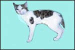
  Tricolored male cat with XY/XX chimerism


back to top

© 2002. All rights reserved.
welcome | home | intro | placentation | glossary | author contact me: kbenirsc@ucsd.edu