 |
| (Clicking
on the thumbnail images below will launch a new window and a larger
version of the thumbnail.) |
| Last updated: |
| March 16, 2004. |
Kobus ellipsiprymnus
(With notes on the Nile Lechwe, K. megaceros)
Order: Artiodactyla
Family: Bovidae
1) General Zoological Data
The ellipsen- or common waterbuck (Kobus e. ellipsiprymnus) is so named because this species has a white ring of hair around its hind which is already visible in fetal life. The species occurs in the savannah zones south of the Sahara desert (Nowak, 1999). Adults weigh up to 300 kg and only males have curved horns with transverse corrugations. The animals prefer an environment near water and have an oily skin that repels water readily. They are gregarious animals that may be found in large herds. Their numbers have been reduced drastically so that this species is now listed as being conservation dependent.
Four other species and several subspecies of waterbuck are recognized: The Nile Lechwe (K. megaceros); The Lechwe (K. leche); the Uganda Kob (K. kob) and the Puku (K. vardonii). The Defassa waterbuck (K. e. defassa) is considered to be a subspecies of the common waterbuck. Several hybrids among the kobs have been listed by Gray (1972), and Kingswood et al. (2000) suggested that chromosomal studies are useful in species assignment, especially for breeding purposes.
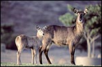 |
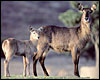
Ellipsen Waterbuck and offspring at San Diego Wild Animal Park. |
Estrus lasts from 12-24 hours and the average interbirth interval is 325 days (Nowak, 1999). Gestation lasts 230+ days with a usually single offspring that weighs up to 5,000 g. The maximum life span recorded is from 17 to 18½ years (Jones, 1993), but Mentis records shorter periods for different species. The general reproductive data have been listed for all species of waterbuck by Mentis (1972).
3) Implantation
No early stages of implantation have been described. There is no reason to believe that this differs much from other ungulates, however. The cotyledons develop by implanting over caruncles into the typical placentomes of Artiodactyla. Contrary to most other African ungulates, waterbucks possess only two rows of caruncles.
4) General Characterization of the Placenta
Hradecky (1983) described the placenta of several kob species. He found the following, to which I added the third:
|
Species
|
#
Cotyledonary rows
|
#
Cotyledons
|
Weight
|
|
K.
leche
|
(incomplete)
|
8
|
|
K. leche |
2 |
13 |
immature |
|
K.
e. ellipsiprymnus
|
(incomplete)
|
6
|
|
|
K. e. ellipsiprymnus
|
2
|
32
|
180
g (immature)
|
|
K.
e. defassa
|
2
|
29
|
2,400
g
|
|
K.
megaceros
|
2
|
24
|
|
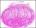
The placenta of this ellipsen waterbuck dam described first was attached to the uterus. It was impossible to detach without causing significant disruption. Placenta and uterus, together, weighed 2,700 g. The fairly mature fetus weighed 4,300 g and had a CR length of 51 cm. The dam had died from trauma and she also had significant dental disease. This common waterbuck's placenta is typical cotyledonary, composed of large and slightly convex cotyledons that are much larger in the horn of implantation (10 x 7 cm) but extend, with smaller cotyledons (2 x 2 cm), into the unoccupied horn. There were 30 cotyledons altogether, arranged in two rows over caruncles, and the umbilical cord measured 9 cm in length. A second placenta also came from a dam with severe dental disease. The very immature male fetus weighed 400 g, with a 20 cm CR length. Both uterine horns had placental tissue attached. The cord was 9 cm long.
The term placenta of a Nile Lechwe weighed 1,650 g, with a male neonate weighing 4,700 g. The placenta measured 45 x 18 cm in greatest dimensions and had only 10 large cotyledons that measured between 5 and 15 cm in diameters; the smaller cotyledons were not enumerated by the prosector. Thus, in total there may have been more cotyledons, as is the case in the other species. Its umbilical cord was 18 cm long and not spiraled. Small amounts of hippomanes were present. The fetus was delivered because of the dam's vaginal prolapse and it breathed for a short while only.
A more recently Red Lechwe (Zambesian) placenta was obtained from a dam that died for unknown reasons. Its immature male fetus weighed 230 g and had an 18 cm CR length. There were 13 cotyledons in 2 rows that were separating because of autolysis. Its placenta was contained mostly in the right uterine horn; some portions extended into the left horn where the cotyledons became very small. The cervix was firmly closed and filled with tenacious mucus. The cotyledons measured up to 3 cm in width and were of the flat type.
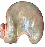 |
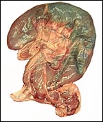
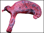
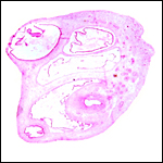
Near term gestation uterus of Ellipsen waterbuck before opening, fetal head at top left. |
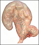 |
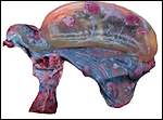
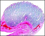
Same uterus from behind. Unoccupied horn at right. |
 |
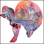
Partially opened uterus with filled allantoic sac (top left and below). The placenta extends into the unoccupied horn at bottom. |
 |
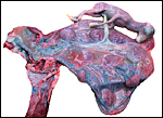
Amnionic cavity has been opened and this exposes the fetus as it lay in utero. Note the short untwisted umbilical cord and twisted neck. |
 |
Close-up of the central portion of this placenta with large cotyledons and cord insertion in the center. |
 |
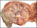
Placenta of Nile Lechwe waterbuck. |
 |
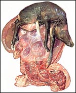
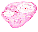
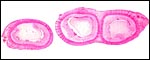
Immature Lechwe uterus with fetus in right horn. |
 |
Same specimen after opening the uterus and detaching some cotyledons from the dark red caruncles. |
 |
Same uterus. Note that the placenta protrudes into the left horn where the cotyledons are very small. |
 |
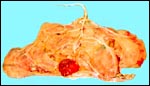
Fully opened Lechwe uterus. |
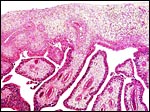
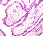
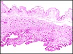
The waterbuck placenta has the usual interface between the filiform villi with their single layer of trophoblastic cover and the endometrial surface epithelium. There is close contact between the epithelia that is obscured in these formalin-fixed sections by shrinkage of the villi away from the endometrium. Binucleate cells are abundant in these specimens. Their nature and further discussion of their product have been discussed in some detail in the chapter on sheep. Detailed information can also be found by Wooding et al. (1997). The binucleate trophoblastic cells are thought to produce placental lactogen. There is no myometrial infiltration by trophoblast.
Over the membranes, the contact between trophoblast and endometrium is less obvious in this specimen. Also, whether areolae exist is difficult to see in these specimens. The trophoblast beneath the chorionic plate, however, has the usual yellow pigmentation.
 |
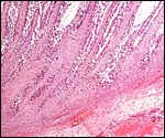
Immature waterbuck placentome with thin, straight villi. |
 |
Attached cotyledon to caruncle of Lechwe waterbuck |
 |
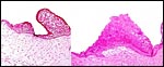
Term cotyledon of Kobus kob to show the filiform villi (Courtesy Dr. P. Hradecky). |
 |
Immature placental surface of common waterbuck placenta with villi arising from the chorion. |
 |
Surface of placenta with pigmented trophoblast beneath chorion. |
 |
Implantation of immature cotyledon on uterus below. |
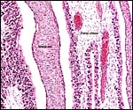 |
Immature villous and maternal interface. |
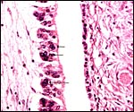 |
Maternal tissue at right, villous surface at left. The arrows point to binucleate or trinucleate trophoblast. |
 |
Villi of delivered, term placenta of K. megaceros. Despite the autolysis, the location of fetal vessels in the central portion of villi is evident. There are few subtrophoblastic capillaries, as are seen in so many other species. |
The umbilical cord of the Ellipsen Waterbuck measured 9 cm in length and 2 cm in width. That of the Nile Lechwe was 18 cm long. Both contained four large blood vessels and a centrally located allantoic duct. Numerous smaller allantoic vessels are also present but they do not markedly surround the allantoic duct as is seen in some other species. There were numerous fine, white granules on both cords' surfaces that, histologically, consist of foci of squamous metaplasia with keratinization. In the Zambesi Lechwe waterbuck added most recently to this chapter, in which the immature fetus was 18 cm long and weighed 230 g, the delicate umbilical cord measured 9 cm in length. It had no twists.
 |
Umbilical cord surface of immature fetus (left) and at term (right) with squamous metaplasia. |
 |
Umbilical cord of Lechwe umbilical cord. Allantoic duct at bottom left. |
 |
Allantoic duct in the center of the immature umbilical cord. Note small allantoic blood vessels in the periphery. |
This has not been studied; nevertheless, the large maternal vessels on the surface are readily seen in the gross specimens above.
8)
Extraplacental membranes
The membranes are covered with trophoblast which is often pigmented. A
large allantoic sac is present which is filled with fetal urine.
 |
Membranes of immature placenta with amnion at left and trophoblast at right showing some pigmentation. |
 |
Term Nile Lechwe waterbuck placental membranes. The amnion is above. |
There is no trophoblast infiltration into the decidua which has remarkably few glands and is relatively fibrous.
10)
Endometrium
The endometrium of pregnancy shows no real decidual transformation and
has relatively few glands whose secretory activity cannot be decided histologically.
The stroma has a fibrous appearance.
11)
Various features
There is no subplacenta, and no other remarkable features are recognized
that have not already been described.
12)
Endocrinology
I have been unable to find any information on endocrine features of kob
gestations. It was surprising, however, that the fetal ovaries of our
specimens showed the stimulation of follicular development that is shown
in the following photographs. On the other hand, the neonatal testis of
the Nile Lechwe showed no stimulation of its interstitial components.
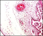
 |
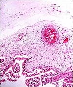
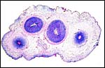
Fetal ovary of immature common waterbuck fetus with cystic follicles. |
 |
The other fetal ovary also showing numerous cysts. |
 |
Fetal testis of mature K. megaceros showing no interstitial cell stimulation. |
The chromosome numbers of K. ellipsiprymnus and that of K. kob were found to be 2n=50, but the Nile Lechwe (K. megaceros) had 52 elements (Wurster & Benirschke, 1968). Later, and more comprehensive, studies by Kingswood et al. (2000) showed considerable chromosomal polymorphism of kobs. Thus, the Ellipsen waterbuck had 50-54 elements, the Kob had 48 elements and K. megaceros had 52 elements. All of these animals, however, had 58 chromosomal arms and the Robertsonian mechanism of chromosomal fusion is the presumed reason for these discrepancies. They pointed out that reproductive isolation is incomplete because of the hybridization among animals and that mixing chromosomally different specimens in breeding colonies is unwise. Thus, Griner (1983) autopsied two hybrid waterbucks in San Diego, and Gray (1972) also referred to numerous hybrids among these different kob species.
Matthee & Davis (2001) remarked in their study on nuclear DNA markers of bovid taxa that the …"problematic Reduncinae (waterbuck, reedbuck) seem to be the earliest-diverging group of the Caprinae/Alcelaphinae/Hippotraginae clade"… Perhaps this explains, in part, the somewhat unusual morphologic features found in the waterbuck that are here listed.
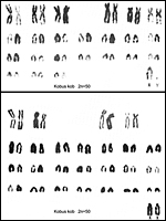 |
Karyotypes of Buffon's kob. From Hsu & Benirschke, 1969. |
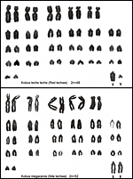 |
Karyotypes of red and Nile lechwes, from Hsu & Benirschke, 1977. |
14)
Immunology
I know of no relevant data.
15)
Pathological features
Griner (1983) autopsied a large number of different species of waterbuck,
including two hybrids. Regrettably he stated nothing about their genitalia.
Malnutrition, bacterial infections, a case of pulmonary hemangiomas and
uterine adenoma were described. In addition, some cases of "white
muscle disease" were seen. Zieger et al. (1998) reported new helminths
and bot fly infestations in waterbuck from Zambia. Anderson & Rowe
(1998) found a prevalence of antibodies against Rift Valley Fever in waterbuck
of Zimbabwe. The susceptibility of waterbuck and other species to trypanosomes
was examined by Moloo et al. (1999).
16)
Physiologic data
I have been unable to find literature of physiological studies on kobs.
There are two unusual features worth mentioning. First, the ovarian stimulation
in fetal ovaries and, second, the small number of caruncular rows in waterbuck.
Hradecky (1983) and my own studies also showed only two rows. This is
also seen in the small number of swollen (?stimulated) caruncles in fetal
uteri shown below.
The low frequency of landing and feeding on waterbuck by Glossina morsitans
was studied by Gikonyo et al. (2000) and explained by their allomones.
 |
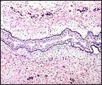
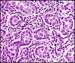
Cross sections through the fetal uterus of a common waterbuck that display the very small number of caruncles in the fetal endometrium as swollen, edematous bodies. |
Numerous cell lines of different Kob species are stored in the "Frozen Zoo" of the Zoological Society of San Diego and can be made available by contacting Dr. Oliver Ryder at oryder@ucsd.edu.
18)
Other remarks - What additional Information is needed?
No endocrine studies are known to me and early stages of implantation
need to be described.
Acknowledgement
The animal photograph in this chapter comes from the Zoological Society
of San Diego. I appreciate also very much the help of the pathologists
at the San Diego Zoo.
References
Anderson E.C. and Rowe, L.W.: The prevalence of antibody to the viruses
of bovine virus diarrhea, bovine herpes virus 1, rift valley fever, ephemeral
fever and bluetongue and to Leptospira sp. in free-ranging wildlife Zimbabwe.
Epidemiol. Infect. 121:441-449, 1998.
Gikonyo, N.K., Hassanali, A., Njagi, P.G. and Saini, R.K.: Behaviour of Glossina morsitans morsitans Westwood (Diptera: Glossinidae) on waterbuck Kobus defassa Ruppel and feeding membranes smeared with waterbuck sebum indicates the presence of allomones. Acta Tropica 77:295-303, 2000.
Gray, A.P.: Mammalian Hybrids. A Check-list with Bibliography. 2nd edition. Commonwealth Agricultural Bureaux Farnham Royal, Slough, England, 1972.
Griner, L.A.: Pathology of Zoo Animals. Zoological Society of San Diego, San Diego, California, 1983.
Hradecky, P.: Placental morphology in African antelopes and giraffes. Theriogenology 20:725-734, 1983.
Hradecky,
P., Mossman, H.W. and Stott, G.G.: Comparative histology of antelope placentomes.
Theriogenology 29:693-729, 1988.
Hsu, T.C. and Benirschke, K.: An Atlas of Mammalian Chromosomes. Springer-Verlag , N.Y. Vol. 3, Folio 143, 1969; and Vol. 10. Folio 505, 1977.
Jones, M.L.: Longevity of ungulates in captivity. Intern. Zoo Yearbk. 32:159-169, 1993.
Kingswood, S.C., Kumamoto, A.T., Charter, S.J., Aman, R.A. and Ryder, O.A.: Centric fusion polymorphism in waterbuck (Kobus ellipsiprymnus). J. Hered. 89:96-100, 1998.
Matthee, C.A. and Davis, S.K.: Molecular insights into the evolution of the family Bovidae: a nuclear DNA perspective. Mol. Biol. Evol. 18:12120-1230, 2001.
Mentis, M.T.: A review of some life history features of the large herbivores of Africa. The Lammergeyer 16:1-89, 1972.
Moloo, S.K., Orinda, G.O., Sabwa, C.L., Minja, S.H. and Masake, R.A.: Study of the sequential tsetse-transmitted Trypanosoma congolense, T. brucei brucei and T. vivax infections to African buffalo, eland, waterbuck, N'Dama and Boran cattle. Vet. Parasitol. 80:197-213, 1999.
Nowak, R.M.: Walker's Mammals of the World. 6th ed. The Johns Hopkins Press, Baltimore, 1999.
Wooding, F.B., Morgan, G. and Adam, C.L.: Structure and function in the ruminant synepitheliochorial placenta: central role of the trophoblast binucleate cell in deer. Microsc. Res. Tech. 38:88-99, 1997.
Wurster, D.H. and Benirschke, K.: Chromosome studies in the superfamily Bovoidea. Chromosoma 25:152-171, 1968.
Zieger, U., Boomker, J., Cauldwell, A.E. and Horak, I.G.: Helminths and bot fly larvae of wild ungulates on a game ranch in central province, Zambia. Onderstepoort J. Vet. Med. 65:137-141, 1998.