 |
| (Clicking
on the thumbnail images will launch a new window and a larger version
of the thumbnail.) |
| Last updated: Oct 5, 2006. |
Delphinaptera leucas
Order: Cetacea
Family: Monodontidae
1) General Zoological Data
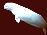
Belugas, also referred to as white whales', are distributed circumpolar and are estimated to number between 62,000 and 80,000 animals (NMFS, 2004). They are thus arctic or subarctic whales. Numerous studies of these animals come from the St. Lawrence River estuary in Canada . They ascend rivers frequently and also migrate south for some distance. Delphinapterus refers to: dolphin a=not pterus feather or wing (no dorsal fin, according to Gotch (1979); she refers to beluga as deriving from belyi (Russian) as white . The animal is also often named belukha'. Males weigh up to 1,500 kg, females 1,360 kg (Nowak, 1999). Weigl (2006) lists maximal life span as 34 years.
 |
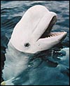
Beluga in aquarium (from Dr. Sam Ridgway). |
 |
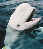
Another white whale' in an aquarium (from Dr. Sam Ridgway). |
2) General Gestational Data
Gestation is 14-14.5 months long and the single newborn weighs 80 kg (Nowak, 1999), while Stewart & Stewart (1989) indicated 35-85 kg.
3) Implantation
Information is available on their implantation or placentas (Mossman (1973).
4) General Characterization of the Placenta
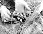
I have had the opportunity of studying four specimens of Beluga whale placentas from the Aquarium for Wildlife Conservation in Brooklyn , N.Y. All of them were bred at the facility and specimens were obtained immediately when the placenta was expelled, about 8 hours after the birth. They were all similar in their appearances and measured and weighed as follows:
Placental weight |
Cord length/ circumference |
Length |
Width |
Dam's age |
10,000 g |
50/7 cm |
274 cm |
60 cm |
21 y. |
6,818 g |
52/6 cm |
250 cm |
46 cm |
11 y. |
9,900 g |
55/6 cm |
270 cm |
57 cm |
10 y. |
5,300 g |
55/6 cm |
234 cm |
50 cm |
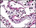
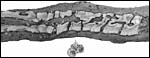
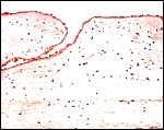
This is a typical thin villous, epithelio-chorial placenta that, so it would appear, extends into both uterine horns. The horn with fetus is much larger in width. The trophoblast sits on a thin basal membrane and is unicellular. No trophoblastic inclusions were identified and there are no binucleated cells. There was no pigmentation, although one animal had hemosiderin deposits. The tips of their placental ends were covered with villi.
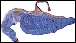 |
One of the mature beluga placentas with typical cord insertion. |
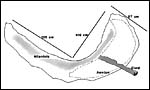 |
Measurements and disposition of placenta depicted above. |
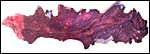 |
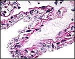
Maternal surface of the placenta. |
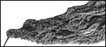 |
Tip of the placenta at end of uterine horn is covered with villi. |
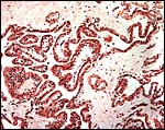
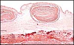
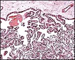 |
General histological appearance of villi arising from the chorion. |
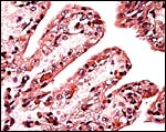
 |
Higher magnification of villous arrangement. Note vascularity beneath trophoblast. |
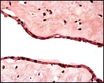
 |
Villous surfaces with capillary bed below trophoblast. |
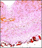
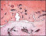
5) Details of fetal/maternal barrier
No implanted placenta has been available and therefore, it is inferred that the interface is similar to that of common dolphins. Occasional endometrial fragments came detached and lay in between the villi.
 |
PAS stain of villous surface to show the (red) basement membrane. |
 |
Higher magnification of PAS stained villus. |
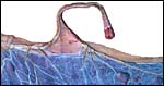
6) Umbilical cord
The umbilical cord of the specimen shown here was 55 cm long and virtually not twisted at all. It had numerous plaques consisting of squamous metaplasia on its surface and there was extensive capillarization beneath these plaques. Some squamous plaques are buried' beneath the cords' surface, as illustrated below. Other cord lengths are listed in the table above. There are four large fetal vessels and numerous small blood vessels with or without musculature; many are clustered around the large allantoic duct. The duct has a very thin, flat single-layered epithelium.
 |
Insertion of umbilical cord to show the numerous foci of squamous metaplasia. |
 |
Marginal cord insertion with numerous squamous pearls' that extend over the vessels onto the amnion. |
 |
There are real plaques of squamous metaplasia on the cord's surface. |
 |
One of the squamous plaques on the umbilical cord. They are underlain by a rich capillary network. |
 |
Many foci of squamous metaplasia are embedded in the cord's surface. |
 |
The Allantoic duct in the cord is accompanied by numerous small blood vessels. |
 |
The allantoic duct is lined by a very flat epithelium. |
7) Uteroplacental circulation
No studies are known to me.
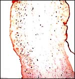
8) Extraplacental membranes
The membranes are thin, have no blood vessels and few squamous plaques, mostly over the vascular branches. There was no decidua.
 |
Cross section of amnion (right) and allantoic membrane (left). There are essentially no allantoic blood vessels. |
 |
This is the surface of the chorion with allantoic epithelium. |
9) Trophoblast external to barrier
There is no known trophoblastic invasion.
10) Endometrium
Although no implanted placenta has been available, inferring from other cetacean, there is no decidua.
11) Various features
The basal body temperature of a captive beluga was ascertained at 2-4 week intervals by Katsumata et al. (2006) in order to detect ovarian cycles. Highest temperatures were at the peak of progesterone levels; the cycle was 36 days long. Details on the anatomy of male genitalia were provided by De Guise et al. (1994a).
12) Endocrinology
I am not aware of any studies.
13) Genetics
Beluga whales have 44 chromosomes that are shown below. These karyotypes were donated by Dr. D. Duffield and were published in Hsu & Benirschke (1977). Jarrell & Arnason (1981) provided banded karyotypes. Remarkably, a true hermaphrodite beluga was found in the St. Lawrence Estuary (De Guise et al., 1994b). The animal had two testes, two ovaries and their respective ducts, but cervix, vagina and vulva were absent. The testes contained spermatozoa and there were several involuted corpora lutea. Hybrids have not been described (Gray, 1972).
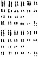 |
Chromosomes of male and female Beluga whale (from Hsu & Benirschke, 1977). |
14) Immunology
The purification of T-lymphocytes from splenic tissue was studied by Bernier et al. (2000), and Philippa et al. (2004) ascertained the prevalence of antibodies against a variety of antigens (adenovirus, herpesvirus etc.) in Canada .
15) Pathological features
In contrast to many other species I have reviewed in these chapters, there is an abundance of neoplasms reported in beluga whales. This is frequently attributed to the pesticides and other residues that have accumulated and that have been analyzed from this species. Whether these are true relationships remains to be established, but the number of cancers identified in belugas is remarkable. Here are some of the reports: De Guise et al. (1994c) reported on 21 tumors identified in 12 of 24 stranded carcasses and reviewed even earlier reports of tumors in belugas and drew the conclusion that there must be relation to carcinogenic contaminants (which are referred to).Martineau et al. (1995a) also reported from the St. Lawrence river estuary on intestinal cancers. Then, Lair et al. (1997a) identified unusual adrenal lesions (hyperplasia, cysts) and also considered xenobiotics as causes. Later (1998b) they reported a uterine adenocarcinoma in a 26 year-old animal with peritoneal spread. Mikaelian et al. (1999), also reporting from the same estuary, found mammary adenocarcinomas in two belugas. Ridgway et al. (2002) reported from San Diego on a poorly differentiated carcinoma after having identified it in the brainstem with MRI. Again from St. Lawrence River , Martineau et al. (2002b) estimated that there are 650 belugas in the estuary of which they examined 129 carcasses (of 263 stranded animals) over 17 years. Cancers were found in 18%, parasites in 22%, protozoal and other infections in 17%. They observed cancer overall in 27% found dead. McKinney et al. (2006) analyzed the organochlorines in the belugas from the same estuary and detailed their varied results. Thus, there may be a definite relationship of xenobiotics and cancers, at least in that river.
In addition to the neoplastic lesions reported, there are many studies of various types of infection occurring in belugas. Thus, candidiasis was successfully treated by Dunn et al. (1982); a herpetic infection occurring 3.5 months after capture occurred in a beluga that cleared up spontaneously (Barr et al., 1989). Mikaelian et al. (2000) reported toxoplasmosis in two free-ranging belugas that stranded perhaps due to this affliction and they found titers in another 27% of stranded animals. Trichinella infection in a beluga was reported by Forbes (2000). Harper et al. (2002) isolated a new helicobacter species from a captive beluga (and other dolphins) with problems of regurgitation and some having esophageal ulcers. A 16 year-old beluga died from a necrotizing dermatitis with ruptured aorta etc. and tuberculous organisms (Bowenkamp et al., 2001). In stranded belugas from the St. Lawrence estuary, Mikaelian et al. (2001) found skin lesions from which they isolated a dermatophilus-like organism. Finally, from the same estuary, De Guise et al. (1995d) reported non-neoplastic lesions in 24 carcasses, such as periodontitis, esophageal ulcers, pneumonia due to parasites and adrenal gland cysts.
16) Physiologic data
Cornell et al. (1988) published the results of hematologic and chemistry values of 31 captive belugas. More recently, Tryland et al. (2006) studied a wide variety of chemistries (urea, creatinine, bilirubin, enzymes, hormones, etc.) from 21 free-ranging belugas. Most interesting data on the composition of milk from belugas and Minke whales were assembled by Urashima et al. (2002). Miller et al. (2002) did ultrastructural comparative studies on spermatozoa of three odontocetes and found significant differences in their structures. Detailed examination of the myoglobin structure and function was published by Stewart et al. (2004).
17) Other resources
None are known to me, although there are many animals in Oceanaria and material should there be available.
18) Other remarks What additional Information is needed?
Time and nature of implantation and a study of an implanted term placenta would be desirable.
Acknowledgement
The animal photographs in this chapter come from Dr. Sam Ridgway, San Diego . Dr. Paul P. Calle of New York provided most of this material for which I am grateful.
References
Barr, B., Dunn, J.L., Daniel, M.D. and Banford, A.: Herpes-like viral dermatitis in a beluga whale (Delphinapterus leucas). J. Wildl. Dis. 25:608-611, 1989.
Benirschke, K. and Calle, P.P.: The placenta of the Beluga whale (Delphinapterus leucas). Verhandl. Ber. Erkrg. Zootiere 36:309-314, 1994.
Bernier, J., de Guise, S., Martineau, D., Beland, P., Beaudet, M. And Fournier, M.: Purification of functional T lymphocytes from splenocytes of the beluga whales (Delphinapterus leucas). Dev. Comp. Immunol. 24:653-662, 2000.
Bowenkamp, K.E., Frasca, S. Jr., Draghi, A 2nd, Tsongalis, G.J., Koerting, C., Hinckley, L., De Guise, S., Mantali, R.J., Goertz, C.E., St. Aubin, D.J. and Dunn, J.L.: Mycobacterium marinum dermatitis and pannicultis with chronic pleuritis in a captive white whale (Delphinapterus leucas) with aortic rupture. J. Vet. Diagn. Invest. 13:524-530, 2001.
Calle, P.P., Cook, R.A., McClave, C. and Palma , S.: Circulating gestational progesterone and estradiol concentrations, parturition, and placental descriptions of two beluga whales (Delphinapterus leucas). Proc. Int. Assoc. Aquat. Anim. Med. Chicago. p.63, 1993.
Coffey, D.J.: Dolphins, Whales and Porpoises. MacMillan , N.Y. 1977.
Cornell, L.H., Duffield, D.S., Joseph, B.E. and Stark, B.: Hematology and serum chemistry values in the beluga (Delphinapterus leucas). J. Wildl. Dis. 24:220-224, 1988.
De Guise, S., Bisaillion, A., Seguin , B. and Lagace, A.: The anatomy of the male genital system of the beluga whale, Delphinapterus leucas , with special reference to the penis. Anat. Histol. Embryol. 23:207-216, 1994a.
De Guise, S., Lagace, A. and Beland, P.: True hemaphroditism in a St. Lawrence beluga whale (Delphinapterus leucas). J. Wildl. Dis. 30:287-290, 1994b.
De Guise, S., Lagace, A. and Beland, P.: Tumors in St. Lawrence beluga whales (Delphinapterus leucas). Vet. Pathol. 31:444-449, 1994c.
De Guise, S., Lagace, A., Beland, P., Girard, C. and Higgins, R.: Non-neoplastic lesions in beluga whales () and other marine mammals from the St. Lawrence Estuary. J. Comp. Pathol. 112:257-271, 1995d.
Dunn, J.L., Buck, J.D. and Spotte, S.: Candidiasis in captive cetaceans. J. Amer. Med. Assoc. 181:1316-1321, 1982.
Forbes, L.B.: The occurrence and ecology of Trichinella in marine mammals. Vet. Parasitol. 93:321-334, 2000.
Gotch, A.F.: Mammals Their Latin Names Explained. Blandford Press, Poole , Dorset, 1979.
Gray, A.P.: Mammalian Hybrids. A Check-list with Bibliography. 2nd edition. Commonwealth Agricultural Bureaux Farnham Royal, Slough , England , 1972.
Harper, C.G., Feng, Y., Xu, S., Taylor , N.S. , Kinsel, M., Dewhirst, F.E., Paster, B.J., Greenwell, M., Levine, G., Rogers , A. and Fox, J.G.: Helicobacter cetorum sp. No., a urease Helicobacter species isolated from dolphins and whales. J. Clin. Microbiol. 40:4536-4543, 2002.
Hsu, T.C. and Benirschke, K.: An Atlas of Mammalian Chromosomes. Vol. 10: Folio 492, 1977.
Jarrell, G.H. and Arnason, U.: Banded karyotypes of a Belukha whale, Delphinapterus leucas. Hereditas 95:37-41, 1981.
Katsumata, E., Furuta, C., Kastumata, H., Watanabe, G. and Taya, K.: Basal body temperature method for detecting ovarian cycle in the captive beluga (Delphinapterus leucas). J. Reprod. Dev. 52:59-63, 2006.
Lair, S., Beland, P., De Guise, S. and Martineau, D.: Adrenal hyperplastic and degenerative changes in beluga whales. J. Wildl. Dis. 33:430-437, 1997a.
Lair, S., De Guise, S. and Martineau, D.: Uterine adenocarcinoma with abdominal carcinomatosis in a beluga whale. J. Wildl. Dis. 34:373-376, 1998b.
Martineau, D., Lair, S., De Guise, S. and Belande, P.: Intestinal adenocarcinoma in two beluga whales (Delphinapterus leucas) from the estuary of the St. Lawrence River . Can Vet. J.: 36:563-565, 1995a.
Martineau, D., Lemberger, K., Dallaire, A., Labelle, P., Lipscomb, T.P., Michel, P. and Mikaelian, I. : Cancer in wildlife, a case study: beluga from the St. Lawrence estuary, Quebec , Canada . Environm. Health Perspect. 110:285-292, 2002b.
McKinney , M.A., De Guise, S., Martineau, D., Beland, P., Lebeuf, M. and Letcher, R.J.: Organohalogen contaminants and metabolites in beluga whale (Delphinapterus leucas) liver from two Canadian populations. Environm. Toxicol. Chem. 25:1246-1257, 2006.
Mikaelian, I. , Labelle, P., Dore, M. and Martineau, D.: Metastatic mammary adenocarcinomas in two beluga whales (Delphinapterus leucas) from the St. Lawrence Estuary, Canada . Vet. Rec. 145:738-739, 1999.
Mikaelian, I. , Boisclair, J., Dubey, J.P., Kennedy, S. and Martineau, D.: Toxoplasmosis in beluga whales (Delphinapterus leucas) from the St. Lawrence estuary: two case reports and a serological survey. J. Comp. Pathol. 122:73-76, 2000.
Mikaelian, I. , Lapointe, J.M., Labelle, P., Higgins, R., Paradis, M. and Martineau, D.: Dermatophilus-like infection in beluga whales, Delphinapterus leucas , from the St. Lawrence estuary. Vet. Dermatol. 12:59-62, 2001.
Miller, D.L., Styer, E.L., Decker, S.J. and Robeck, T.: Ultrastructure of the spermatozoa from three odontocetes: a killer whale (Orcinus orca), a Pacific white-sided dolphin (Lagenorhynchus obliquidens) and a beluga (Delphinapterus leucas). Anat. Histol. Embryol. 31:158-168, 2002.
Mossman, H.W. and Duke, K.L.: Comparative Morphology of the Mammalian Ovary. University of Wisconsin Press, Madison , Wisconsin , 1973.
Nowak, R.M.: Walker 's Mammals of the World. 6 th ed. The Johns Hopkins Press, Baltimore, 1999.
Philippa, J.D., Leighton, F.A., Daoust, P.Y., Nielsen, O., Paogliarulo, M., Schwantje, H., Shury, T., Van Herwijnen, R., Martina, B.E., Kuiken, T., van de Bildt, M.W. and Osterhaus, A.D.: Antibodies to selected pathogens in free-ranging terrestrial carnivores and marine mammals in Canada . Vet. Rec. 155:135-140, 2004.
Ridgway, S.H., Marino, L. and Lipscomb, T.P.: Description of a poorly differentiated carcinoma within the brainstem of a white whale (Delphinapterus leucas) from magnetic resonance images and histologic analysis. Anat. Rec. 268:441-449, 2002.
Stewart, J.M., Blakely, J.A., Karpowicz, P.A., Kalanxhi, E., Thatcher, B.J. and Martin, B.M.: Unusually weak oxygen binding, physical properties, partial sequence, autooxidation rate and a potential phosphorylation site of beluga whales (Delphinapterus leucas) myoglobin. Comp. Biochem. Physiol. B Biochem. Mol. Biol. 137:401-412, 2004.
Stewart, B.E. and Stewart, R.E.A.: Delphinapterus leucas . Mamm. Species 336:1-8, 1989)
Tryland, M., Thoresen, S.I., Kovacs, K.M. and Lydersen, C.: Serum chemistry of free-ranging white whales (Delphinapterus leucas) in Svalbard . Vet. Clin. Pathol. 35:199-203, 2006.
Urashima, T., Sato, H., Munakata, J., Nakamura, T., Arai, I. , Saito, T., Tetsuka, M., Fukui , Y., Ishikawa, H., Lydersen, C. and Kovacs, K.M.: Chemical characterization of the oligosaccharides in beluga (Delphinapterus leucas) and Minke whale (Balaenoptera acutorostrata) milk. Comp. Biochem. Physiol. B Biochem. Mol. Biol. 132:611-624, 2002.
Weigl, R.: Longevity of Mammals in Captivity; From the Living Collections of the World. [Kleine Senckenberg-Reihe 48]. E. Schweizerbart'sche Verlagsbuchhandlung (Nägele und Obermiller), Stuttgart, 2005.