(Clicking on the thumbnail images below will launch a new window and a larger version of the thumbnail.)
 
Last updated: July 23, 2005.
Aardvark
Orycteropus afer

Order: Tubulidentata
Family: Orycteropodidae

1) General zoological data


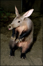
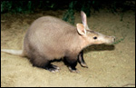
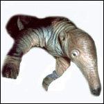
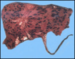
This single species of this mammalian Order lives in the sub-Saharan countries of Africa and is primarily nocturnal. The fossil records show that it extended once to the Mediterranean region. Its precise taxonomic relationship to other mammals has been a problem for classification. Perhaps this, as well as its unusual architecture (teeth, skin, tongue, etc.), is reason enough for a separate Order status among the mammals. Current genetic studies to clarify its taxonomic position are discussed under genetics below. Adults weigh 40-100 kg, most are around 50-70 kg (Nowak, 1999). The teeth lack enamel and grow continuously. Aardvarks ("earth-pigs") burrow deep holes and consume large quantities of ants and termites. They have their single young in deep tunnels, are solitary, and have a very characteristic appearance. They are allegedly good swimmers. Aardvarks have poor eyesight but can hear and smell exquisitely well. They are solitary, males are slightly larger than females. They have a very long tongue and extremely tough skin. Externally, it is hard to determine their sex. Aardvarks do well in captivity. They can be trained and have repeatedly reproduced in different zoological gardens (see Dulaney, 1987). An interesting finding of aardvarks is that they do not have a clavicle.

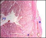
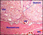
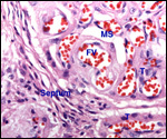
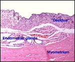
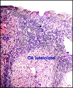
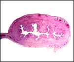
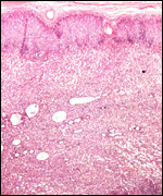
2) General gestational data

The gestation is 7-9 months long, according to Hayssen et al. (1993). Twins are very uncommon. Newborns weigh approximately 2 kg. In captive births, the newborns are being "marked" frequently by the scent gland at the mother's tail (Dulaney, 1987). Sexual maturity is reached at 2 years of age.

   
  Aardvark in defensive position.
     
  Usual appearance of aardvarks.
     
  Neonatal aardvark.
     
     
  3) Implantation  
     
  Fetal aardvark within maternal uterus; it certainly has the appearance of an unicornuate uterus. San Diego Zoo.
     
 
As can be seen from the specimen shown above, the aardvark fetus that I was able to examine was located in an apparently unicornuate uterus. Most authorities have been quoted as stating that the aardvark has a bicornuate or bifid uterus (v.d. Horst, 1949; Hayssen et al., 1993; others). This is of interest, especially when one observes the single fetal uterus shown below that came from one of our gestations. One of the few photographs of v.d. Horst's paper on a very young gestation shows the fundus to be Y-shaped, clearly different from our specimens. Moreover, the excellent description of Turner (1876) of a pregnant uterus and his review of the older anatomical papers leave no doubt that a small secondary horn exists in aardvarks. In the drawing he provided, there was an extremely small secondary horn, which may have been overlooked by the dissector of our specimen. But Turner is quite clear also that the fetal membranes do not extend into the small secondary horn.

The fetus of the specimen shown above weighed 2,100 g and was at term. The mother died from severe wounds with secondary infection that led to sepsis.

The earliest implantation of aardvarks was studied and described in great detail by v.d. Horst (1949). He found that the uterus had two openings into the vagina and that only one horn was pregnant, similar to the description of Turner (1876). Moreover, he described that there were five corpora lutea. This is inconsistent with my findings of a single corpus luteum and many luteinized corpora atretica in the ovaries associated with a pregnant uterus. On superficial study, the atretica follicles may well have looked like corpora lutea. Regrettably, mostly drawings accompany this article, to which Mossman (1987), however, gives much credence.

The first attachment of the blastocyst is apparently antimesometrial, with destruction of the superficial endometrium and the development of a large yolk sac. A large allantoic sac surrounds the yolk sac at that time and persists to term (as four compartments, according to Mossman). Of the yolk sac placentation, only a small permanent attachment remains to term. The ultimate shape of the placenta is nearly zonary but very little of "free membranes" remain. These are located over cervical os and the tubal orifice.

4) General Characterization of the Placenta

There are still several uncertainties about the characteristics of the aardvark placentation and about some aspects of its general anatomy. Therefore, a more detailed description of the placenta follows to draw attention to them so that perhaps these gaps may be filled in future studies. One minor but important observation to be made would be to confirm, with certainty, that the structure of the uterus is always bicornuate or duplex, aside from the deficient knowledge we have of the placenta.

The aardvark placenta is largely zonary, well described and illustrated by Turner (1876). He already remarked upon its unusually wide character, especially when compared with the zonary placentas of cats and other carnivores. The fetal surface vessels are shown to extend over the "free membranes" which are those portions overlying the orifices of cervix and fallopian tube. The placenta observed by Dulaney (1987) weighed 280 g. Other specimens differ in size and weight (see Taverne & Bakker-Slotboom, 1970). The aardvark placenta we first observed was zonary and probably endotheliochorial in character. This was affirmed by Mossman (1987) who reviewed my placental slides. That placenta was from the term delivery of a 15 year-old animal and weighed 400 g. It measured 28 x 17 and 15 x 9 cm in greatest dimensions and was nearly uniformly only 1 cm in thickness. It appeared to have been made up of two adjoining lobes with a somewhat zonary macroscopic structure. As can be seen from the next photograph, it had many dark regions of pigmented tissue. This spotty pigmentation was also described in some detail by Taverne & Bakker-Slotboom (1970) who observed the delivered placenta of a term gestation. There were virtually no "free membranes". The mother had died from large infected wounds and she had also suffered some small myocardial infarcts.

A second specimen, also of a full-term placenta with surviving female fetus, weighed 740 g and measured 28 x 18 x 0.2 cm. The neonate weighed 2,400 g. The specimen of Taverne & Bakker-Slotboom (1970) measured 36 x 22 cm wide. The villous tissue of the aardvark placenta has very many small lobular compartments that are divided by maternal connective tissue septa. The villi are short and plump. Turner (1876) found this to account for the "velvety" structure of the anteater placenta. The placenta has, histologically, a labyrinthine character.

   
  There are essentially two lobes and a small relatively villus-free region near the left center. The dark spots are hemosiderin-rich regions of "areolar" nature.
     
 
5) Details of fetal/maternal barrier
     
  Cross-section of implanted aardvark placenta. FV=fetal vessel; S=septum. Note that fibrous septa come from the concentration of endometrial vessels and partition the placenta partially.
     
  Implantation of term placenta on uterus. Note the absence of superficial endometrium, only a few glands remain. MV=maternal vein at base of septum; MA=maternal artery.
     
  Villi with fetal vessels (FV - NOT capillaries!), covered by single large trophoblastic cells, next to maternal septum. MS=maternal space, bloodless, but probably a vessel.
     
 
The fine structure of the trophoblast/endometrial relation requires future electronmicroscopic study. At present it is not clearly evident that this placenta has a truly endothelio-chorial relationship of mother to fetus. But that is the current assumption. In the earliest specimen described (v.d. Horst, 1949), a syndesmochorial placentation is obvious and this is also true in some areas of term placenta relationship. That is to say, trophoblast directly abuts the endometrial stroma at the site of attachment. In contrast though, in the very dense villous (labyrinthine areas), the single layer of thick trophoblast appears to abut maternal spaces that are empty in this specimen. They may be lined by maternal endothelium. That is impossible to decide in paraffin sections. While v.d. Horst's specimen was so early that the fetal circulation was not yet established, his blood-filled spaces were clearly maternal. But that is not so in the mature placenta shown above. In that illustration, the fetal vessels are filled with blood; one can easily trace them to the chorionic surface vessels. There are small, thin-walled blood vessels in the maternal septa, but they cannot be traced into the villous parenchyma. No syncytium is present and no cytotrophoblast can be differentiated histologically. Many trophoblastic cells are large and have a plump cytoplasm. v.d. Horst (1949) speaks of "giant cells", but these are absent in mature placentas.

Thinner chorionic membranes are present at the edges of the villous tissue compartments. These have a columnar, only minimally villous epithelium. They correspond to the pigmented regions seen in the macroscopic picture and presumably are located over the mouths of glands, the "areolar" regions shown further below.

6) Umbilical cord

The umbilical cord of this specimen measured 46 cm in length and had a marginal attachment. There were only a few spirals of the cord. It contained four large blood vessels in one cross section, but only three in another. I believe that four vessels is the norm, as this was also the number observed by Turner (1876). He remarked that the vessels separate several inches above the placenta. The lengths of the umbilical cords of his two specimens were similar, 22.5 inches in one, and the other slightly shorter. In addition to these structures, there are many small blood vessels and a large allantoic duct. The cord's surface has a thick, irregular, keratinized squamous epithelium. Taverne & Bakker-Slotboom (1970) described the cord of the placenta they described in great detail. They also show an excellent diagram that leads one to believe that the "cyst" shown below is the beginning of the allantoic cavity which lies close to the surface of the placenta and is surrounded by four segments of cord blood vessels. Their cord was 67 cm long, had surface squamous metaplasia and elastic tissue in the larger blood vessels. A remnant of vitelline duct and obliterated vitelline vessels were also found.

   
  Cross-section of cord. Small duct in center, squamous metaplasia on surface, A=artery; V=vein. At right is a large artifactual cyst (perhaps the beginning of the allantoic sac). Many tiny vessels are seen as red spots.
     
 
7) Uteroplacental circulation


I know of no publications. One remarkable feature described by v.d. Horst (1949), however, is the presence of a "vascular" zone in the lower endometrium that was restricted to the implantation zone. This feature has a remote similarity to the vascularization seen in Xenarthra. In contrast to these animals, however, it does not eventuate in the "lamellar" basal placental region of the Xenarthran placenta (Please see chapters on armadillo and anteater). The maternal septa that extend into the placental tissue divide it into the many small lobules. They contain small maternal blood vessels. These septa are invariably located over the presence of a collection of larger endometrial blood vessels.

8) Extraplacental membranes

   
  Amnion and chorionic membrane at the edge of the villous tissue. These are directly adjacent to the villous portions.
     
 
There is no decidua capsularis. The free membranes are thin and have an amnion with a thin squamoid epithelium as its lining. The chorionic membrane is lined by columnar epithelium and, underlying this, there is proteinaceous debris. The chorion contains many small blood vessels. These regions also present a different, much taller trophoblast with pigment in the cytoplasm.
   
  These slides correspond to the grossly dark/black-appearing regions shown on the gross photograph. They contain much hemosiderin in the columnar epithelium and are in relatively avillous regions of the membranes.
     
  These slides correspond to the grossly dark/black-appearing regions shown on the gross photograph. They contain much hemosiderin in the columnar epithelium and are in relatively avillous regions of the membranes. The explanation for them follows here.
     
 
In examining the slides of the aardvark placenta that I had sent to Harlan Mossman, he responded about his findings on 1/29/1980 as follows:

" The most interesting thing about these full-term aardvark membranes is the pigmented areas. I believe that they are large "absorptive" areas corresponding to areolae, and that they were associated with uterine glands. Many areolae and similar areas in other mammals are not only involved in absorption of gland secretion but also phagocytosis of sloughed uterine cells and extravasated erythrocytes. If the erythrocytes are phagocytosed about as fast as they extravasate, then there is no accumulation of a blood mass such as occurs in a typical hematome. This happens in the domestic cat and bobcat where a structurally typical carnivore hematome is present, except that there is little or no free mass of maternal blood. The pigmented spots appear black in the photographs, but the little pigment I could see in the slides is not black. I therefore think it is hemosiderin, or at least some breakdown product of hemoglobin. Am I right? - I notice that many of the dark spots have light centers; also that there are two large apparently avillous areas, and that these have relatively sharp and distinct boundaries which are often pigmented. One of these could have been associated with the cervical region, the other with the tubouterine junction area, as the aardvark has a duplex uterus; or there could be an antimesometrial avillous area, as in Tragulus."

9) Trophoblast external to barrier

There is no trophoblastic infiltration into the endometrium other than the erosion the trophoblast makes upon implantation. There is no infiltration of trophoblast into the myometrium either, but it appears that some of the maternal blood vessels at the base of the septa contain a few trophoblastic cells in their walls.

10) Endometrium
   
  Endometrium adjacent to placental implantation. There is little obvious decidual transformation of the endometrial stroma.
     
 
The post partum endometrium is thin, appears hyalinized and has numerous hemosiderin-laden macrophages. v.d. Horst (1949) already described the hemosiderin in the endometrium and believed it to stem from a former pregnancy in the uterus of the young pregnancy that he observed. There is only a very small amount of true decidua formed, despite the fact that the placenta is generally classified as being "deciduate". Turner (1876), however, declared this to be most likely a non-deciduate placenta, which comes closest to the truth.

11) Various features

No other relevant data on gestation or placentation are available.

12) Endocrinology

No endocrine measurements have been reported. Therefore, only an "indirect" evaluation is possible from the state of ovary, endometrium, adrenals and testes of mother and offspring. These are unusual for such a "primitive" animal. The maternal ovary has a single corpus luteum during pregnancy, although v.d. Horst (1949) described five corpora lutea with his singleton gestation. He supplied no pictures and this feature is, therefore, very difficult to evaluate. Considering the findings made in our specimen, however, I am convinced that he mistook some of the massively luteinized corpora atretica for corpora lutea. Numerous other follicles have striking luteinization of the theca, but it is most remarkable for the corpora atretica, as shown next.

   
  Maternal ovary at the end of gestation. Numerous luteinized corpora atretica were present, in addition to one corpus luteum. There are also numerous primordial ova.
     
 
The fetal uterus and ovaries of the term pregnancy I examined were available. The fetal cervix was remarkably dilated, but the uterus was unicornuate as shown below very clearly. This contrasts markedly with the description of Turner (1876), v.d. Horst (1949), and the remarks of Mossman (1987) who found bicornuate uteri. There were large numbers of Graafian follicles in both fetal ovaries with striking theca luteinization.
   
  Complete cross-section of fetal uterus of term aardvark. It is unicornuate here.
     
 
The other unusual feature is the appearance of the fetal adrenal glands. While they were not strikingly large, they had a pronounced "fetal zone" of their cortices. The definitive cortex was wide, but I found the fetal zone to be unusually broad and striking. It is nearly as large as that of the human fetus. It would thus be nice to know whether it also produces DHEA or similar estrogen precursors, and also, what hormones drive this zone. In a four-day old animal that died from enterocolitis, this zone was involuting very much like that of human neonates. It is also striking how much testicular interstitial cell stimulation in the neonatal testis is present. Like the fetal ovary that shows endocrine stimulation. This indicates that some gonadotropic stimulus must have existed in utero. Another unusual feature of the neonatal histology is the very large number of megakaryocytes in the splenic parenchyma.
   
  Cross-section through one-half of the fetal adrenal gland at birth. The darker definitive cortex measures 1/3 of the width, the remainder is "fetal zone".
     
  This adrenal gland is from a 4-day-old neonate. The fetal zone is partially involuting. Its definitive cortex has become much broader.
     
  Neonatal testis (left) and ovary (below left). Both show gonadotropic stimulation (interstitial cells and theca interna).
     
  Neonatal testis (upper left) and ovary (left). Both show gonadotropic stimulation (interstitial cells and theca interna).


13) Genetics

Aardvarks have 20 large chromosomes with relatively small-appearing X and Y chromosomes, because of the large size of the autosomes (Benirschke, et al., 1970). The total DNA content as measured by microspectrophotometry, is 1.67 times that of human lymphocytes, an unusually large quantity. The X chromosome is only 5% of the haploid set. Pathak et al. (1980) added to this exploration of the genetics by reporting banding studies of chromosomes of a female animal. They were unable to account for the 1.6% DNA content by heterochromatin, as is the case in some other species and called for confirmation of the DNA content.

Chromosome complement of aardvark: 2n-20, with thymidine replication chromosomes in second rows. (From Benirschke, et al., 1970).


In order to clarify the taxonomic status of aardvarks, a number of studies were performed. Thus, Dene et al. (1983) examined the myoglobin sequence and compared it with that of other mammalia. This led them to suggest that the aardvark is one of the oldest lineage in Eutheria. Arnason et al. (1999) studied the mtDNA of aardvarks and found its closest similarities in ancient xenarthran relations. They estimated that the species diverged from other stock some 90 Million Years Ago (MYA). Murphy et al. (2001) did a "comprehensive molecular phylogenetic analysis of 64 species" and grouped the aardvark with elephants, manatees, hyraxes, tenrecs and elephant shrews, among the Afrotheria, one of four subdivisions of Mammalia. Very similar groupings were arrived at in the study of Madsen et al. (2001). The hypothesis is that these southern animals arose in Gondwanaland. But, none of these studies is really definitive and they disagree in several yet unresolved aspects. Suffice it to say, this is a very unusual animal from many points of view.

Hybrids have not been described.

14) Immunology

There are no data.

15) Pathological features

Griner (1983) described four autopsies. One adult died with sepsis and endocarditis, three juvenile had malnutrition or pneumonia. One of our specimens had sepsis from large wounds (sinus tracts) in the musculature. This placenta contained small metastatic abscesses.

   
  Term placenta with abscess (right) due to maternal sepsis. MS=maternal space; FV=fetal villous vessel; T=trophoblast.
     
 
16) Physiologic data


There are no data.

17) Other resources

Cell strains are available from CRES at the San Diego Zoological Society. We maintain aardvarks at present, as do a number of other zoos.

18) What additional Information is needed?

The endocrine features are unusual. We need neonatal and maternal endocrine studies to delineate the reasons for the ovarian, testicular and adrenal stimulation of the fetus, and the ovarian stimulation in the mother.

Acknowledgement

Most of the animal photographs in these chapters come from the Zoological Society of San Diego. I appreciate also very much the help of the pathologists at the San Diego Zoo.


References

Benirschke, K., Wurster, D.H., Low, R.J. and Atkin, N.B.: The chromosome complement of the aardvark, Orycteropus afer. Chromosoma 31:68-, 1970.

Cell strains from CRES at: http://www.sandiegozoo.org/conservation/cres_home.html. Please direct your inquiries to Dr. Oliver Ryder (oryder@ucsd.edu).

Dene, H., Goodman, M., Walz, D.A. and Romero-Herrera, A.E.: The phylogenetic position of aardvark (Orycteropus afer) as suggested by its myoglobin. Hoppe Seylers Z. Physiol. Chem. 364:1585-1595, 1983.

Dulaney, M.W.: a mother-reared second-generation Aardvark Orycteropus afer at the Cincinnati Zoo. Int. Zoo Yearbook 26:281-283, 1987.

Griner, L.A.: Pathology of Zoo Animals. Zoological Society of San Diego, San Diego, California, 1983.

Hayssen, V., van Tienhoven, A. and van Tienhoven, A.: Asdell's Patterns of Mammalian Reproduction: a Compendium of Species-specific Data. Comstock/Cornell University Press, Ithaca, 1993.

Horst, C.J.v.d.: An early stage of placentation in Aard Vark, Orycteropus. Proc. Zool. Soc. London 119:1-18, 1949.

Madsen, O., Scally, M., Douady, C.J., Kao, D.J., deBry, R.W., Adkins, R., Amrine, H.M., Stanhope, M.J., deJong, W.W. and Springer, M.S.: Parallel adaptive radiations in two major clades of placental mammals. Nature 409:610-614, 2001.

Mossman, H.W.: The fetal membranes of the aardvark. Mitteil. Naturforsch. Gesellsch., Bern. Neue Folge 14:119-128, 1957.

Mossman, H.W.: Vertebrate Fetal Membranes. MacMillan, Houndmills, 1987.

Tavern, M.A.M. and Bakker-Slotboom, M.F.: Observation on the delivered placenta and fetal membranes of the aardvark, Orycteropus afer (Pallas, 1766). Bijd. Dierk. Amsterdam 40:154-162, 1970.

Turner, W.: On the placentation of the Cape ant-eater (Orycteropus capensis). J. Anat. Physiol. 10:693-706, 1876.


back to top

© 2005. All rights reserved.
welcome | home | intro | placentation | glossary | author | contact