(Clicking on the thumbnail images below will launch a new window and a larger version of the thumbnail.)
 
Last updated:
February 16, 2005.

Urial Sheep
Ovis vignei


Order:
Artiodactyla
Family:
Bovidae

1) General Zoological Data

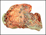
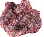
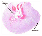
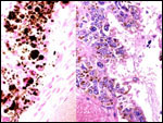
The principal reason for adding this chapter on urial sheep to the more general chapter on domestic sheep is first, the much different genetic background of urial cytogenetics, second, that it gives me a chance to detail my studies of the large quantity of yellow pigment found in the trophoblast of most ruminant placentas, and finally, that urial are becoming very endangered and the possibility of future cloning should be contemplated. For that reason, knowledge of placentation may be useful. The reader should also make reference to the earlier general chapter on sheep placentation.

The name Urial derives from the Punjabi, denoting a Himalayan wild sheep (Gotch, 1979). The animals originate from India , Kazakhstan and neighboring countries (Nowak, 1999). They are now an endangered species and are said to hybridize with Ovis orientalis in a narrow region of northern Iran (Nadler et al., 1971; Valdez et al., 1978). There is much controversy in general, however, on the origin of the many different types of sheep, and the numerous different cytogenetic findings also suggest that interbreeding among ovine species occurs (Gray, 1972). Robertsonian fusion mechanisms of chromosomes are largely responsible for this speciation, as was shown i.a. by Bunch et al. (1976). They considered that a “prezygotic selection” was the mechanism of chromosome reduction from an ancestral stock with 2n=60. Hiendleder et al. (1998) found by mtDNA study of sheep that the origin of domestic sheep was from two different forms of wild sheep that did not include the urial or argali sheep. Adult male urials weigh around 60 kg.

The domestic sheep, however, is generally presumed to have derived from Ovis musimon , the mouflon, and a species that is now distributed across Mediterranean islands. Domestication is thought to have begun approximately 10,000 years ago. There are six well-defined species and numerous subspecies of sheep distributed throughout Asia and North America . The domestic sheep now occurs worldwide and this is partly the reason for the dwindling populations of wild sheep - because of hybridization. Because some sheep species and/or subspecies are significantly endangered, they now require protection and have been listed as CITES I species, as is the urial, O. vignei (Nowak, 1999). Longevity varies with species type and with race. For mouflons, the maximal life span is given as 19-20 years (Puschmann, 1989). Jones (1993) gives a maximal life span for urials as 13 years 10 months.

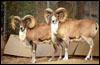
Transcaspian urial sheep at San Diego Zoo.
   
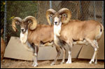
Transcaspian urial at San Diego Zoo.
   

2) General gestational data

Keisler (1999) has provided a detailed review of the reproductive parameters of domestic sheep and goats, but much less information is available on urials. There are about 800 different "breeds" of domestic sheep, with many different physical characteristics. Depending on the breed, puberty occurs between 5 and 12 months. Breeding occurs throughout the year in domestic sheep and is somewhat dependent on photoperiods. The duration of estrus is between 3 and 72 hrs. (average 29 hrs.); it is influenced by the presence of a male. The estrous cycle averages 16 days.

While the length of gestation in wild sheep species varies between 150 and 180 days, the domestic sheep has an average length of gestation of 148 days. Brown (1936) gave the length of gestation for urials as approximately 6 months. Most sheep produce singletons, but some races regularly have twins, even triplets. This certainly is true of urials (Zuckerman, 1953). Singletons weigh about 1.5 kg (Alexander, 1964).

The placental weight is difficult to ascertain, as the membranes are voluminous and much of the structure is of little importance to fetal growth. Kleemann et al. (2001) provided weights of around 600 g for domestic sheep. These authors were interested in the relation of placental and fetal sizes after progesterone treatment in early gestation. Whether these weights reflect a direct relation to the provision of nutrients to the fetus can be questioned. It is for that reason that investigators have weighed what they considered to be the more meaningful weight - that of blotted cotyledons. This is discussed in detail subsequently for domestic sheep (Alexander, 1964).

3) Implantation

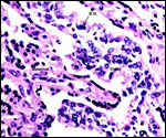
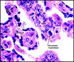
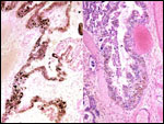
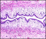
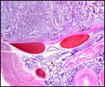
Within three to four days, the fertilized domestic sheep ovum reaches the uterus as a morula. The blastocyst develops by day 6 and sheds its zona pellucida by day 8 or 9. The blastocyst enlarges rapidly after the tenth day (Assheton, 1906). It then becomes "sticky" and, on about day 17-18, it attaches with cytoplasmic protrusions to the endometrial surface. Naturally, this attachment is only on the caruncles of the endometrium. The placenta remains diffuse for the first month or so after its mesometrial attachment (King et al., 1982). Lawn et al. (1969) did an electronmicroscopic study of goat and domestic sheep placentas. Their youngest sheep gestation was at 42 days of pregnancy; the cotyledons were then just beginning to develop their villous structures. The trophoblast was apposed to an intact endometrium that had the appearance of producing a protein secretion. Microvilli were abundant and occasional binucleated trophoblastic cells were present. Interdigitation of the cells progressed thereafter and the endometrial epithelium began to transform. Moreover, multinucleated giant cells became apparent. The syncytial cytoplasm then acquired characteristic cytoplasmic granules.

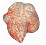
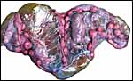
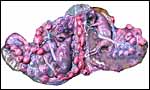
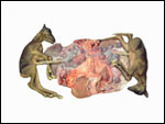
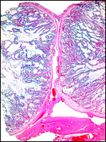
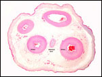
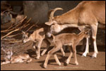
Uterus of urial containing mid-gestation twins, one in each uterine horn.


4) General characteristics of placenta

The cotyledons of ruminant placentas are the fetal portions of the placentomes. The latter term, placentome, additionally encompasses the maternal caruncle. The domestic sheep placenta is a polycotyledonary organ that usually possesses some 70 to 100 cotyledons when pregnant with a singleton. In these immature urial twin placentas shown here, there were only 35 cotyledons per twin. There were no inter-twin vascular anastomoses of the fetal blood vessels.

An additional uterus has since become available from a mid-term pregnancy. In that bicornuate uterus were male and female twins (310 and 290 g). The dam weighed 43.5 kg and had died from trauma. There were four rows of cotyledons in each horn and varying from 4x2cm to 2x1 cm in size. There were 45 cotyledons with the male and 42 cotyledons with the female. The cords measured 10 cm each and had numerous granules of squamous metaplasia on the amnionic side. This specimen in shown in the next three pictures.

Mid-term urial uterus with twins.


Each twin occupies on horn of the uterus; there are no vascular anastomoses. Each has 45 or 42 cotyledons. The corpora lutea were on the left side.


The nature of cotyledons and their chorioallantoic vascularization are especially nicely shown in this attached placenta.


In between the cotyledons is the intercotyledonary allantochorion, which has an additional and different absorptive function. It is covered by simple trophoblast and modified only over the mouths of endometrial glands. These regions are referred to as the "areolae".

In between the areolae there are the "arcades" with focal hematomas and presumed absorption of blood elements by trophoblast. This leads to the interesting and intense pigmentation of the large trophoblastic cells that line the underside of the chorion in the cotyledons (v.i.). Wimsatt (1950) makes the point that, more than in other ruminants, sheep placentas are characterized by more massive destruction of the endometrial tissue. Large hematomas of maternal blood form under the chorionic plate and this blood is said to be digested by the trophoblast, even more so in the intercotyledonary regions. The site of maternal bleeding has not been clearly demonstrated, as Wimsatt (1950) stated in much detailed discussion. Some degeneration of the maternal septal tips can be seen, but defects in the capillaries have not been identified. The septa are later repaired and it may be that, early in gestation when the tips degenerate, bleeding occurs because of focal congestion. But that point is far from clear and is not apparently related to trophoblastic damage inflicted upon maternal structures.

After the initial expansion of the blastocyst on about day 10, the elongated chorionic sac fills both uterine horns in singleton gestations. This sac is entirely covered with trophoblast on the outside. This large allantochorionic cavity contains an unusually large, elongated allantoic sac/membrane that apposes the chorion to the end of the outer chorionic sac and apposes the amnion directly over its central portions. The fluid pressure from within the allantois is believed to press the intercotyledonary membrane into close contact with the endometrium. The yolk sac is short-lived and plays no major role in ovine placentation and it cannot be found at term.


Diagram of an opened sheep placenta from a singleton gestation. The largest compartment is the allantoic sac. Inside is the smaller amnionic cavity with the fetus. Dark spots are the "placentomes" - cotyledons.
     
  I have had two urial uteri with twin conceptuses available for study. Both dams had been traumatized and were euthanized. In one set of male twins, one placenta filled one uterine horn, the other gestation was in the other uterine horn, without there being any vascular interconnections. The fetuses weighed 1850 g each and were 39 cm in CR-length. The uterus, including placenta, but without fetuses, weighed 1700 g. Each cotyledon was approximately 3 cm in greatest diameter. In the other gestation studied, the twins weighed 590 and 560 g, and had crown-rump lengths of 20.5 cm.
     
 
Opened uterus exposing the larger set of urial twins, one in each uterine horn. The large allantoic sac is best seen on the right, within which is the smaller amnionic sac that contains the fetus.
     
 
The amnions are now opened as well and this exposes the male twins and some of the cotyledons.
     
 
Urial twin placenta with 35 cotyledons of each fetal sac.
     
 
The younger urial twin gestational sac with macerated fetuses.
     
 
One cotyledon of the larger urial twins to show the location of the pigmentation and the "concave" character of cotyledons. Hematomas are seen under the chorionic plate.


Two adjacent cotyledons in implanted new pregnancy. Myometrium below.


Mossman (1987) differentiated between three types of ruminant cotyledons: the "flat" kind of certain deer, the "convex" kind of some other cervidae and giraffes, and the "concave" kind of sheep and caprines. The concave cotyledonary urial placenta has concave cotyledons that implant upon the predetermined number of uterine caruncles. In between the cotyledons, the chorion is simple and lacks villi. The trophoblast intertwines with the uterine endometrial tissue in the caruncular regions, to form the "placentome". Following death of a pregnant animal, it is easy to peel the placenta away from the endometrium, without causing significant damage to either structure. Sheep and urials have a large allantoic sac that is filled with urine. Urine is led to it through the allantoic duct within the umbilical cord, passing from the dome of the fetal bladder. Some urine must also pass through the urethra into the amnionic cavity. Urials possess multiply-branched villi, in contrast to some other ruminants. They appear as folds in reconstructions.

Alexander (1964) sought to explore the relation between fetal weight and cotyledonary weights in Merino sheep gestations; Corredale sheep had slightly different values and were not described in detail. He extensively discussed the differences between different breeds, and emphasized the need to be mindful of the existence of wide variations. He suggested that it is necessary to collect normative data for breeds and altitude when studies are undertaken to ascertain influences of altitude etc. on fetal development. The number of cotyledons was poorly related to birth weight, in contrast to their combined weights. It is important to note that not all uterine caruncles are used for cotyledonary development and this feature apparently varies widely. Moreover, it appears that some caruncles disappear with aging. Male fetuses occupy more caruncles than do female fetuses. Alexander (1964) separated the cotyledons (without intervening membranes) from the caruncles by traction; after blotting them dry, he weighed them. There was an excellent correlation between fetal weight and cotyledonary mass at different stages of gestation. Male fetuses and their cotyledons were slightly heavier. Those of twins were slightly smaller. The average number of uterine caruncles was 73 in Merino sheep (from 64 to 145). Also, the number of caruncles was generally similar in both uterine horns. Older ewes had a slightly heavier cotyledonary mass which varied from 115 to 130 g. Individual cotyledons weighed about 1.6-1.8 g. There were, however, enormous differences in their weights, from 0.1 to12 g of fetal cotyledons and 0.1- 45 g of complete placentomes. The fetal part of a complete cotyledon was 40% and it was darker than the maternal part. Alexander made another important observation: He found that whereas in immature cotyledons the maternal tissue (caruncle) surrounds the cotyledon (which is concave, according to Mossman), towards term the opposite is true. It is of further interest to contemplate that most of these investigators considered only the cotyledonary weight although it had been long appreciated that the intercotyledonary region serves also as an important exchange (nutritive) region. Perhaps it should be included in the weights when an association with fetal growth is sought.


5) Details of barrier structure

The nature of the interface between maternal epithelium and trophoblast was described in the electronmicroscopic study of domestic sheep placentas by Lawn et al. (1969). This interface comprises also an area in which maternal blood is extravasated "to supply the fetus with iron". Wimsatt (1950) had studied this region in great detail and it will be discussed again below.
The endometrial crypt is lined with epithelium that has a striking microvillous surface. This, in turn, is closely adherent to the trophoblast with which its microvilli interdigitate. The maternal epithelium is continuous, is not interrupted, and it is often multinucleated. The microvilli of trophoblast and endometrium interdigitate diffusely and thus, this placenta is here epitheliochorial (-bichorial to be correct), as was first clearly defined by Ludwig (1962). Because of the degeneration occurring in some endometrial epithelium during the course of gestation, areas of syndesmochorial relation also exist. Mossman (1987), however, correctly pointed out that there is little vascular supply in those areas and, from a functional view; these areas are probably of little importance. Thus, one should consider that the sheep placenta is principally an epitheliochorial one.

Lawn et al. (1969) paid special attention to the development of the well-known binucleate cells of the ruminant placenta. This cell has spawned an enormous amount of literature. These investigators traced the cells' development from early nuclear division (without cytoplasmic division) to their accumulation of numerous cell organelles. Among these, the Golgi complex appeared first and was then succeeded by a large number of granules. In the mature cell, the granules fill the cellular cytoplasm, nearly crowding out all other components. It was also shown that these cells never completely bordered the endometrial epithelium - a rim of cytoplasm always separated the two cell types. All ruminants have binucleate trophoblast, but their number varies greatly, from species to species. Urials have relatively few binucleate cells. Contrary to earlier investigators, Lawn et al. (1969) correctly identified the binucleate cells as having derived from trophoblast. The organelles, as well as some of the endometrial crypt lining cells, stain intensely with the PAS method. They are now known to contain placental lactogen. Wooding et al. (1997) later showed their fusion with endometrium to make trinucleate cells, and produced evidence for their production of placental lactogen.

The original classification of the sheep placenta as being an epitheliochorial organ then is not considered to be entirely correct. (Please see also the chapter on Cretan Goat for additional considerations.) Autoradiographic studies by Wooding et al. (1981; 1997) probably now supersede the older information. Relative to its cotyledons, the ovine/caprine placenta must now be considered as a focally syndesmochorial organ. The intercotyledonary regions remain epitheliochorial in the mature placenta, as had been previously determined.

The most controversial aspect perhaps of ungulate placentation has been the origin of the syncytium that lines the crypts of the cotyledons. Similarly, the nature of, or the relation of the syncytium to the binucleated cells has been interpreted differently. Binucleate cells are clearly trophoblastic in origin and they produce the placental lactogen. But the autoradiographic study by Wooding et al. (1981) investigated the origin of syncytial cells. It appeared to me that this convincingly showed the syncytium to have a trophoblastic origin and that it is not derived by fusion with endometrial epithelium, as was once thought (Wooding et al., 1993). Presumably, the binucleate cells initiate the process of fusion and subsequent migration within the trophoblastic layer, and they continue to do migrate during the entire course of pregnancy. Moreover, the fact that there are no mitoses in the syncytium, while they do occur in the cellular trophoblast, supports this notion. Thus, the syncytium is considered to develop much like other fusion-induced cells. Wooding (1984; 1993) later showed that some fusion with maternal epithelium also produces trinucleate hybrid cells. These cells are quite specific, and secretion of the lactogen-containing granules (perhaps to maintain gestation) was described in his paper. Wooding emphasized that there is no immunologic reaction as the result of maternal epithelial erosion and this fusion ("hybridization" of cells). This may be an erroneous conclusion, as will be seen shortly. Thus, in addition to the binucleate cells, numerous giant cells are found deep in the placentome. According to Lawn et al. (1969) they originate from the endometrial epithelium in later gestation; more likely they are part of the "syncytium". In addition to these developments, the endothelium of the maternal capillaries becomes markedly hypertrophied.


Urial placental villi in between the maternal endometrial septa. B.N.= binucleate cell.
     
 
Higher magnification of terminal villi of urial placenta between maternal septa.
     
 
There is a layer of heavily pigmented trophoblast beneath the chorionic membrane of the older urial twin placenta. Left: Prussian blue reaction is negative.
     
 
The layer of heavily pigmented trophoblast beneath the chorionic membrane at higher magnification. Left: Prussian blue reaction is negative.


To reiterate, for many years, the nature and origin of these giant cells has been disputed in the literature. Wimsatt (1950) considered them to be trophoblastic in origin after earlier believing them to be endometrial. A later electronmicroscopic study by Davis and Wimsatt (1966) reaffirmed the trophoblast derivation of the syncytial (giant) cells. These authors then suggested the movement of binucleate cells into the crypts where the giant cells develop. Wooding (1983) stated that the binucleate cells comprise as many as 1/5th of the trophoblastic population of ruminant placentomes but this is not the case in urials where I find far fewer. Another 1/5th of the binucleate cells was "migrating" and, in domestic sheep, they were closer to the maternal tissues than the remainder of trophoblast. They arose early in gestation and decreased significantly towards term. A syncytium with uterine epithelium develops and it is later replaced by cellular endometrial epithelium. One or two mitoses per 100 cells are present in the trophectoderm, but none in the syncytial giant cells.

Lee et al. (1986) studied binucleate cells with an antibody (SBU-3) that was found to interact commonly with most ruminants (deer, sheep, and cow). Binucleate cell cytoplasm stained with this antibody, both in the placentomes and in the intercotyledonary regions. These investigators, however, did not find this immunological marker to be expressed in the syncytial giant cells. This has to make one reconsider the true origin and nature of the syncytial giant cells. I believe that their origin from trophoblastic binucleate cells is not completely verified. Additional studies are needed.

Another area of special study to Wimsatt (1950) was the subchorial hematoma and the pigmentation. At the top of the villous tree and, perhaps to a lesser extent in the "arcades" of the intercotyledonary membrane, significant amounts of maternal blood are extravasated during the course of pregnancy. The liberated red cells are here lysed and phagocytosed by trophoblast. As a result, large quantities of yellow ("nonferruginous") pigment accumulate. These hematomas are well developed in early and late pregnancy, and bleeding may occur intermittently throughout gestation. The precise vascular location of the origin of these maternal hemorrhages is still uncertain. The blood may derive from peripheral capillaries of the septal tips in which Wimsatt observed degenerative changes. Others have suggested that the hematomas are the result of trophoblastic invasion which is, however, less evident at these sites than elsewhere in the cotyledons.

My interest has been to determine the nature of this very large amount of pigment in the urial placenta. It is yellow-brown, may be finely granular or composed of large brown plates with a seemingly crystalline character. It presents primarily within the trophoblast that lines the undersurface of the chorionic membrane of the cotyledon and also between cotyledons. The pigment occurs near the regions of hemorrhage and Wimsatt (1950), as well as others, have described phagocytosis of red cells by trophoblast. I have not been able to observe this unequivocally. Attempts at staining with the Prussian blue reaction for hemosiderin have been uniformly negative. The ancient "Gmelin reaction" for hematoidin is also negative (concentrated sulfuric acid should change the brown color to red, violet to green. It has remained brown). Bleaching the material, as is used to remove melanin, removes all color from the cells. The bleach is performed by potassium permanganate solution, followed by oxalic acid and wash. Extraction with common solvents has been impossible as well. It is reasonable to assume that the pigment should derive from hemoglobin, but the exact nature is elusive. It is neither hemosiderin nor is it characteristic hematoidin (which is identical or extremely similar to bilirubin [Rich, 1925]). Additional studies of the pigment were undertaken in nilgai placentas (see there).


6) Umbilical cord

The urial umbilical cord attaches mesometrially. It has four large allantoic blood vessels (2A, 2V) and a large, patent allantoic duct. In addition to the four large blood vessels, there are numerous smaller allantoic vessels. They are especially congregated around the large allantoic duct. In the larger set of twins, the cords were 19 and 13 cm long. They also were covered with fine granules and they were not twisted. In the new set of twins the cords measured 10 cm each.

Arvy & Pillery (1976) gave the length of the domestic sheep cords as being only 7 cm, while Reynolds (1952) found it to be 10 cm. Reynolds studied the compression of Wharton's jelly by forcefully injecting the cord vessels and found that Wharton's jelly was only slightly compressible in sheep and goats, much less so than was found for human cords.


The urial umbilical cord surface has many plaques of squamous metaplasia (verrucae). The allantoic duct is in the center. Note numerous small vessels.
     
 
Allantoic duct of urial umbilical cord.


7) Uteroplacental circulation

In his study of sheep placentation, Wimsatt (1950) injected the fetal artery with colored dyes and thus characterized the villous vascular tree. A single artery injects blood centrally into the cylindrical main stem villus; it then divides, sending a single vessel into the flattened villous ramifications, where a plexiform capillary network lines the surface of the villi with protrusions into the trophoblast. A complex network of venules drains the villous structures, the veins having larger caliber and being less muscular.

Makowski (1968) illustrated the injected maternal/fetal vasculature of individual cotyledons. He found that there was no evidence of a true counter-current blood flow. The maternal arterioles apparently have a sphincter-like arrangement at their bases. This is presumed to dictate the blood flow through the maternal plates. There was no evidence of a neural supply to the vessels.

     
  8) Extraplacental membranes
     
 
Diagram of the intercotyledonary membranes attached to the uterus.
     
 
Implantation site of urial twin placenta. Bottom is myometrium, above are endometrial glands.
     
 
Intercotyledonary region of urial placenta. Endometrial glands below, pigmented trophoblastic surface of membranes above.
     
 
Intercotyledonary membranes of urial placenta. Allantoic epithelium is at the top, vascularized chorioallantoic membrane beneath, then follows the pigmented tall trophoblastic cell layer over the areolae.


There are no "free" membranes in the urial placenta. Instead, between the cotyledons the intercotyledonary chorion is a relatively simple membrane. It is avillous and covered on the outside with a simple layer of cylindrical trophoblastic epithelium that has a microvillous surface. The trophoblastic epithelial cells have a varied morphology, as is evident in the photograph. They are pressed against the endometrium by pressure from within the allantoic cavity. In late stages, the exocoelomic space is completely obliterated. The allantoic sac, which is of endodermal origin, fuses with the chorionic sac as early as on the 22nd day of ovine development (Davies & Wimsatt, 1962). It is very large and elongate. Its columnar epithelium possesses only very short microvilli and, in the mature placenta, it is relatively flat. Davies (1952) was perhaps the first to draw attention to the accumulation of large amounts of fructose in the allantoic fluid. The urial sheep sacs contained no hippomanes.

Wimsatt (1964) paid special attention to this region in domestic sheep and observed that, soon after implantation, the endometrium of these areas is lost and the trophoblast apposes the connective tissue of the uterus. The placental relation then is temporarily of a syndesmochorial kind. In later stages (after 100 days) the epithelium of the endometrium has regenerated and then the trophoblast has the usual epitheliochorial relation again.

A small amount of acellular debris separates the trophoblast from the endometrial epithelium. The trophoblastic epithelium of these areas also accumulates various electron-dense inclusions in advanced gestation. They are of uncertain nature and not comparable to the lactogen granules. They possibly relate to the small extravasations of maternal blood in the "arcades". The intercotyledonary membrane region is also characterized by the "areolae", an old description dating back to Eschricht (reviewed by Ludwig, 1968). Numerous studies have suggested that the areolae are situated above the mouths of several endometrial glands and that uterine "milk" secreted here serves a nutritive function for the fetus, at least in sheep gestations. It has not been studied in urials but is likely to be similar. The areolae increase in size with gestation, up to a 3mm diameter, and were beautifully studied by Wimsatt (1964). He showed PAS positive material to increase in the course of gestation at these locations, and found several modifications of the histologic appearance of the trophoblastic epithelium over time. With the secretion of the glands, a "pit" forms and the trophoblastic epithelium becomes focally elevated. Wimsatt also segregated three "epochs" in the development of the areolae, which are not discussed further. These are presumably related to an increased nutritive uptake or the production of uterine milk. Ludwig (1968) suggested the term "enteroid" for these areolae, while he called the cotyledonary regions as subserving a "nephropneumatoid" function.

Wimsatt (1964) was also the first author to study the vasculature of the intercotyledonary membranes in some detail. Interestingly, here the arteries are located in such a manner that they are situated above the veins, the latter being closer to the trophoblast. That is the same arrangement as is found in primate placentas. The capillaries are "more copious" in the vicinity of the areolae than elsewhere in the chorion.

The structure of the domestic sheep amnion has been studied by Bautzmann & Schroder (1955). Their investigations of the amnion from birds and reptiles had disclosed the presence of a smooth musculature in the amnion. This is absent in sheep and other mammals. The ovine amnionic epithelium, and that of urials, is flat and single-layered, with occasional "warts" or verrucae. They are composed of squamous proliferations and are similar to the verrucae of squamous metaplasia in the amnionic epithelium of the umbilical cord. These authors described in detail also the collagenous fibers and the glassy membrane of the amnion. These layers were further delineated in the comprehensive studies of human placental membranes by Bourne (1962). Additional studies on these epithelial proliferations in hoofed animals and whales were published by Naaktgeboren & Zwillenberg (1961). They found them to exist in essentially all hoofed animals and whales with the exception of the pig. In domestic sheep, they were initially white but became yellow as gestation advanced. The allantoic sac of my urial fetuses contained brownish, cloudy fluid but no hippomanes. There was no evidence of inflammation.

     
  9) Trophoblast external to barrier

There is no infiltration of trophoblast beyond the caruncles, and there is no subplacenta.
     
  10) Endometrium

There is no decidual transformation of the endometrial stroma in the ovine uterus as that which is found in primates. Endometrial glands are few and scattered below the cotyledons, but they are mainly located in the intercotyledonary regions.
     
  11) Various features

There is no subplacenta.
     
  12) Endocrinology

Keisler (1999) provided detailed information on endocrine signals of domestic ewes. To the best of my knowledge, it has not been studied in any detail in urials. I assume, however, that it is generally similar. Nine hours after onset of estrus in the domestic sheep, large amounts of pituitary LH are released. Ovulation occurs 21-26 hours later. Three days following ovulation, progesterone levels can be detected and the uterus produces PGF2a episodically. This leads to the dissolution of the corpus luteum and a fall in progesterone levels. If conception takes place, "corpus luteum rescue" occurs with the first maternal recognition of pregnancy. The conceptus secretes various proteins (interferons) that inhibit PGF2a production. Later in gestation, a variety of pregnancy-specific proteins and placental lactogen are produced. These increase to term; the progesterone secretion by the corpus ceases to be essential after 60 days. The placental production of progesterone is then adequate to maintain pregnancy.

The onset of labor has been of particular interest to investigators. It is mediated via the fetal hypothalamic-pituitary-adrenal axis. Likewise, the placental ability to produce prostaglandins increases towards term. Fetal hypophysectomy or adrenalectomy leads to postmaturity. Fetal ACTH production increases two weeks before parturition with marked increase of cortisol production by the fetus approximately 3-4 days before delivery. There is extensive literature on these investigations, some of which is cited by Keisler (1999); other publications can be found in the summary by Liggins (1969).

Progesterone appears to have a significant effect on fetal growth when administered in the first few days of gestation according to Kleemann et al. (2001). They found fetal size and weight to increase with minor effect on placental weights. These authors also made extensive measurements of fetal body parts and of the placental surface and its components that must be read in the original publication.


13) Genetics

Domestic sheep have 54 chromosomes that have been well delineated in numerous studies. Urials, however, have 58 chromosomes with one pair of large metacentric elements (Nadler et al., 1971). Areas of sympatry with O. ammon (2n=54) were found by these investigators in northern Iran and the question of reduced hybrid fertility was discussed but it was not resolved. The chromosomal variations are all the result of Robertsonian fusion mechanisms among acrocentric chromosomes (Bunch et al., 1976). The animals displayed in this chapter are all composed of specimens with 2n=58.

Long & Williams (1980) studied the frequency of chromosomal errors in domestic sheep embryos and in unfertilized eggs. They found 4.7% trisomies in 89 fertilized eggs and one mosaic diploid/haploid specimen. Several eggs or embryos had a "cracked" zona pelludica.

Domestic sheep have hybridized with many other taxa (Gray, 1972), especially other ovine forms. Of greatest interest have been the many attempts to hybridize sheep (2n=54) and goat (2n=60). Occasional offspring have been achieved (2n=57) but that has been difficult, and hemolytic disease has often caused fetal demise. In a large series of experiments, Dent et al. (1971) showed with electronmicroscopy that the trophoblast of the hybrids is normal and implantation appears to proceed normally at first. There begins, however, an extensive accumulation of platelets in the maternal blood vessels and endothelial swelling to the point of occlusion with hemorrhage ensuing between 34 and 38 days. These authors suggested that this is akin to an immunological rejection of the fetal hybrid implant. The hybrids are only exceptionally fertile (Bunch et al., 1976). Dent et al. (1971) indicated that fertilization occurs regularly in such attempts at hybridization but that placentation usually fails before the second month of gestation. The platelet accumulation in such hybrid placentas beneath the uterine epithelium of the placentomes on day 34 is the apparent cause of failure. "The maternal blood vessels showed the presence of large numbers of platelets, either alone or mixed with isolated red blood cells". On day 38 the platelets were very prominent, but absent in normal goat placentas. Subsequently, the uterine epithelium became necrotic and hemorrhage occurred focally. They interpreted these lesions as resulting from antigen-antibody interaction, akin to that seen in skin graft rejection. Embryo transfers of mouflon into sheep, however, are successful.

The mitochondrial study of Hiendleder et al. (1998) suggested that domestic sheep derived from two ancestral wild stocks, but this did not include argali and urial sheep. The lowest number of chromosomes in sheep known is that of the Siberian snow sheep (Ovis nivicola) with 2n=52 (Bunch et al. 1998). They found Severtzov's sheep (Ovis ammon severtzovi) to possess 2n=56.


Karyotype of male domestic sheep – 2n=54.


Karyotype of female Urial – 2n=58.

   
  14) Immunology

The domestic sheep has been used to study the onset of the production and the intricate nature of early antibodies by the fetus (Silverstein et al., 1963). When the fetus is stimulated with specific antigens, it responds by producing different antibodies at different gestational times. Moreover, skin grafts placed upon fetuses from other fetuses were rejected in a typical manner; it was discovered then, however, that plasma cells did not participate in the rejection process as the fetus was not yet capable to produce them (Silverstein, et al. 1963).


15) Pathological features

Griner (1983) lists trauma as the number one cause of mortality in urials at the San Diego Zoo, with occasional pulmonary nematodes being secondary.

An important disease of domestic sheep is scrapie, a CNS degenerative disorder that is caused by a variant prion protein. It is so important because, in recent years, infected animal derivatives were fed to cattle with the occurrence of bovine spongiform encephalopathy (BSE). Subsequently, through ingestion of beef, humans have developed a variant of Creutzfeldt-Jakob Disease (vCJD) traced to this source. Houston et al. (2000) showed that blood from a domestic sheep that was fed BSE-infected brain of cattle transmitted this prion to sheep via transfusion of the blood from the challenged sheep.

Domestic sheep and goats commonly harbor Chlamydia psittaci organisms in their generative tracts. This infection often leads to abortion or birth of weak lambs. In subsequent pregnancies, the infection recurs in some 5-10%, with some immunity resulting (Wilsmore et al., 1990). In addition, vaginal shedding of organisms occurs. The placenta contains numerous organisms, and areas of inflammation; areas of necrosis are also found. Pregnant women attending the delivery of infected ewes have repeatedly developed severe febrile illnesses, aborted and developed placental lesions characteristic of this infection (Hyde & Benirschke, 1997). Numerous other infectious diseases affect the domestic sheep. They have been summarized in the veterinary literature, e.g. by Smith et al. (1972). Among these is brucellosis due to infection with Brucella ovis. It causes mainly epididymitis in rams but may affect the female generative tract and lead to abortion with placental inflammation.


16) Physiological data

The domestic sheep has been used for numerous physiological studies; many are designed to better understand human prenatal development, but there are no such studies in urials. Thus, Barron (1951) reviewed in some detail the transfer of oxygen from mother to fetus. He and others have studied this transfer at high altitude and compared it with data found at sea level. Liggins (1969) reviewed the endocrine signals for the onset of labor and parturition. Kleemann et al. (2001) showed that progesterone administration in very early pregnancy enhances fetal growth. Alexander (1964) removed up to 84 caruncles from Merino sheep prior to mating in order to assess the impact on placentation. While fertility was not affected, the ewes in which sixty or more caruncles had been removed delivered prematurely and the lambs had reduced birth weights. The caruncle removal was compensated by an increased size of the cotyledons. Worthington et al. (1981) carried out similar reductions with electrocautery. They found a similar effect on fetal growth. Creasy et al. (1972) embolized fetal cotyledons with microspheres between days 96 and 125 and found significant reduction in fetal size. The affected cotyledons showed hyalinization and necrosis with marked reduction in size. This was followed by similar experiments of Duncan et al. (2000). These investigators examined the consequences of interference with placentation on fetal brain development. They identified significant damage to the white matter of the CNS in the growth-restricted lambs. There are many other, similar studies that probe fetal CNS development by restricting placental performance. For instance, Dawes & Mott (1964) altered oxygen supply to fetal lambs and studied their responses. Keunen et al. (1997) found no CNS damage with cord occlusion of up to ten minutes, but Ball et al. (1994) found that seizures took place when there was severe cord occlusion for 90 minutes. Ikeda et al. (1998) sought to explain the CNS lesions after cord occlusion by brain lipid peroxidation. Mallard et al. (1992) found primarily hippocampal lesions after occlusion, while Clapp et al. (1988) found mostly white matter damage after intermittent occlusion of the cord's vessels.

Gilbert et al. (1996) showed that technetium crossed the ovine placenta more rapidly than the smaller sodium ion. Ovine urine production was studied by Ross, et al. (1988) and by many other investigators who are listed by these authors. Ross et al. found in this carefully executed protocol (days after surgical canulations, standing ewes, 4-hour experiment, careful electrolyte measurements) that the amount of allantoic fluid was greater than that of amniotic fluid; that fetal urination is greater into the allantoic sac (through urachus) than the amnionic sac (through urethra); that transmembranous flow probably exists; that lung fluid participates in amnionic fluid composition and swallowing in disposing of it. These are difficult experiments whose interpretation is complicated by the presence of two large fluid-filled sacs in the ovine placenta. They are not directly transferable to human amnionic fluid metabolism for that reason. With Barron we catheterized the urachus in an instrumented sheep that was suspended in a water bath. We found that urine production decreased markedly over a few hours when all urine from the urachus was collected externally. Its osmolality also increased and much fructose was contained in the urine. Urination increased rapidly when water was injected into the allantoic cavity. We conjectured that the water was rapidly absorbed by the allantoic circulation and rehydrated the water-deprived fetus. Matsumoto et al. (2000) ligated the fetal esophagus and urachus and were unable to produce polyhydramnios, as had been their aim. This suggested to them that transmembranous transport must be large.

     
  17) Other resources

Cell strains of various sheep species, including urials, are available from CRES at the Zoological Society of San Diego by contacting Dr. Oliver Ryder at: oryder@ucsd.edu.
     
  18) Other remarks - What additional Information is needed?

More knowledge is needed on umbilical cords. Mossman (1987) pointed out that more information is needed on the insertion of the cords of multiple gestations. It can be seen in above photographs for urial twins. Because there is still some uncertainty as to the true origin of the syncytial giant cells, additional studies are warranted.
     
  Acknowledgement

The animal photographs in this chapter come from the Zoological Society of San Diego. I appreciate also very much the help of the pathologists at the San Diego Zoo.
     
  References

Alexander, G.: Studies of the placenta of the sheep (Ovis aries L.). Placental size. J. Reprod. Fertil. 7:289-305, 1964.
     
  Alexander, G.: Studies on the placenta of the sheep (L.). Effect of surgical reduction in the number of caruncles. J. Reprod. Fertil. 7:307-322, 1964.

Arvy, L. and Pilleri, G. The Cordon Ombilical (Funis umbilicalis). Verlag Hirnanatom. Instit., Ostermundigen, Switzerland, 1976.

Assheton, R.: VI. The morphology of the ungulate placenta, particularly the development of that organ in the sheep, and notes upon the placenta of the elephant and hyrax. Phil. Trans. Roy. Soc. London, Series B 198:143-220, 1906.

Ball, R.H., Parer, J.T., Caldwell, L.E. and Johnson, J.: Regional blood flow and metabolism in ovine fetuses during severe cord occlusion. Amer. J. Obstet. Gynecol. 171:1549-1555, 1994.

Barron, D.H.: Some aspects of the transfer of oxygen across the syndesmochorial placenta of the sheep. The Yale J. Biol. Med. 24:169-190, 1951.

Bautzmann, H. and Schroder, R.: Vergleichende Studien uber Bau und Funktion des Amnions. Das Amnion der Säuger am Beispiel des Schafes (Ovis aries). Z. Zellforsch. 43:48-63, 1955.

Bourne, G.L.: The Human Amnion and Chorion. Lloyd-Luke, London, 1962.

Brown, C.E.: Rearing wild animals in captivity and gestation periods. J. Mammal. 17:10-13, 1936.

Bunch, T.D., Foote, W.C. and Spillett, J.J.: Sheep-goat hybrid karyotypes. Theriogenology 6:379-385, 1976.

Bunch, T.D., Vorontsov, N.N., Lyapunova E.A. and Hoffman R.S.: Chromosome number of Severtzov's sheep (Ovis ammon severtzovi): G-banded karyotype comparisons with ovis. J. Hered. 89:266-269, 1998.

Bunch, T.D., Foote, W.C. and Spillett, J.J.: Translocations of acrocentric chromosomes and their implications in the evolution of sheep (Ovis). Cytogenet. Cell Genet. 17:122-136, 1976.

Clapp, J.F., Peress, N.S., Wesley, M. and Mann, L.I.: Brain damage after intermittent partial cord occlusion in the chronically instrumented fetal lamb. Amer. J. Obstet. Gynecol. 159:504-509, 1988.

Creasy, R.K., Barrett, C.T., Swiet, M.de, Kahanpää, K.V. and Rudolph, A.M.: Experimental intrauterine growth retardation in the sheep. Amer. J. Obstet. Gynecol. 112:566-573, 1972.

     
  Davies, J.: Correlated anatomical and histochemical studies on the mesonephros and placenta of the sheep. Amer. J. Anat. 91:263-299, 1952.

Davies, J. and Wimsatt, W.A.: Observations on the fine structure of the sheep placenta. Acta anat. 65:182-223, 1966.

Dawes, G.S. and Mott, J.C.: Changes in O2 distribution and consumption in foetal lambs with variation in umbilical blood flow. J. Physiol. 170:524-540, 1964.

Dent, J., McGovern, P.T. and Hancock, J.L.: Immunological implications of ultrastructural studies of goat x sheep hybrid placentae. Nature 231:115-117, 1971.

Duncan, J.R., Cock, M.L., Harding, R. and Rees, S.M.: Relation between damage to the placenta and the fetal brain after late-gestation placental embolization and fetal growth restriction in sheep. Amer. J. Obstet. Gynecol. 183:1013-1022, 2000.

Gilbert, W.M., Newman, P.S., Eby-Wilkens, E. and Brace, R.A.: Technetium Tc 99m rapidly crosses the ovine placenta and intramembranous pathway. Amer. J. Obstet. Gynecol. 175:1557-1562, 1996.

Gotch, A.F.: Mammals - Their Latin Names Explained. Blandford Press, Poole, Dorset, 1979.

Gray, A.P.: Mammalian Hybrids. Second edition. A Check-List with Bibliography. Commonwealth Agricultural Bureaux, Farnham Royal, Slough, UK, 1972.

Griner, L.A.: Pathology of Zoo Animals. Zoological Society of San Diego, San Diego, California, 1983.

Hiendleder, S., Mainz, K., Plante, Y. and Lewalski, H: Analysis of mitochondrial DNA indicates that domestic sheep are derived from two different ancestral maternal sources: no evidence for contribution from urial and argali sheep J. Hered. 89:113-120, 1998.

Houston, F., Foster, J.D., Chong, A., Hunter, N. and Bostock, C.J.: Transmission of BSE by blood transfusion in sheep. Lancet 356:999-1000 (and 955-956 for Comments), 2000.

Hyde, S.R. and Benirschke, K.: Gestational psittacosis: Case report and literature review. Modern Pathol. 10:602-607, 1997

Ikeda, T., Murata, Y., Quilligan, E.J., Parer, J.T., Doi, S. and Park, S-D.: Brain lipid peroxidation and antioxidant levels in fetal lambs 72 hours after asphyxia by partial umbilical cord occlusion. Amer. J. Obstet. Gynecol. 178:474-478, 1998.

Jones, M.L.: Longevity of ungulates in captivity. Intern. Zoo Yearbk. 32:159-169, 1993.

Keisler, D.H.: Sheep and Goats. In, Encyclopedia of Reproduction. E. Knobil and J.D. Neill, eds. Vol. 4, pp. 479-492. Academic Press, San Diego, 1999.


Keunen, H., Blanco, C.E., Reempts, J.L.H.van and Hasaart, T.H.M.: Absence of neuronal damage after umbilical cord occlusion of 10, 15, and 20 minutes in midgestation fetal sheep. Amer. J. Obstet. Gynecol. 176:515-520, 1997.

King, G.J., Atkinson, B.A. and Robertson, H.A.: Implantation and early placentation in domestic ungulates. J. Reprod. Fertil. Suppl. 31:17-30, 1982.

Kleemann, D.O., Walker, S.K., Hartwich, K.M., Fong, L., Seamark, R.F., Robinson, J.S. and Owens, J.A.: Fetoplacental growth in sheep administered progesterone during the first three days of pregnancy. Placenta 22:14-23, 2001.

Lawn, A.M., Chiquoine, A.D. and Amoroso, E.C.: The development of the placenta in the sheep and goat: An electron microscopic study. J. Anat. 105:557-578, 1969.

Lee, C.S., Gogolin-Ewens, K. and Brandon, M.R.: Comparative studies on the distribution of binucleate cells in the placentae of deer and cow, using monoclonal antibody, SBU-3. J. Anat. 147:163-179, 1986.

Liggins, G.C.: The foetal role in the initiation of parturition in the ewe. CIBA Fndt. Symp. On Foetal Anatomy. G.E.W. Wolstenholm & M. O'Connor, eds. Pp. 218-231, 1969.

Long, S.E. and Williams, C.V.: Frequency of chromosomal abnormalities in early embryos of the domestic sheep (Ovis aries). J. Reprod. Fertil. 58:197-201, 1980.

Ludwig, K.S.: Zur Feinstruktur der materno-fetalen Verbindung im Placentom des Schafes (Ovis aries L.). Experientia 18:212-213, 1962.

Ludwig, K.S.: Zur vergleichenden Histologie des Allantochorion. Rev. Suisse Zool. 75:819-831, 1968.

Makowski, E.L.: Maternal and fetal vascular nets in placentas of sheep and goats. Amer. J. Obstet. Gynecol. 100:283-288, 1968.

Mallard, E.C., Gunn, A.J., Williams, C.E., Johnston, B.M. and Gluckman, P.D.: Transient umbilical cord occlusion causes hippocampal damage in the fetal sheep. Amer. J. Obstet. Gynecol. 167:1423-1430, 1992.

Matsumoto, L.C., Cheung, C.Y. and Brace, R.A.: Effect of esophageal ligation on amniotic fluid volume and urinary flow rate in fetal sheep. Amer. J. Obstet. Gynecol. 182:699-705, 2000.

Mossman, H.W.: Vertebrate Fetal Membranes. MacMillan, Houndmills, 1987.

Naaktgeboren, C. and Zwillenberg, H.H.L.: Untersuchungen uber die Auswuchse am Amnion und an der Nabelschnur bei Walen und Huftieren, mit besonderer Berucksichtigung des europäischen Hausrindes. Acta morphol. Neerlando-Scand. 4:31-60, 1961.

     
  Nadler, C.F., Lay, D.M. and Hassinger, J.D.: Cytogenetic analyses of wild sheep populations in northern Iran. Cytogenetics 10:137-152, 1971.

Nowak, R.M.: Walker's Mammals of the World. 6th ed. The Johns Hopkins Press, Baltimore, 1999.

Puschmann, W.: Zootierhaltung. Vol. 2, Säugetiere. VEB Deutscher Landwirtschaftsverlag Berlin, 1989.

Reynolds, S.R.M.: The proportion of Wharton's jelly in the umbilical cord in relation to distention of the umbilical arteries and vein, with observations on the folds of Hoboken. Anat. Rec. 119:365-377, 1952.

Rich, A.R.: The formation of bile pigments. Physiol. Rev. 5:182-24 19.

Ross, M.G., Ervin, M.G., Rappaport, V.J., Youssef, A., Leake, R.D. and Fisher, D.A.: Ovine fetal urine contribution to amniotic and allantoic compartments. Biol. Neonat. 53:98-104, 1988.

Silverstein, A.M., Prendergast, R.A. and Kraner, K.L.: Homograft rejection in the fetal lamb: The role of circulating antibody. Science 142:1172-1173, 1963.

Silverstein, A.M., Thorbecke, G.J., Kraner, K.L. and Lukes, R.J.: Fetal response to antigenic stimulus. III. y-globulin production in normal and stimulated lambs. J. Immunol. 91:384-395, 1963.

Smith, H.A., Jones, T.C. and Hunt, R.D.: Veterinary Pathology. Lea & Febiger, Philadelphia, 1972.

Valdez, R., Nadler, C.F. and Bunch, T.D.: Evolution of wild sheep in Iran. Evolution 32:56-72, 1978.

Wilsmore, A.J., Izzard, K.A., Wilsmore, B.C. and Dagnall, G.I.R.: Breeding performance of sheep infected with Chlamydia psittaci (ovis) during their preceding pregnancy. Vet. Rec. 130:68-70, 1990.

Wimsatt, W.A.: New histological observations on the placenta of the sheep. Amer. J. Anat. 87:391-458, 1950.

Wooding, F.B.P.: Role of binucleate cells in fetomaternal cell fusion at implantation in the sheep. Amer. J. Anat. 170:233-250, 1950.

Wooding, F.B.P.: Frequency and localization of binuclear cells in the placentome of ruminants. Placenta 4:527-540, 1983.

     
  Wooding, F.B.P., Flint, A.P.F., Heap, R.B. and Hobbs, T.: Autoradiographic evidence for migration and fusion of cells in the sheep placenta: Resolution of a problem in placenta classification. Cell Biol. Internat. Reports 5:821-827, 1981.

Wooding, F.B., Morgan, G. and Adams, C.L.: Structure and function in the ruminant synepitheliochorial placenta: central role of the trophoblast binucleate cell in deer. Microsc. Res. Tech. 38:88-99, 1997.

Worthington, D., Piercy, W.N. and Smith, B.T.: Effects of reduction of placental size in sheep. Obstet. Gynecol. 58:215-221, 1981.

Zuckerman, S.: The breeding seasons of mammals in captivity. Proc. Zool. Soc. London 122:827-950, 1953.


back to top 

© 2005. All rights reserved.
welcome | home | intro | placentation | glossary | author contact me: kbenirsc@ucsd.edu