(Clicking on the thumbnail images below will launch a new window and a larger version of the thumbnail.)
 
Last updated:
July 4, 2004.

Francois' Langur
Trachypithecus francoisi

Order: Primates
Family: Cercopithecidae (Colobidae)

1) General Zoological Data

Langurs have been placed in a variety of different genera, but for this species, the current agreed-upon genus is Trachypithecus (Nowak, 1999), while others have formerly named the genus Semnopithecus. This endangered species lives in Southern China, North Vietnam and in Laos (Nadler 1996) but is threatened with habitat loss and from poaching. It is a typical leaf-eater with subdivided stomach. There are colonies in several zoological parks and, at the Endangered Primate Rescue Center (EPRC) at Cuc Phuong, Vietnam, confiscated animals breed well and are then released into natural habitats (Nadler,1994). A productive colony exists at the San Diego Zoo with longevities to about 30 years. Singleton births are the norm in langurs, including this species. Detailed management protocols, diet, mortality, housing and other aspects on keeping this species have been described by Robinson (2004).

   
  Adult Francois' langur at San Diego Zoo.
     
  Adult Francois' langur at San Diego Zoo's breeding colony.
     
  Francois' langur at San Diego Zoo.


2) General Gestational Data

There is virtually no published information on cycle length (guessed at 28 days), gestational length (about 200+ days), and on any other aspects of gestational physiology.

3) Implantation

As is true for most Cercopithecidae, notably the langurs, Francois' langur has a relatively superficial implantation in the uterus simplex. It then develops a secondary lobe on the abembryonic pole of the placenta and thus comes to possess a bilobed placenta with large vessels coursing in the membranes from the larger lobe to the usually smaller secondary lobe. Remarkably, the intervening membranes do not have atrophic villi as is true of human placental membranes, despite the fact that extravillous trophoblast covers the entire chorionic plate of langur placentas. No early implantation studies have been published on Francois' langur but the closely related Dusky Leaf Monkey (Presbytis obscura) has been studied in some detail by Burton (1980). These specimens nicely demonstrate the superficial nature of implantation.

4) General Characterization of the Placenta

Two term placentas of the colony at San Diego Zoo were available. Both were bilobed and weighed 119 g and 70 g respectively. In one, the larger lobe measured 7x2x1 cm, the smaller lobe was 6x5x1 cm; in the other placenta the lobes were 7x1 and 5x0.5 cm in size. The two lobes were connected by 2 pairs of large bridging blood vessels in both placentas. The 8 cm (16 cm in the other) umbilical cord inserted centrally on the bigger lobe and possessed 3 blood vessels. It had no spirals in one, while a right-handed twist was found in the other. As is the case for many cercopithecidae (Benirschke & Miller, 1982), the first placenta had many small old infarcts; none were found in the other placenta.

     
  Bilobed, term placenta of Francois' langur. The cord inserts on the larger lobe; some connecting vessels are seen in the bottom center.
     
  Maternal surface of bilobed placenta with intervening membranes.
     
  A small amount of retroplacental clot is often attached to delivered primate placentas without this connoting a clinically meaningful "abruptio" placentae.
     
  Numerous small infarcts (yellow) are present in the serially sectioned placental tissue.


5) Details of fetal/maternal barrier

This is a typical hemochorial, villous placenta. In the term placentas examined here it is impossible to depict or even to delineate the cytotrophoblast cover of the villi. Although they undoubtedly exist, they become sparse and very thin with advancing gestation. Electronmicroscopy would be needed to identify these "Langhans cells". What one sees in these villous surfaces is a continuous cover with syncytiotrophoblast that often produces "buds" which may be deported to the maternal lung by the intervillous circulation. These cells possess no mitoses. The villous core is composed of connective tissue cells, capillaries and a few macrophages. The latter are usually referred to as Hofbauer cells. The syncytium has a brush border (microvilli) on the surface that facilitates exchange with nutrients etc. from the maternal blood in the intervillous space.

     
  Upper portion of langur placenta with a branching main stem villous coming from the chorionic plate. The large amount of blood is within the intervillous space.
     
  Another main stem villus entering the main placental villous mass from the chorion on top.
     
  Higher magnification of villous tissue with fetal blood vessel at arrows. Red cells in intervillous space are actually in a much thinner space. The larger area was created by fixation shrinkage of villi. The large blue cell/nuclear masses are syncytiotrophoblast.


6) Umbilical cord

The cord of one placenta was 8 cm long and 0.7 cm in diameter. It was not spiraled and contained three blood vessels as well as a tiny duct remnant. The other cord was spiraled to the right and measured 16 cm in length and also possessed a remnant of allantoic duct between its two arteries.

     
  Umbilical cord with two arteries (right) and vein at left. At arrow is the tiny remains of a former allantoic duct. The surface is covered with amnionic epithelium.


7) Uteroplacental circulation

I am not aware of any studies conducted on these physiological events.

8) Extraplacental membranes

There are the two sets of two large interlobar blood vessels in the chorion and a layer of decidua capsularis on the outside. The membranes lack the atrophied villi within the decidua capsularis, however, that are found in human placental membranes. The abundant maternal arterioles of the decidua capsularis were normal in both cases. No vitelline sac or allantois is present. The amnion is avascular, has a thin layer of supportive connective tissue upon which a single layer of epithelium is placed.

     
  Cross section of membranes with decidua capsularis and maternal arterioles. Dark cells in the center are the extravillous trophoblast ("X-cells"), to the left of which is the chorionic membrane. The amnion (it would be at left) is artifactually absent.


9) Trophoblast external to barrier

The decidua basalis is infiltrated with extravillous trophoblastic cells that are readily distinguished from decidual cells by their purple-staining cytoplasm. It may be difficult for the newcomer to differentiate these often multinucleated cells from decidual stroma; silver impregnation is here helpful as it stain the crisp borders of the decidual cells' cytoplasm but not that of the extravillous trophoblast. In addition to these cells, occasional typical syncytial cells are found in the decidua basalis (see also Burton, 1980).

The same extravillous trophoblast also infiltrates the spiral arteriolar walls and changes them, much as is seen in human gestations. Such altered blood vessels are shown below. One of these has a mural thrombus and is suspect as perhaps being causally related to the infarcts.

     
  Implantation site of Francois' monkey placenta with fresh blood from its separation. There is little fibrinoid associated with the implantation.


10) Endometrium

Normal decidual transformation is present, similar to that found in most other higher primate placentas. Some extravillous trophoblast is found in between the decidual stromal cells and vessels are altered by this trophoblast. Selenka (1903) has described in some detail the extensive decidual reaction of langur implantation.

11) Various features

The mural infarct in a basal decidual blood vessel is unusual and it may be related to the placental infarcts, as their usual cause (in humans) of atherosis was absent.

     
  This small mural thrombus is present in a maternal spiral arteriole. At right, the arterial wall is infiltrated with some extravillous trophoblast.
     
  Maternal decidual arteriole with large extravillous trophoblast that has transformed this artery much as is seen in apes and humans. At top left is decidua basalis, also infiltrated by cytotrophoblast.


12) Endocrinology

I am not aware of any publications that define endocrine parameters in Francois' langurs. A few studies have been done on Douc langurs and are summarized under that chapter.

13) Genetics

Francois' langurs, as most other leaf-eating primates, possess 44 chromosomes that have been studied in detail with chromosome painting by Nai et al. (1999). They detected numerous homologous chromosomes and segments when comparing the langur karyotype with human chromosomes. They also suggested that there is more commonality among Asian langurs than with those of the African species.

I have not found any reports purporting hybridization with related species.

14) Immunology

I know of no reports.

15) Pathological features

Streicher (2001) has summarized her experience with confiscated Vietnamese langurs. Dehydration and injuries predominated. Most animals also had endoparasites, and occasional lice were encountered.

While it is true that most cercopithecid monkeys have some infarcts in their placenta, these do not appear to affect the fetal development measurably. Unlike these infarcts in human placentas, where they are tightly correlated with preeclampsia, no such clinical signs have been observed in Francois' langurs. Rarely even has albuminuria been detected in pregnancy of captive monkeys. The human disease preeclampsia affects primarily primigravidae and the placental lesions are associated with, if not due to, alterations of the spiral arterioles, the so-called atherosis (Benirschke & Kaufmann, 2000). No such lesions have ever been demonstrated in these monkeys. Also, while one may observe retroplacental blood clot in the delivered monkey placentas, these do not have the appearance of "abruptio placentae" which produces a characteristic lesion in human deliveries. In addition, infection with Helicobacter is present in the wild.

     
  Edge of an infarct (right) with ghost villi and much fibrin deposition in the intervillous space.


16) Physiologic data

No reproductive physiological studies are known to me, however, the NIH Medline Service lists many studies on CNS structure.

17) Other resources

Numerous cell strains from the breeding colony at San Diego Zoo, and some from Cuc Phuong, Vietnam are deposited in the "Frozen Zoo" at CRES and can be made available by contacting Dr Oliver Ryder at oryder@ucsd.edu.

18) Other remarks - What additional Information is needed?

Because of the severe reduction of leaf-eating primates, especially in Vietnam, more breeding colonies should be established. Moreover, 
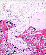
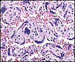
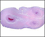
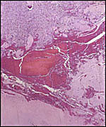
since they all have extremely similar placentas and chromosome morphology, some of the more readily bred species might be considered for effort in embryo transfer of the least common species, if not cloning from the cells placed in storage. This also necessitates better knowledge of endocrine parameters and early implantational stages, perhaps to be ascertained by ultrasound.

Acknowledgement

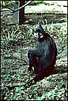
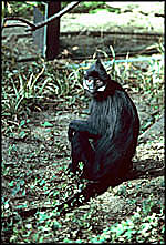
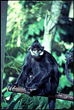
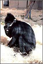
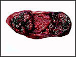
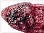
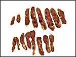
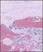
The animal photographs in this chapter come from the Zoological Society of San Diego. I appreciate also very much the help of the pathologists at the San Diego Zoo.

References

Benirschke, K. and Kaufmann, P.: The Pathology of the Human Placenta, 4th ed. Springer-Verlag, NY, 2000.

Benirschke, K. and Miller, C.J.: Anatomical and functional differences in the placenta of primates. Biol. Reprod. 26:2-53, 1982.

Burton, G.J.: Early placentation in the dusky leaf monkey (Presbytis obscura). Placenta 1:187-195, 1980.

Nadler, T.: Zur Haltung von Delacour-und Tonkinlanguren (Trachypithecus delacouri und Trachypithecus francoisi) im Gebiet ihres natuerlichen Lebensraumes. Zool. Garten 64:379-398, 1994.

Nadler, T.: Verbreitung und Status von Delacour-, Tonkin- und Goldschopflanguren (Trachypithecus delacouri, Trachypithecus francoisi und Trachypithecus poliocephalus) in Vietnam. Zool. Garten 66:1-12, 1996.

Nai, W.H., Liu, RQ., Chen,Y.Z. and Wang, J.H.: Chromosome homologies between human and Francois' monkey (Semnopithecus francoisi) established by chromosome painting. Y Chuan Bao 26:474-479, 1999. (In Chinese).

Nowak, R.M.: Walker's Mammals of the World. 6th ed. The Johns Hopkins Press, Baltimore, 1999.

Robinson, R.: Breeding and management of Francois's langurs at Belfast zoo. Int. Zoo News 51:200-208, 2004.

Selenka, E.: Zur vergleichenden Keimesgeschichte der Primaten. Studien über Entwicklungsgeschichte der Tiere. Heft 10. Menschenaffen, L.5, pp. 329-372, F. Keibel, Wiesbaden, 1903.

Streicher, U.: Health problems in confiscated leaf eating monkeys. Verh. Ber. Erkg. Zootiere 40:207-211, 2001 (Meeting in Rotterdam).


back to top
© 2004. All rights reserved.
welcome | home | intro | placentation | glossary | author contact me: kbenirsc@ucsd.edu